A three branches aortic arch variant with a bi-carotid trunk and a retro-esophageal right subclavian artery
Nurru L. Mligiliche*, Nithila D. Isaac
Medical Education – Anatomy, Weill Cornell Medical College in Qatar, Doha, Qatar.
- *Corresponding Author:
- Nurru L. MLIGILICHE, MD, PhD
Medical Education – Anatomy, Weill Cornell Medical College in Qatar, P.O. Box 24144, Doha, Qatar.
Tel: +974 492-8316
Fax: +974 492-8333
E-mail: nul2001@qatar-med.cornell.edu
Date of Received: September 29th, 2008
Date of Accepted: January 12th, 2009
Published Online: January 18th, 2009
© IJAV. 2009; 2: 11–14.
[ft_below_content] =>Keywords
anatomical variations, anomalous, aortic arch, bi-carotid trunk
Introduction
Anatomical variations of the aortic arch and great vessels are well documented as seen from autopsies, anatomical and clinical studies. Most of the anomalies of the aortic arch and its branches arise as a result of altered development of primitive aortic arches of the embryo during the early gestation period [1]. Similarly, variations in the origin of great vessels of the aortic arch and the pulmonary trunk may occur as a consequence of irregular and imperfect development of aorticopulmonary septum of the truncus arteriosus [2].
The most common pattern in the origin of the great vessels of the aortic arch as described in standard anatomical texts, is where the brachiocephalic artery is the first and the largest vessel arising from the aortic arch, followed by the left common carotid artery and then the left subclavian artery. This pattern occurs in 65-80% of cases [3,4]. Adachi reported that, 11% of cases have a common trunk for the left common carotid artery and the brachiocephalic artery, thus only two branches arising from the arch [3]. The third most common pattern constitutes, the left vertebral artery arising as a branch of the aortic arch, proximal to the left subclavian artery. Numerous other variations of the branching pattern of the aortic arch are found in less than 1% of cases [3,4].
We report observations of a rare anatomical variation in the branching pattern of the left-sided aortic arch in a female cadaver encountered during routine anatomy dissection.
Case Report
The anomalous origin of the retro-esophageal right subclavian artery referred here, was found in a 100-years old Caucasian female body donor. The donor’s medical records were not available for review and the cause of her death was stated as “respiratory arrest”.
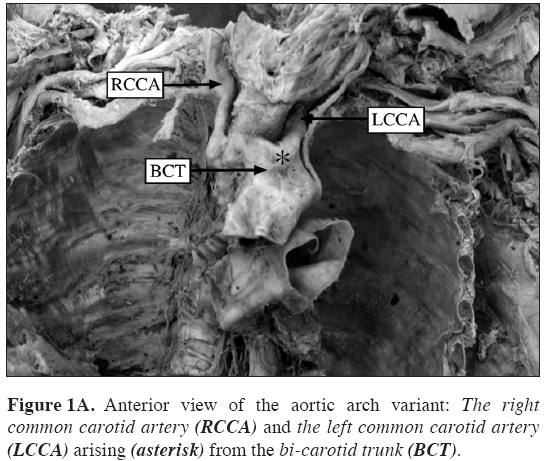
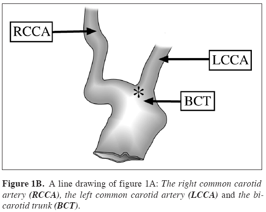
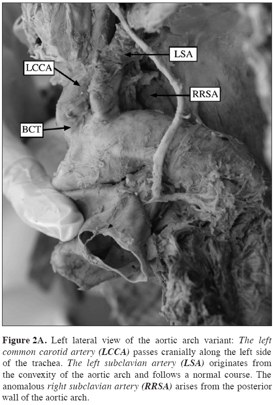
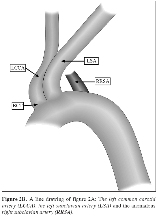
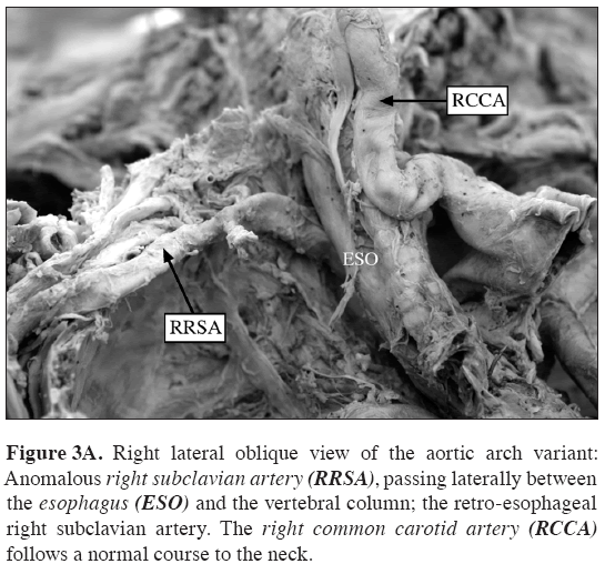
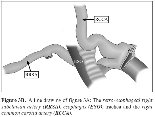
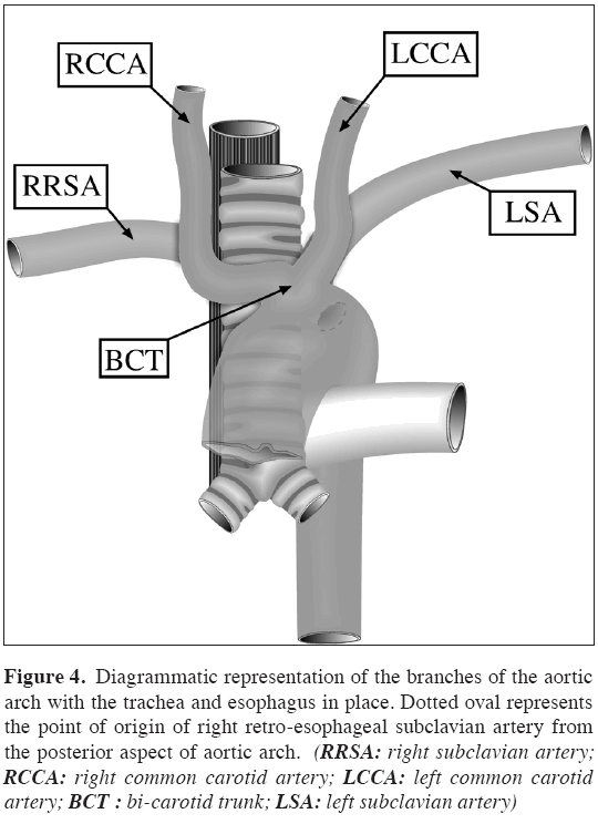
During routine dissection of the heart and the great vessels, she was noted to have hardened, atherosclerotic vascular walls and an unusual pattern of the branches originating from aortic arch. Thorough inspection, revealed a left-sided aortic arch with three branches, the first branch from the convexity of the arch being a wide vessel giving origin to the right and left common carotid arteries, i.e., a bi-carotid trunk (Figures 1A and 1B). From the point of origin (*), the right common carotid artery passed laterally for a short distance crossing the anterior surface of the trachea, and then making a sharp turn to ascend along its right side to the neck. The left common carotid artery passed cranially along the left side of the trachea in the normal fashion (Figures 2A and 2B). The second branch of the aortic arch was the left subclavian artery, which followed a normal course to reach the upper limb (Figures 2A and 2B). The anomalous right subclavian artery was the last branch of the aortic arch, taking origin more posteriorly (Figures 2A, 2B, 3A and 3B), about 3 cm from the origin of the left subclavian artery. It reached the right upper limb by crossing obliquely behind the esophagus as it reached the thoracic inlet (Figures 3A, 3B and 4). It was noted that, at the crossing point the posterior surface of the esophagus was slightly grooved by the artery.
Figure 2a: Left lateral view of the aortic arch variant: The left common carotid artery (LCCA) passes cranially along the left side of the trachea. The left subclavian artery (LSA) originates from the convexity of the aortic arch and follows a normal course. The anomalous right subclavian artery (RRSA) arises from the posterior wall of the aortic arch.
Figure 3a: Right lateral oblique view of the aortic arch variant: Anomalous right subclavian artery (RRSA), passing laterally between the esophagus (ESO) and the vertebral column; the retro-esophageal right subclavian artery. The right common carotid artery (RCCA) follows a normal course to the neck.
Figure 4: Diagrammatic representation of the branches of the aortic arch with the trachea and esophagus in place. Dotted oval represents the point of origin of right retro-esophageal subclavian artery from the posterior aspect of aortic arch. (RRSA: right subclavian artery; RCCA: right common carotid artery; LCCA: left common carotid artery; BCT : bi-carotid trunk; LSA: left subclavian artery)
Despite the anomalies mentioned above, it was also noted that each vessel originating from the aortic arch, had a normal caliber and a usual area of distribution. The left vertebral artery arose from the left subclavian artery about 4 cm distal to its origin, and the right vertebral artery commenced from the right subclavian artery about 7 cm distal to its origin. No other congenital vascular anomalies were found, and there were no noticeable divergence in the heart’s internal and external morphology.
Discussion
Whenever anatomical variations are encountered in dissection, autopsy or medical examination, an attempt is usually made to try to elucidate the underlying developmental factors. Our current observations of a three branches aortic arch variant with a bi-carotid trunk and a retro-esophageal right subclavian artery may have resulted from an altered development of primitive aortic arches. Poultsides et al. described the embryological basis of anomalies of the common carotid arteries [5].
He summarized that, during early gestation period, the third pair of aortic arches give rise to the left and right common carotid arteries, both vessels originating from embryonic ventral aorta as common vascular trunk, as a result, persistence of this stage of development gives rise to a vascular pattern called the common origin of carotid arteries [5].
Anatomical variations of the origin of the left common carotid artery are more common than of the right common carotid artery, because, the left common carotid artery arises often from the brachiocephalic trunk or from a common stem with the right common carotid. Those cases where the right subclavian artery is a separate branch of the aorta, the right and left common carotid arteries most frequently arise by a common trunk [4].
The origin of the retroesophageal right subclavian artery as the last branch of the aortic arch is often seen as a congenital aortic arch anomaly. The reported frequency of this anatomical variation is about 0.4-2% [4]. The possible embryological basis of anomalous origin of the right subclavian artery is the early involution of the right fourth aortic arch and the cranial part of the right dorsal aorta. Consequently, the right subclavian artery develops from the right seventh dorsal intersegmental artery and from the distal segment of the right dorsal aorta [6]. As development continues and the arch of aorta forms, a differential growth shifts the origin of the right subclavian artery closer to the left subclavian artery. Bergman et al., state that, the right subclavian artery may arise directly from the arch of the aorta, as the first, second, third, fourth or fifth branch. In 1899, Holzapfel studied 133 autopsy cases of anomalous right subclavian artery arising as the last branch of the aortic arch. He reported that, in 80% of cases the right subclavian artery passed behind the esophagus to reach the groove on the right first rib, in 15% of cases between the trachea and oesophagus, and in front of trachea in 5% of cases [4].
The branching pattern of the aortic arch described in this case report, is generally considered as asymptomatic [4]. However, surgical reports from clinical cases showed that, anomalous arteries are more prone to atherosclerotic degeneration. In elderly patients an anomalous right subclavian artery occasionally becomes tortuous and it can compress the trachea or the oesophagus causing dysphagia lusoria [7-9]. Moreover, an anomalous right subclavian artery may be associated with a diverticulum of the aorta or, it may give rise to aneurysms [10,11]. In the current observations, the posterior wall of the esophagus was found slightly compressed at the region where the anomalous right subclavian artery crossed it.
Observations on a similar type of anatomical variation of the branching pattern of the aortic arch have rarely been reported in literature. Awareness of vascular variations, especially in this region, is imperative in diagnostic procedures and in planning surgical interventions during clinical practice.
Acknowledgements
We would like to thank Mr. Charles Msuya and Mr. Marion Martin for their excellent technical support.
References
- Stewart JR, Kincaid OW, Edwards JE. An atlas of vascular rings and related malformations of the aortic arch system. Springfield – Illinois, Charles C. Thomas. 1964; 3–13, 52–72.
- Berguer R, Kieffer E. Surgery of the arteries to the head. New York, Springer-Verlag. 1992; 6.
- Adachi B. Das Arteriensystems der Japaner. Bd 1. Tokyo, Kenkyu-sha publishing Co. 1928; 22–43.
- Bergman RA, Afifi AK, Miyauchi R. Illustrated encyclopaedia of human anatomic variation. http://www.anatomyatlases.org/AnatomicVariants/Cardiovascular/Text/Arteries/Aorta.shtml (accessed June 2008).
- Poultsides GA, Lolis ED, Vasquez J, Drezner AD, Venieratos D. Common origins of carotid and subclavian arterial systems: report of a rare aortic arch variant. Ann. Vasc. Surg. 2004; 18: 597–600.
- Moore K, Persaud TVN. The developing human: Clinically oriented embryology. 7th Ed., Philadelphia, Elsevier Science. 2003; 364–366.
- Stone WM, Brewster DC, Moncure AC, Franklin DP, Cambria RP, Abbott WM. Aberrant right subclavian artery: varied presentations and management options. J. Vasc. Surg. 1990; 11: 812–817.
- Kieffer E, Bahnini A, Koskas F. Aberrant subclavian artery: surgical treatment in thirty-three adult patients. J. Vasc. Surg. 1994; 19: 100-109.
- Azakie A, McElhinney DB, Dowd CF, Stoney RJ. Percutaneous stenting for symptomatic stenosis of aberrant right subclavian artery. J. Vasc. Surg. 1998; 27: 756–758.
- Brown DL, Chapman WC, Edwards WH, Coltharp WH, Stoney WS. Dysphagia lusoria: aberrant right subclavian artery with a Kommerell’s diverticulum. Am. Surg. 1993; 59: 582–586.
- Bahnson HT, Blalock A. Aortic vascular rings encountered in the surgical treatment of congenital pulmonic stenosis. Ann. Surg.1950; 131: 356–362.
Nurru L. Mligiliche*, Nithila D. Isaac
Medical Education – Anatomy, Weill Cornell Medical College in Qatar, Doha, Qatar.
- *Corresponding Author:
- Nurru L. MLIGILICHE, MD, PhD
Medical Education – Anatomy, Weill Cornell Medical College in Qatar, P.O. Box 24144, Doha, Qatar.
Tel: +974 492-8316
Fax: +974 492-8333
E-mail: nul2001@qatar-med.cornell.edu
Date of Received: September 29th, 2008
Date of Accepted: January 12th, 2009
Published Online: January 18th, 2009
© IJAV. 2009; 2: 11–14.
Abstract
We report observations from a female cadaver presenting a rare variation of the branches of a left-sided aortic arch. Three branches originated from the convexity of the arch in the following order: A bi-carotid trunk giving origin to the right and left common carotid arteries, a left subclavian artery and a right subclavian artery. The anomalous right subclavian artery ran obliquely from the left to the right, between the oesophagus and the vertebral column to reach the right upper limb.
-Keywords
anatomical variations, anomalous, aortic arch, bi-carotid trunk
Introduction
Anatomical variations of the aortic arch and great vessels are well documented as seen from autopsies, anatomical and clinical studies. Most of the anomalies of the aortic arch and its branches arise as a result of altered development of primitive aortic arches of the embryo during the early gestation period [1]. Similarly, variations in the origin of great vessels of the aortic arch and the pulmonary trunk may occur as a consequence of irregular and imperfect development of aorticopulmonary septum of the truncus arteriosus [2].
The most common pattern in the origin of the great vessels of the aortic arch as described in standard anatomical texts, is where the brachiocephalic artery is the first and the largest vessel arising from the aortic arch, followed by the left common carotid artery and then the left subclavian artery. This pattern occurs in 65-80% of cases [3,4]. Adachi reported that, 11% of cases have a common trunk for the left common carotid artery and the brachiocephalic artery, thus only two branches arising from the arch [3]. The third most common pattern constitutes, the left vertebral artery arising as a branch of the aortic arch, proximal to the left subclavian artery. Numerous other variations of the branching pattern of the aortic arch are found in less than 1% of cases [3,4].
We report observations of a rare anatomical variation in the branching pattern of the left-sided aortic arch in a female cadaver encountered during routine anatomy dissection.
Case Report
The anomalous origin of the retro-esophageal right subclavian artery referred here, was found in a 100-years old Caucasian female body donor. The donor’s medical records were not available for review and the cause of her death was stated as “respiratory arrest”.
During routine dissection of the heart and the great vessels, she was noted to have hardened, atherosclerotic vascular walls and an unusual pattern of the branches originating from aortic arch. Thorough inspection, revealed a left-sided aortic arch with three branches, the first branch from the convexity of the arch being a wide vessel giving origin to the right and left common carotid arteries, i.e., a bi-carotid trunk (Figures 1A and 1B). From the point of origin (*), the right common carotid artery passed laterally for a short distance crossing the anterior surface of the trachea, and then making a sharp turn to ascend along its right side to the neck. The left common carotid artery passed cranially along the left side of the trachea in the normal fashion (Figures 2A and 2B). The second branch of the aortic arch was the left subclavian artery, which followed a normal course to reach the upper limb (Figures 2A and 2B). The anomalous right subclavian artery was the last branch of the aortic arch, taking origin more posteriorly (Figures 2A, 2B, 3A and 3B), about 3 cm from the origin of the left subclavian artery. It reached the right upper limb by crossing obliquely behind the esophagus as it reached the thoracic inlet (Figures 3A, 3B and 4). It was noted that, at the crossing point the posterior surface of the esophagus was slightly grooved by the artery.
Figure 2a: Left lateral view of the aortic arch variant: The left common carotid artery (LCCA) passes cranially along the left side of the trachea. The left subclavian artery (LSA) originates from the convexity of the aortic arch and follows a normal course. The anomalous right subclavian artery (RRSA) arises from the posterior wall of the aortic arch.
Figure 3a: Right lateral oblique view of the aortic arch variant: Anomalous right subclavian artery (RRSA), passing laterally between the esophagus (ESO) and the vertebral column; the retro-esophageal right subclavian artery. The right common carotid artery (RCCA) follows a normal course to the neck.
Figure 4: Diagrammatic representation of the branches of the aortic arch with the trachea and esophagus in place. Dotted oval represents the point of origin of right retro-esophageal subclavian artery from the posterior aspect of aortic arch. (RRSA: right subclavian artery; RCCA: right common carotid artery; LCCA: left common carotid artery; BCT : bi-carotid trunk; LSA: left subclavian artery)
Despite the anomalies mentioned above, it was also noted that each vessel originating from the aortic arch, had a normal caliber and a usual area of distribution. The left vertebral artery arose from the left subclavian artery about 4 cm distal to its origin, and the right vertebral artery commenced from the right subclavian artery about 7 cm distal to its origin. No other congenital vascular anomalies were found, and there were no noticeable divergence in the heart’s internal and external morphology.
Discussion
Whenever anatomical variations are encountered in dissection, autopsy or medical examination, an attempt is usually made to try to elucidate the underlying developmental factors. Our current observations of a three branches aortic arch variant with a bi-carotid trunk and a retro-esophageal right subclavian artery may have resulted from an altered development of primitive aortic arches. Poultsides et al. described the embryological basis of anomalies of the common carotid arteries [5].
He summarized that, during early gestation period, the third pair of aortic arches give rise to the left and right common carotid arteries, both vessels originating from embryonic ventral aorta as common vascular trunk, as a result, persistence of this stage of development gives rise to a vascular pattern called the common origin of carotid arteries [5].
Anatomical variations of the origin of the left common carotid artery are more common than of the right common carotid artery, because, the left common carotid artery arises often from the brachiocephalic trunk or from a common stem with the right common carotid. Those cases where the right subclavian artery is a separate branch of the aorta, the right and left common carotid arteries most frequently arise by a common trunk [4].
The origin of the retroesophageal right subclavian artery as the last branch of the aortic arch is often seen as a congenital aortic arch anomaly. The reported frequency of this anatomical variation is about 0.4-2% [4]. The possible embryological basis of anomalous origin of the right subclavian artery is the early involution of the right fourth aortic arch and the cranial part of the right dorsal aorta. Consequently, the right subclavian artery develops from the right seventh dorsal intersegmental artery and from the distal segment of the right dorsal aorta [6]. As development continues and the arch of aorta forms, a differential growth shifts the origin of the right subclavian artery closer to the left subclavian artery. Bergman et al., state that, the right subclavian artery may arise directly from the arch of the aorta, as the first, second, third, fourth or fifth branch. In 1899, Holzapfel studied 133 autopsy cases of anomalous right subclavian artery arising as the last branch of the aortic arch. He reported that, in 80% of cases the right subclavian artery passed behind the esophagus to reach the groove on the right first rib, in 15% of cases between the trachea and oesophagus, and in front of trachea in 5% of cases [4].
The branching pattern of the aortic arch described in this case report, is generally considered as asymptomatic [4]. However, surgical reports from clinical cases showed that, anomalous arteries are more prone to atherosclerotic degeneration. In elderly patients an anomalous right subclavian artery occasionally becomes tortuous and it can compress the trachea or the oesophagus causing dysphagia lusoria [7-9]. Moreover, an anomalous right subclavian artery may be associated with a diverticulum of the aorta or, it may give rise to aneurysms [10,11]. In the current observations, the posterior wall of the esophagus was found slightly compressed at the region where the anomalous right subclavian artery crossed it.
Observations on a similar type of anatomical variation of the branching pattern of the aortic arch have rarely been reported in literature. Awareness of vascular variations, especially in this region, is imperative in diagnostic procedures and in planning surgical interventions during clinical practice.
Acknowledgements
We would like to thank Mr. Charles Msuya and Mr. Marion Martin for their excellent technical support.
References
- Stewart JR, Kincaid OW, Edwards JE. An atlas of vascular rings and related malformations of the aortic arch system. Springfield – Illinois, Charles C. Thomas. 1964; 3–13, 52–72.
- Berguer R, Kieffer E. Surgery of the arteries to the head. New York, Springer-Verlag. 1992; 6.
- Adachi B. Das Arteriensystems der Japaner. Bd 1. Tokyo, Kenkyu-sha publishing Co. 1928; 22–43.
- Bergman RA, Afifi AK, Miyauchi R. Illustrated encyclopaedia of human anatomic variation. http://www.anatomyatlases.org/AnatomicVariants/Cardiovascular/Text/Arteries/Aorta.shtml (accessed June 2008).
- Poultsides GA, Lolis ED, Vasquez J, Drezner AD, Venieratos D. Common origins of carotid and subclavian arterial systems: report of a rare aortic arch variant. Ann. Vasc. Surg. 2004; 18: 597–600.
- Moore K, Persaud TVN. The developing human: Clinically oriented embryology. 7th Ed., Philadelphia, Elsevier Science. 2003; 364–366.
- Stone WM, Brewster DC, Moncure AC, Franklin DP, Cambria RP, Abbott WM. Aberrant right subclavian artery: varied presentations and management options. J. Vasc. Surg. 1990; 11: 812–817.
- Kieffer E, Bahnini A, Koskas F. Aberrant subclavian artery: surgical treatment in thirty-three adult patients. J. Vasc. Surg. 1994; 19: 100-109.
- Azakie A, McElhinney DB, Dowd CF, Stoney RJ. Percutaneous stenting for symptomatic stenosis of aberrant right subclavian artery. J. Vasc. Surg. 1998; 27: 756–758.
- Brown DL, Chapman WC, Edwards WH, Coltharp WH, Stoney WS. Dysphagia lusoria: aberrant right subclavian artery with a Kommerell’s diverticulum. Am. Surg. 1993; 59: 582–586.
- Bahnson HT, Blalock A. Aortic vascular rings encountered in the surgical treatment of congenital pulmonic stenosis. Ann. Surg.1950; 131: 356–362.


