A unique variation of superficial palmar arch
Jiji PJ1*, Sujatha D'Costa1, Soubhagya R. Nayak1, Latha V. Prabhu1, C. Ganesh Kumar1 and Prakash2
1Department of Anatomy, Centre for Basic Sciences,Kasturba Medical College, Manipal University, Karnataka, India
2Department of Anatomy, Vyedehi Institute of Medical Sciences and Research Centre, Karnataka, India
- *Corresponding Author:
- Jiji PJ, MSc
Sr. Grade Lecturer, Department of Anatomy, Centre for Basic Sciences, Kasturba Medical College, Manipal University, Mangalore, Karnataka, 575004, India
Tel: +91 824 2211746
E-mail: pj.jiji@gmail.com
Date of Received: March 20th, 2009
Date of Accepted: July 10th, 2009
Published Online: September 7th, 2009
© IJAV. 2009; 2: 105–107.
[ft_below_content] =>Keywords
superficial palmar arch, arteria radialis indicis, first dorsal metacarpal artery, radial artery, arterial pattern
Introduction
The general pattern of arterial supply of the hand consists of two systems for the volar aspect and a single system for the dorsal aspect. The volar supply is arranged into a superficial and a deep group i.e., superficial palmar arch (SPA) and deep palmar arch. The superficial palmar arch is mainly fed by the ulnar artery, passing superficial to the flexor retinaculum, then curving laterally to form an arch, lying just deep to the palmar aponeurosis. About one third of the SPAs is formed by the ulnar artery alone; a further third is completed by the superficial palmar branch of the radial artery; and a third by the arteria radialis indicis, often a branch of the arteria princeps pollicis, or by the median artery [1].
Coleman and Anson in a study of SPA in 650 specimens, while describing the complete arch group (80%), mentioned an unreported type-f (2%) in which the SPA is formed by an ulnar artery joined by a large vessel from the deep palmar arch at the base of the thenar eminence. The present case belongs to this ulnar-deep palmar arch type of SPA [2].
Case Report
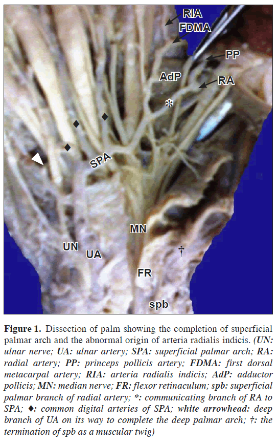
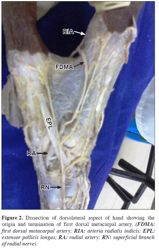
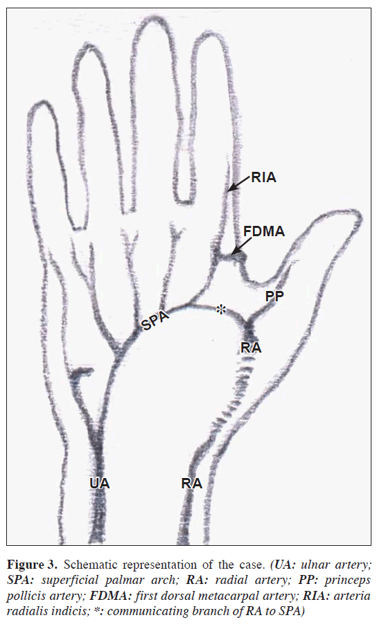
During regular dissections for undergraduate medical students, we encountered a unique variation in the superficial palmar arch as it was completed by one of the large terminal branches of radial artery. The superficial palmar branch of radial artery was very thin and ended as muscular artery supplying the abductor pollicis brevis. The origin of the arteria radialis indicis was also peculiar that it was arising from midway between the communication of the radial artery with SPA and further reinforced by the termination of the first dorsal metacarpal artery (FDMA) as it reached the volar aspect 1 cm distal to the transverse head of adductor pollicis just proximal to the metacarpophalangeal joint (Figure 1). The course of radial artery was normal on the dorsum as it passed deep through the anatomical snuffbox. Just before leaving the dorsum by penetrating between the two heads of the first interosseus, it gave FDMA that after leading a fascial course lateral to the 3rd dorsal digital nerve of index finger joined arteria radialis indicis by winding round the first dorsal interosseus muscle (Figure 2). Proceeding further deep to the oblique head of adductor pollicis the radial artery gave the deep palmar arch and later emerged between the oblique and transverse heads of adductor pollicis, then bifurcated into princeps pollicis artery and a communicating branch to SPA. The deep palmar arch was completed by the deep branch of ulnar artery (Figure 1). Schematic representation of the present case is given (Figure 3).
Figure 1: Dissection of palm showing the completion of superficial palmar arch and the abnormal origin of arteria radialis indicis. (UN: ulnar nerve; UA: ulnar artery; SPA: superficial palmar arch; RA: radial artery; PP: princeps pollicis artery; FDMA: first dorsal metacarpal artery; RIA: arteria radialis indicis; AdP: adductor pollicis; MN: median nerve; FR: flexor retinaculum; spb: superficial palmar branch of radial artery; *: communicating branch of RA to SPA; ♦: common digital arteries of SPA; white arrowhead: deep branch of UA on its way to complete the deep palmar arch; †: the termination of spb as a muscular twig)
Discussion
The superficial palmar arch is a dominant vascular structure of the palm but variations in its formation are numerous and well studied. The deep palmar arch is less variable than the superficial and was complete in most of specimens. The occurrence of ulnar-deep palmar arch type of SPA was found to be 2% by Coleman and Anson, and 2.2% by Gellman et al. But Ikeda et al. reported this as 14.2% in their studies [2,3,4]. In contrast to these studies, a dorsally arising small radial artery branch to the SPA, coined as dorsalis pollicis artery by Agur and Lee [5], was found in 33% of the left hands and in 20% of the right hands in a study by Fazan et al. [6]. This most common variation in their study was found arising on the dorsal surface of the first dorsal interosseous muscle, at the same level of origin of the princeps pollicis artery, which passed into the palm to reach the ulnar artery. However, its arterial diameter was significantly smaller than that of the radial artery. McCormack et al., also reported a small vessel arising dorsally from the radial artery passing into the palm to join the ulnar artery in 51% of the hands studied [7].
The first dorsal metacarpal artery arises from the radial artery just before it passes between the heads of the first interosseus muscle to enter the palm. Since FDMA often has a fascial course on the dorsal surface of the index head of first interosseus muscle, this artery can be easily injured in an intervention over the carpometacarpal joint of the thumb, when approached from the dorsum of this joint [8]. The radial aspect of the thenar eminence is a good donor source for innervated and vascularized free or island flap transfer for reconstruction of various skin defects of the volar side of the fingers. Reverse dorsal digital and metacarpal flaps use the dorsal skin of the digital or metacarpal areas, and they are based on the arterial branches anastomosing the volar and dorsal arterial networks of the fingers [9]. The dorsal skin flap raised on FDMA is particularly useful for reconstruction of the thumb following injuries [1,10].
Since SPA is the center of attraction for most of the procedures and traumatic events in the hand, the hand surgeon needs to refer to the existence and healthy function of the arch before surgical procedures such as, arterial repairs, vascular graft applications, and free and/or pedicled flaps depending on radial or ulnar artery, in order to maintain or not to harm the perfusion of the hand and digits [11–14]. Even while making incisions to evacuate pus from the hand, special attention should be paid to the superficial position of termination of ulnar artery and SPA [15].
References
- Johnson D, Ellis H, Collins P. Wrist and hand. In: Standring S, Ellis H, Healy JC, Johnson D, Williams A, eds. Gray’s Anatomy. 39th Ed., Edinburgh, Churchill Livingstone. 2005; 929.
- Coleman SS, Anson BJ. Arterial patterns in the hand based upon the study of 650 specimens. Surg Gynecol Obstet. 1961; 113: 409–424.
- Gellman H, Botte MJ, Shankwiler J, Gelberman RH. Arterial patterns of the deep and superficial palmar arches. Clin Orthop Relat Res. 2001; 383: 41–46.
- Ikeda A, Ugawa A, Kazihara Y, Hamada N. Arterial patterns in the hand based on a three-dimensional analysis of 220 cadaver hands. J Hand Surg Am. 1988; 13: 501–509.
- Agur AMR, Lee MJ. Grant’s Atlas of Anatomy. 9th Ed., Baltimore, Williams & Wilkins. 1991; 434.
- Fazan VP, Borges CT, Da Silva JH, Caetano AG, Filho OA. Superficial palmar arch: an arterial diameter study. J Anat. 2004; 204: 307–311.
- McCormack LJ, Cauldwell EW, Anson BJ. Brachial and antebrachial arterial patterns: a study of 750 extremities. Surg Gynecol Obstet. 1953; 96: 43–54.
- Wilgis EFS, Kaplan EB. The blood and the nerve supply of the hand. In: Morton Spinner, ed. Kaplan’s Functional and Surgical Anatomy of the Hand. 3rd Ed., Philadelphia, J.B. Lippincott Company. 1984; 206.
- Pelissier P, Casoli V, Bakhach J, Martin D, Baudet J. Reverse dorsal digital and metacarpal flaps: a review of 27 cases. Plast Reconstr Surg. 1999; 103: 159–165.
- Sherif MM. First dorsal metacarpal artery flap in hand reconstruction. Anatomical study. J Hand Surg Am. 1994; 19: 26–31.
- Parks BJ, Arbelaez J, Horner RL. Medical and surgical importance of the arterial blood supply of the thumb. J Hand Surg Am. 1978; 3: 383–385.
- Pierer G, Steffen J, Hoflehner H. The vascular blood supply of the second metacarpal bone: anatomic basis for a new vascularized bone graft in hand surgery. An anatomical study in cadavers. Surg Radiol Anat. 1992; 14: 103–112.
- Richards RS, Dowdy P, Roth JH. Ulnar artery palmar to palmar brevis: cadaveric study and three case reports. J Hand Surg Am. 1993; 18: 888–892.
- Khan K, Riaz M, Small JO. The use of the second dorsal metacarpal artery for vascularized bone graft. An anatomical study. J Hand Surg Br. 1998; 23: 308–310.
- Lockhardt RD, Hamilton GF, Fyfe FW. Anatomy of the human body. In: Vascular system Systemic arteries. London, Feber & Feber Ltd. 1959; 612–619.
Jiji PJ1*, Sujatha D'Costa1, Soubhagya R. Nayak1, Latha V. Prabhu1, C. Ganesh Kumar1 and Prakash2
1Department of Anatomy, Centre for Basic Sciences,Kasturba Medical College, Manipal University, Karnataka, India
2Department of Anatomy, Vyedehi Institute of Medical Sciences and Research Centre, Karnataka, India
- *Corresponding Author:
- Jiji PJ, MSc
Sr. Grade Lecturer, Department of Anatomy, Centre for Basic Sciences, Kasturba Medical College, Manipal University, Mangalore, Karnataka, 575004, India
Tel: +91 824 2211746
E-mail: pj.jiji@gmail.com
Date of Received: March 20th, 2009
Date of Accepted: July 10th, 2009
Published Online: September 7th, 2009
© IJAV. 2009; 2: 105–107.
Abstract
We present a unique variation in the arterial pattern of superficial palmar arch in which it was completed by one of the large terminal branches of radial artery. The origin of the arteria radialis indicis was also peculiar that it was arising from the communicating branch of the radial artery and further reinforced by the first dorsal metacarpal artery that joined it after reaching the volar aspect. Pertinent anatomical knowledge regarding the variations of the palmar arch is significant for the purposes of microvascular repairs and re-implantations.
-Keywords
superficial palmar arch, arteria radialis indicis, first dorsal metacarpal artery, radial artery, arterial pattern
Introduction
The general pattern of arterial supply of the hand consists of two systems for the volar aspect and a single system for the dorsal aspect. The volar supply is arranged into a superficial and a deep group i.e., superficial palmar arch (SPA) and deep palmar arch. The superficial palmar arch is mainly fed by the ulnar artery, passing superficial to the flexor retinaculum, then curving laterally to form an arch, lying just deep to the palmar aponeurosis. About one third of the SPAs is formed by the ulnar artery alone; a further third is completed by the superficial palmar branch of the radial artery; and a third by the arteria radialis indicis, often a branch of the arteria princeps pollicis, or by the median artery [1].
Coleman and Anson in a study of SPA in 650 specimens, while describing the complete arch group (80%), mentioned an unreported type-f (2%) in which the SPA is formed by an ulnar artery joined by a large vessel from the deep palmar arch at the base of the thenar eminence. The present case belongs to this ulnar-deep palmar arch type of SPA [2].
Case Report
During regular dissections for undergraduate medical students, we encountered a unique variation in the superficial palmar arch as it was completed by one of the large terminal branches of radial artery. The superficial palmar branch of radial artery was very thin and ended as muscular artery supplying the abductor pollicis brevis. The origin of the arteria radialis indicis was also peculiar that it was arising from midway between the communication of the radial artery with SPA and further reinforced by the termination of the first dorsal metacarpal artery (FDMA) as it reached the volar aspect 1 cm distal to the transverse head of adductor pollicis just proximal to the metacarpophalangeal joint (Figure 1). The course of radial artery was normal on the dorsum as it passed deep through the anatomical snuffbox. Just before leaving the dorsum by penetrating between the two heads of the first interosseus, it gave FDMA that after leading a fascial course lateral to the 3rd dorsal digital nerve of index finger joined arteria radialis indicis by winding round the first dorsal interosseus muscle (Figure 2). Proceeding further deep to the oblique head of adductor pollicis the radial artery gave the deep palmar arch and later emerged between the oblique and transverse heads of adductor pollicis, then bifurcated into princeps pollicis artery and a communicating branch to SPA. The deep palmar arch was completed by the deep branch of ulnar artery (Figure 1). Schematic representation of the present case is given (Figure 3).
Figure 1: Dissection of palm showing the completion of superficial palmar arch and the abnormal origin of arteria radialis indicis. (UN: ulnar nerve; UA: ulnar artery; SPA: superficial palmar arch; RA: radial artery; PP: princeps pollicis artery; FDMA: first dorsal metacarpal artery; RIA: arteria radialis indicis; AdP: adductor pollicis; MN: median nerve; FR: flexor retinaculum; spb: superficial palmar branch of radial artery; *: communicating branch of RA to SPA; ♦: common digital arteries of SPA; white arrowhead: deep branch of UA on its way to complete the deep palmar arch; †: the termination of spb as a muscular twig)
Discussion
The superficial palmar arch is a dominant vascular structure of the palm but variations in its formation are numerous and well studied. The deep palmar arch is less variable than the superficial and was complete in most of specimens. The occurrence of ulnar-deep palmar arch type of SPA was found to be 2% by Coleman and Anson, and 2.2% by Gellman et al. But Ikeda et al. reported this as 14.2% in their studies [2,3,4]. In contrast to these studies, a dorsally arising small radial artery branch to the SPA, coined as dorsalis pollicis artery by Agur and Lee [5], was found in 33% of the left hands and in 20% of the right hands in a study by Fazan et al. [6]. This most common variation in their study was found arising on the dorsal surface of the first dorsal interosseous muscle, at the same level of origin of the princeps pollicis artery, which passed into the palm to reach the ulnar artery. However, its arterial diameter was significantly smaller than that of the radial artery. McCormack et al., also reported a small vessel arising dorsally from the radial artery passing into the palm to join the ulnar artery in 51% of the hands studied [7].
The first dorsal metacarpal artery arises from the radial artery just before it passes between the heads of the first interosseus muscle to enter the palm. Since FDMA often has a fascial course on the dorsal surface of the index head of first interosseus muscle, this artery can be easily injured in an intervention over the carpometacarpal joint of the thumb, when approached from the dorsum of this joint [8]. The radial aspect of the thenar eminence is a good donor source for innervated and vascularized free or island flap transfer for reconstruction of various skin defects of the volar side of the fingers. Reverse dorsal digital and metacarpal flaps use the dorsal skin of the digital or metacarpal areas, and they are based on the arterial branches anastomosing the volar and dorsal arterial networks of the fingers [9]. The dorsal skin flap raised on FDMA is particularly useful for reconstruction of the thumb following injuries [1,10].
Since SPA is the center of attraction for most of the procedures and traumatic events in the hand, the hand surgeon needs to refer to the existence and healthy function of the arch before surgical procedures such as, arterial repairs, vascular graft applications, and free and/or pedicled flaps depending on radial or ulnar artery, in order to maintain or not to harm the perfusion of the hand and digits [11–14]. Even while making incisions to evacuate pus from the hand, special attention should be paid to the superficial position of termination of ulnar artery and SPA [15].
References
- Johnson D, Ellis H, Collins P. Wrist and hand. In: Standring S, Ellis H, Healy JC, Johnson D, Williams A, eds. Gray’s Anatomy. 39th Ed., Edinburgh, Churchill Livingstone. 2005; 929.
- Coleman SS, Anson BJ. Arterial patterns in the hand based upon the study of 650 specimens. Surg Gynecol Obstet. 1961; 113: 409–424.
- Gellman H, Botte MJ, Shankwiler J, Gelberman RH. Arterial patterns of the deep and superficial palmar arches. Clin Orthop Relat Res. 2001; 383: 41–46.
- Ikeda A, Ugawa A, Kazihara Y, Hamada N. Arterial patterns in the hand based on a three-dimensional analysis of 220 cadaver hands. J Hand Surg Am. 1988; 13: 501–509.
- Agur AMR, Lee MJ. Grant’s Atlas of Anatomy. 9th Ed., Baltimore, Williams & Wilkins. 1991; 434.
- Fazan VP, Borges CT, Da Silva JH, Caetano AG, Filho OA. Superficial palmar arch: an arterial diameter study. J Anat. 2004; 204: 307–311.
- McCormack LJ, Cauldwell EW, Anson BJ. Brachial and antebrachial arterial patterns: a study of 750 extremities. Surg Gynecol Obstet. 1953; 96: 43–54.
- Wilgis EFS, Kaplan EB. The blood and the nerve supply of the hand. In: Morton Spinner, ed. Kaplan’s Functional and Surgical Anatomy of the Hand. 3rd Ed., Philadelphia, J.B. Lippincott Company. 1984; 206.
- Pelissier P, Casoli V, Bakhach J, Martin D, Baudet J. Reverse dorsal digital and metacarpal flaps: a review of 27 cases. Plast Reconstr Surg. 1999; 103: 159–165.
- Sherif MM. First dorsal metacarpal artery flap in hand reconstruction. Anatomical study. J Hand Surg Am. 1994; 19: 26–31.
- Parks BJ, Arbelaez J, Horner RL. Medical and surgical importance of the arterial blood supply of the thumb. J Hand Surg Am. 1978; 3: 383–385.
- Pierer G, Steffen J, Hoflehner H. The vascular blood supply of the second metacarpal bone: anatomic basis for a new vascularized bone graft in hand surgery. An anatomical study in cadavers. Surg Radiol Anat. 1992; 14: 103–112.
- Richards RS, Dowdy P, Roth JH. Ulnar artery palmar to palmar brevis: cadaveric study and three case reports. J Hand Surg Am. 1993; 18: 888–892.
- Khan K, Riaz M, Small JO. The use of the second dorsal metacarpal artery for vascularized bone graft. An anatomical study. J Hand Surg Br. 1998; 23: 308–310.
- Lockhardt RD, Hamilton GF, Fyfe FW. Anatomy of the human body. In: Vascular system Systemic arteries. London, Feber & Feber Ltd. 1959; 612–619.


