A variant oblique fissure of left lung
Ajay Ratnakarrao Nene*, Krishna Swami Gajendra and Manchiraju Venkata Ramananda Sarma
Department of Anatomy, Ganni Subba Lakshmi Medical College, Andhra Pradesh, India.
- *Corresponding Author:
- Ajay Ratnakarrao Nene, MS
Associate Professor, Department of Anatomy, Ganni Subba Lakshmi Medical College, NH-5, Lakshmipuram, Rajahmundry, Dist - East Godavari, Andhra Pradesh, 533296, India.
Tel: +91 (883) 2404167
E-mail: ajay.nene@gmail.com
Date of Received: May 14th, 2010
Date of Accepted: August 8th, 2010
Published Online: August 19th, 2010
© IJAV. 2010; 3: 125–127.
[ft_below_content] =>Keywords
left lung,oblique fissure,accessory fissure,upper lobe,lower lobe
Introduction
The anatomical knowledge of the fissures and the lobes of the lung is important for accurate interpretation on x-rays and CT scans. The left lung is divided into two lobes by a deep oblique fissure. During life, this fissure begins posteriorly at the level of fifth thoracic vertebra. It passes anteroinferiorly in a spiral course, to meet the inferior margin close to the sixth costochondral junction. It extends into the lung almost to the hilus and separates the inferior and superior lobes. The superior lobe forms the apex and anterior margin of the lung. The inferior lobe makes up the diaphragmatic and greater part of the posterior surface [1]. Here we present a variant oblique fissure of left lung dividing it unevenly.
The knowledge of anatomical variations of lobes of the lung is important for identifying bronchopulmonary segments. Many a times the radiologists may misinterpret an x-ray or a CT scan. Anatomical knowledge of such variations is useful for lobectomies and surgical resections involving individual segments.
Case Report
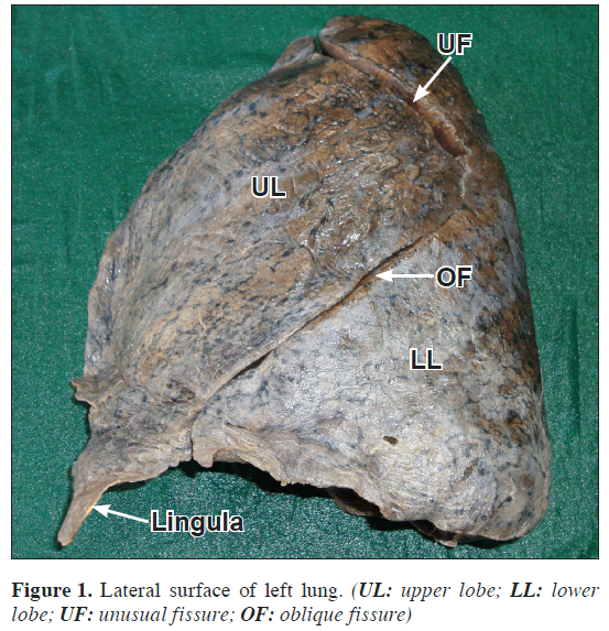
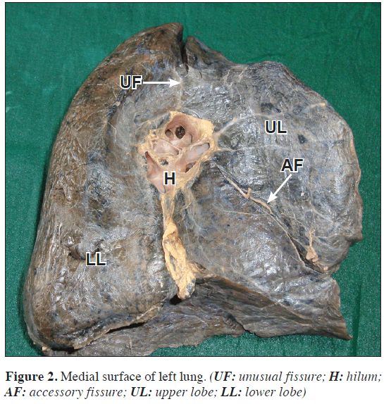
During routine dissection classes of thorax for the undergraduate students, a left lung was found displaying variation in the pattern of oblique fissure. Also an accessory fissure was noted on medial surface. The fissures and lobes were studied and appropriate measurements were taken. The specimen was photographed (Figures 1, 2).
The left lung displayed a variant complete fissure which originated at the hilus, ran vertically upwards and cut the anterior border 1 cm in front of the apex. The fissure then continued backwards, downwards and laterally for about 4.5 cm and then turned sharply to meet the oblique fissure, whose proximal part was conspicuously absent, at 3.5 cm from lung apex. The oblique fissure continued its forward, downward and medialward course to transect the inferior border 3 cm behind the lingula. It then continued its upward, medialward and slightly backward course to reach the inferior aspect of the hilus.
Owing to its course on upper part of medial and lateral surface of the lung, the variant fissure divided the lung unevenly. The apical and posterior segments which classically belonged to upper lobe now were seen to be parts of the lower lobe thus reducing the number of bronchopulmonary segments in the upper lobe to three, while the lower lobe had seven segments. The peculiar course of the fissure divided the left lung into two lobes in such a way that the upper lobe was placed anterior to rather than anterosuperior to the lower lobe. An accessory fissure was also noted in the territory of upper lobe, running from the front of hilus on the medial surface, passing downwards and forwards to meet the anterior border 2 cm above the lingula.
Discussion
During the development, as the lung grows, the spaces or fissures that separate individual bronchopulmonary buds/segments become obliterated except along two planes, evident in the fully developed lungs as oblique or horizontal fissures. Absence or incomplete oblique or horizontal fissure could be due to obliteration of these fissures either completely or partially. Accessory fissure could be the result of non-obliteration of spaces which normally are obliterated [2]. The accessory fissure may be of varying depth occurring between bronchopulmonary segments. The inferior accessory fissure which demarcates the medial basal segment, and superior fissure which demarcates the superior segment, are the most common accessory fissures detected on CT scan [3]. However, no data was found referring to the type of the variant fissure found in the present case and also its continuation with an incomplete oblique fissure.
The presence of fissures in the normal lungs enhances uniform expansion, and their position could be used as reliable landmark in specifying lesions within the thorax, in general and within the lungs in particular [4]. Sometimes especially in infant, accessory fissures of varying depth can be seen in unusual locations of the lung, delimiting abnormal lobes which corresponding to the normal bronchopulmonary segments [5]. From a radiological point of view, an accessory or anomalous fissure is important as it can be mistaken for a lung lesion [6].
Knowledge of an accessory fissure is helpful for clinicians in order to differentiate it from other normal anatomical and pathological structures. Interpretations of various radiographic appearances of interlobar fluid are important for clinicians. In x-ray, incomplete fissure always give an atypical appearance of pleural effusion. Many a times the accessory fissure fail to be detected on CT scans, because of their incompleteness, thick sections and orientation in relation to a particular plane [7].
In many diseases, segmental localization is a must, and the knowledge of accessory or variant fissure is of much clinical importance to the clinician. Preoperative planning and strategy for segmental resection or pulmonary lobectomy may also change during presence of such accessory or variant fissure, as can be appreciated in the present case, where the apical and posterior segments belong to lower rather than the upper lobe – a matter of great clinical and surgical importance.
An incomplete fissure is also a cause for postoperative air leakage [8]. Often these accessory fissures act as a barrier to spread of infection, creating a sharply marginated pneumonia, which can wrongly be interpreted as atelectasis or consolidation [9].
The knowledge of anatomy of fissures of lung may help clarifying confusing radiographic findings like extension of fluid into an incomplete major fissure or spread of various diseases through different pathways [10].
Considering the clinical and surgical importance of such variations, from anatomical point of view, one can opine that prior anatomical knowledge and high index of suspicion for probable variations in the fissures, lobes and bronchopulmonary segments in the lung may be important for clinicians, surgeons and radiologists.
References
- Romanes GJ. Cunningham’s manual of practical anatomy. Vol.2. New York, Oxford Medical Publications. 1996; 32–33.
- Larsen WJ. Human Embryolgy. New York, Churchill Livingstone. 1993; 111–130.
- Tarver RD. How common are incomplete pulmonary fissures, and what is their clinical significance? AJR Am J Roentgenol. 1995; 164: 761.
- Kent EM, Blades B. The surgical anatomy of the pulmonary lobes. J Thorac Surg. 1942; 12: 18–30.
- Rosse C, Gaddum-Rosse P. Hollinshed’s Textbook of Anatomy. Philadelphia; Lippincot-Raven. 1997; 441–461.
- Aldur MM, Denk CC, Celik HH, Tascioglu AB. An accessory fissure in the lower lobe of the right lung. Morphologie. 1997; 81; 5–7.
- Ariyurek OM, Gulsun M, Demirkazik FB. Accessory fissures of the lung: evaluation by high-resolution computed tomography. Eur Radiol. 2001; 11: 2449–2253.
- Walker WS, Craig SR. A proposed anatomical classification of the pulmonary fissures. J R Coll Surg Edinb. 1997; 42: 233–234.
- Godwin JD, Tarver RD. Accessory fissures of the lung. AJR Am J Roentgenol. 1985; 144: 39–47.
- Dandy WE Jr. Incomplete pulmonary interlobar fissure sign. Radiology. 1978; 128: 21–25.
Ajay Ratnakarrao Nene*, Krishna Swami Gajendra and Manchiraju Venkata Ramananda Sarma
Department of Anatomy, Ganni Subba Lakshmi Medical College, Andhra Pradesh, India.
- *Corresponding Author:
- Ajay Ratnakarrao Nene, MS
Associate Professor, Department of Anatomy, Ganni Subba Lakshmi Medical College, NH-5, Lakshmipuram, Rajahmundry, Dist - East Godavari, Andhra Pradesh, 533296, India.
Tel: +91 (883) 2404167
E-mail: ajay.nene@gmail.com
Date of Received: May 14th, 2010
Date of Accepted: August 8th, 2010
Published Online: August 19th, 2010
© IJAV. 2010; 3: 125–127.
Abstract
The left lung classically has one fissure, the oblique fissure and two lobes namely the upper and lower lobes. Variations of lobar pattern and fissures have been described by many authors on imaging techniques whereas there are fewer studies in gross anatomy. In the present case which was detected incidentally, we report a variant oblique fissure of left lung. Such abnormal fissures are clinically important for identifying broncho-pulmonary segments. Anatomical knowledge of variations of fissures and lobes may be important for surgeons performing lobectomies, radiologists interpreting x-rays and CT scans and also to academicians in the medical field.
-Keywords
left lung,oblique fissure,accessory fissure,upper lobe,lower lobe
Introduction
The anatomical knowledge of the fissures and the lobes of the lung is important for accurate interpretation on x-rays and CT scans. The left lung is divided into two lobes by a deep oblique fissure. During life, this fissure begins posteriorly at the level of fifth thoracic vertebra. It passes anteroinferiorly in a spiral course, to meet the inferior margin close to the sixth costochondral junction. It extends into the lung almost to the hilus and separates the inferior and superior lobes. The superior lobe forms the apex and anterior margin of the lung. The inferior lobe makes up the diaphragmatic and greater part of the posterior surface [1]. Here we present a variant oblique fissure of left lung dividing it unevenly.
The knowledge of anatomical variations of lobes of the lung is important for identifying bronchopulmonary segments. Many a times the radiologists may misinterpret an x-ray or a CT scan. Anatomical knowledge of such variations is useful for lobectomies and surgical resections involving individual segments.
Case Report
During routine dissection classes of thorax for the undergraduate students, a left lung was found displaying variation in the pattern of oblique fissure. Also an accessory fissure was noted on medial surface. The fissures and lobes were studied and appropriate measurements were taken. The specimen was photographed (Figures 1, 2).
The left lung displayed a variant complete fissure which originated at the hilus, ran vertically upwards and cut the anterior border 1 cm in front of the apex. The fissure then continued backwards, downwards and laterally for about 4.5 cm and then turned sharply to meet the oblique fissure, whose proximal part was conspicuously absent, at 3.5 cm from lung apex. The oblique fissure continued its forward, downward and medialward course to transect the inferior border 3 cm behind the lingula. It then continued its upward, medialward and slightly backward course to reach the inferior aspect of the hilus.
Owing to its course on upper part of medial and lateral surface of the lung, the variant fissure divided the lung unevenly. The apical and posterior segments which classically belonged to upper lobe now were seen to be parts of the lower lobe thus reducing the number of bronchopulmonary segments in the upper lobe to three, while the lower lobe had seven segments. The peculiar course of the fissure divided the left lung into two lobes in such a way that the upper lobe was placed anterior to rather than anterosuperior to the lower lobe. An accessory fissure was also noted in the territory of upper lobe, running from the front of hilus on the medial surface, passing downwards and forwards to meet the anterior border 2 cm above the lingula.
Discussion
During the development, as the lung grows, the spaces or fissures that separate individual bronchopulmonary buds/segments become obliterated except along two planes, evident in the fully developed lungs as oblique or horizontal fissures. Absence or incomplete oblique or horizontal fissure could be due to obliteration of these fissures either completely or partially. Accessory fissure could be the result of non-obliteration of spaces which normally are obliterated [2]. The accessory fissure may be of varying depth occurring between bronchopulmonary segments. The inferior accessory fissure which demarcates the medial basal segment, and superior fissure which demarcates the superior segment, are the most common accessory fissures detected on CT scan [3]. However, no data was found referring to the type of the variant fissure found in the present case and also its continuation with an incomplete oblique fissure.
The presence of fissures in the normal lungs enhances uniform expansion, and their position could be used as reliable landmark in specifying lesions within the thorax, in general and within the lungs in particular [4]. Sometimes especially in infant, accessory fissures of varying depth can be seen in unusual locations of the lung, delimiting abnormal lobes which corresponding to the normal bronchopulmonary segments [5]. From a radiological point of view, an accessory or anomalous fissure is important as it can be mistaken for a lung lesion [6].
Knowledge of an accessory fissure is helpful for clinicians in order to differentiate it from other normal anatomical and pathological structures. Interpretations of various radiographic appearances of interlobar fluid are important for clinicians. In x-ray, incomplete fissure always give an atypical appearance of pleural effusion. Many a times the accessory fissure fail to be detected on CT scans, because of their incompleteness, thick sections and orientation in relation to a particular plane [7].
In many diseases, segmental localization is a must, and the knowledge of accessory or variant fissure is of much clinical importance to the clinician. Preoperative planning and strategy for segmental resection or pulmonary lobectomy may also change during presence of such accessory or variant fissure, as can be appreciated in the present case, where the apical and posterior segments belong to lower rather than the upper lobe – a matter of great clinical and surgical importance.
An incomplete fissure is also a cause for postoperative air leakage [8]. Often these accessory fissures act as a barrier to spread of infection, creating a sharply marginated pneumonia, which can wrongly be interpreted as atelectasis or consolidation [9].
The knowledge of anatomy of fissures of lung may help clarifying confusing radiographic findings like extension of fluid into an incomplete major fissure or spread of various diseases through different pathways [10].
Considering the clinical and surgical importance of such variations, from anatomical point of view, one can opine that prior anatomical knowledge and high index of suspicion for probable variations in the fissures, lobes and bronchopulmonary segments in the lung may be important for clinicians, surgeons and radiologists.
References
- Romanes GJ. Cunningham’s manual of practical anatomy. Vol.2. New York, Oxford Medical Publications. 1996; 32–33.
- Larsen WJ. Human Embryolgy. New York, Churchill Livingstone. 1993; 111–130.
- Tarver RD. How common are incomplete pulmonary fissures, and what is their clinical significance? AJR Am J Roentgenol. 1995; 164: 761.
- Kent EM, Blades B. The surgical anatomy of the pulmonary lobes. J Thorac Surg. 1942; 12: 18–30.
- Rosse C, Gaddum-Rosse P. Hollinshed’s Textbook of Anatomy. Philadelphia; Lippincot-Raven. 1997; 441–461.
- Aldur MM, Denk CC, Celik HH, Tascioglu AB. An accessory fissure in the lower lobe of the right lung. Morphologie. 1997; 81; 5–7.
- Ariyurek OM, Gulsun M, Demirkazik FB. Accessory fissures of the lung: evaluation by high-resolution computed tomography. Eur Radiol. 2001; 11: 2449–2253.
- Walker WS, Craig SR. A proposed anatomical classification of the pulmonary fissures. J R Coll Surg Edinb. 1997; 42: 233–234.
- Godwin JD, Tarver RD. Accessory fissures of the lung. AJR Am J Roentgenol. 1985; 144: 39–47.
- Dandy WE Jr. Incomplete pulmonary interlobar fissure sign. Radiology. 1978; 128: 21–25.


