Additional muscle slips from the bicipital aponeurosis and a long communicating branch between the musculocutaneous and the median nerves
Kumar Mr Bhat*, Vinay Kulakarni and Chandni Gupta
Department of Anatomy, Kasturba Medical College, Manipal University, Manipal, India.
- *Corresponding Author:
- Dr. Kumar MR Bhat
Associate Professor, Department of Anatomy Kasturba Medical College Manipal University Manipal, 576104, India.
Tel: +91 (820) 2922327
E-mail: kumar.mr@manipal.edu
Date of Received: April 15th, 2011
Date of Accepted: January 17th, 2012
Published Online: October 12th, 2012
© Int J Anat Var (IJAV). 2012; 5: 41–43.
[ft_below_content] =>Keywords
pronator teres,flexor carpi radialis,musculocutaneous nerve,communicating branch,additional head
Introduction
Pronator teres usually originates as two heads, the superficial humeral head from medial epicondyle and the deep ulnar head from the coronoid process. The median nerve passes between the two heads, and the ulnar artery passes deep to the deep head of the pronator teres in the cubital fossa. Flexor carpi radialis, another superficial flexor of the forearm arises from common flexor origin and runs obliquely in the forearm. Here we report the presence of additional heads for the pronator teres and the flexor carpi radialis arising from the bicipital aponeurosis.
Musculocutaneous nerve, a branch of the lateral cord of the brachial plexus in the axilla pierces the coracobrachialis muscle and supplies the coracobrachialis, biceps and brachialis muscles and continues as lateral cutaneous nerve of the forearm. Usually, there will be no communication between the musculocutaneous and the median nerves. Here we report a rare and long communicating branch between these two nerves.
Case Report
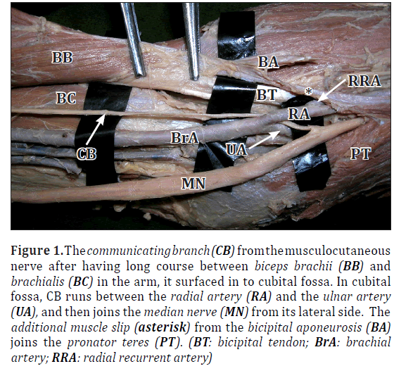
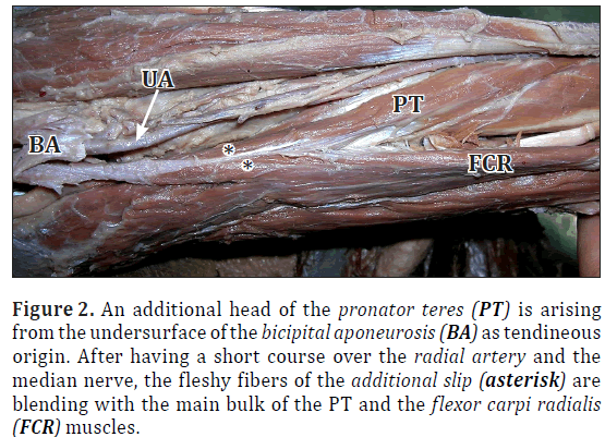
Out of 48 upper limbs dissected, in the right cubital fossa of a 72-year-old male cadaver, we observed an additional head of pronator teres and flexor carpi radialis (2.08%). This additional muscle was originated as tendineous slip from the undersurface of the aponeurotic extension of the biceps brachii muscle near its tendo-aponeurotic bifurcation in the cubital fossa (Figure 1). This variant muscle slip, in addition to pronator teres’ humeral and ulnar heads, had oblique course from medial to lateral under the superficial fascia over the radial artery, radial recurrent artery and the median nerve in the cubital fossa. After its tendineous origin, on its midway between the course, before joining to main bulk of the pronator teres, it became a fleshy muscle belly and initially merging with the main bulk of the pronator teres (Figure 1) and then these muscle fibers split into two bands, one to continue with the pronator teres and another to join the flexor carpi radialis muscle (Figure 2). The Median nerve, radial artery and the radial recurrent artery were passing deep to the muscular bridge formed by this additional muscle band in the cubital fossa. Further, we did not find additional nerve fiber supplying this accessory muscle slips separately. Therefore, these fibers might have been getting their innervations from the main motor branch for the pronator teres and the flexor carpi radialis from the median nerve, which was ramifying from the median nerve after it joins with the communicating branch from the musculocutaneous nerve.
Figure 1: The communicating branch (CB) from the musculocutaneous nerve after having long course between biceps brachii (BB) and brachialis (BC) in the arm, it surfaced in to cubital fossa. In cubital fossa, CB runs between the radial artery (RA) and the ulnar artery (UA), and then joins the median nerve (MN) from its lateral side. The additional muscle slip (asterisk) from the bicipital aponeurosis (BA) joins the pronator teres (PT). (BT: bicipital tendon; BrA: brachial artery; RRA: radial recurrent artery)
Figure 2: An additional head of the pronator teres (PT) is arising from the undersurface of the bicipital aponeurosis (BA) as tendineous origin. After having a short course over the radial artery and the median nerve, the fleshy fibers of the additional slip (asterisk) are blending with the main bulk of the PT and the flexor carpi radialis (FCR) muscles.
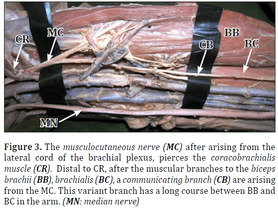
In the same upper limb, we also observed a long communicating branch arising from the musculocutaneous nerve high in the arm after its usual muscular and cutaneous branches. After the origin, this variant branch had a long course in the arm between the biceps brachii and brachialis muscles (Figure 3). In the cubital region, it became superficial, medial to the tendon of the biceps and passed between the radial and ulnar arteries before joining the median nerve (Figure 1). There were no branches arising from this communicating branch throughout its course. However, such additional muscle slips and the variant communicating branch were not found on the left upper limb.
Figure 3: The musculocutaneous nerve (MC) after arising from the lateral cord of the brachial plexus, pierces the coracobrachialis muscle (CR). Distal to CR, after the muscular branches to the biceps brachii (BB), brachialis (BC), a communicating branch (CB) are arising from the MC. This variant branch has a long course between BB and BC in the arm. (MN: median nerve)
Discussion
Pronator teres muscle may contain additional muscle slips from the medial epicondyle, supracondylar process of the humerus, Struthers’ ligament [1], tendineous insertion of the biceps brachii, medial epicondyle [2], lateral aspect of the brachialis muscle and lateral intermuscular septum [3]. However, the current variation, additional muscular slip from the bicipital aponeurosis which is not only extending to the pronator teres but also to the flexor carpi radialis has not been recorded earlier. It has been observed that, the presence of such accessory slips are the most predisposing factors for median nerve entrapment [4]. In the present variation, the addition slip was tendineous in origin and was superficial to the superficial head of the pronator teres and was forming a muscular bridge/tunnel through which the radial artery, ulnar artery and the median nerve were passing, thus may cause neurovascular compression.
During the initial stages of the upper limb development, the confined somites are individually migrating to the developing limb bud, later several segments fuses to form the specific muscle. Topographic and temporal molecular regulation leads to patterning of the muscles via differential growth and also by apoptosis. The imbalance in this process may lead to absence of a muscle or unusual presence, orientation of the muscles which may be the reasons for the presence of additional slips from the bicipital aponeurosis [5].
The knowledge of such muscular variations may be important during surgeries of the arm and elbow and may also explain the uncommon neurovascular symptoms due to their close and unusual association with the neurovascular bundles in this area. In addition, these variations in the musculature may also cause diagnostic perplexities while interpreting MRI or CT scans.
Communication between musculocutaneous and median nerves
The presence of short communication between musculocutaneous nerve and median nerve in the arm/axilla has been reported several times before [6–8]. It has been shown that, such communications were seen in 53.6% of the dissections (84.6% were proximal and 7.7% distal) [9]. In another study, communicating branch was proximal (45%) and distal (35%) to the point of entry of the musculocutaneous nerve into coracobrachialis muscle [10]. Interestingly, the whole musculocutaneous nerve found to be fused to the median nerve in the axilla itself [1,11]. In another observation, a communicating branch was found to join the median nerve at the level of insertion of deltoid [12]. However, the present finding shows a long communicating branch from the musculocutaneous nerve arising in the axilla; and after having long course between the biceps and the brachialis in the arm, it was joining the median nerve in the cubital fossa. Further, this variant branch –before joining the median nerve in the cubital fossa– was running between the ulnar and radial arteries near their origin from the brachial artery. Such long course, its relation to cubital vessels and communication to median nerve in the cubital fossa has not been reported previously. Since no cutaneous or muscular branches were found arising from this communicating branch, this nerve may contain nerve roots of lateral cord that might have been failed to join the median nerve via lateral root of the median nerve. Further, the presence of additional heads for pronator teres and flexor carpi radialis muscles from bicipital aponeurosis may also be the reason for the presence of long communicating branch which may supply these additional muscle slips via median nerve in the forearm.
Knowledge of the musculocutaneous nerve ultrasound appearance facilitates localization and successful block. Thus, awareness of these variant branches from musculocutaneous nerve may help to nerve puncture during ultrasound-guided regional anesthesia [13] and may prove valuable in traumatology of the elbow joint, as well as in plastic and reconstructive repair operations.
References
- Jelev L, Georgiev GP. Unusual high-origin of the pronator teres muscle from a Struthers’ ligament coexisting with a variation of the musculocutaneous nerve. Rom J Morphol Embryol. 2009; 50: 497–499.
- Shiraishi N, Matsumura G. Identification of two accessory muscle bundles with anomalous insertions in the flexor side of the right forearm. Okajimas Folia Anat Jpn. 2007; 84: 35–42.
- Pai MM, Nayak SR, Vadgaonkar R, Ranade AV, Prabhu LV, Thomas M, Sugavasi R. Accessory brachialis muscle: a case report. Morphologie. 2008; 92: 47–49.
- Nebot-Cegarra J, Reina-de la Torre F, Pérez-Berruezo J. Accessory fasciculi of the human pronator teres muscle: incidence, morphological characteristics and relation to the median nerve. Ann Anat. 1994; 176: 223–227.
- Mooney EK, Loh C. Hand Embryology Gross Morphologic Overview of Upper Limb Development. http://emedicine.medscape.com/article/1287982-overview (accessed January 2011)
- Basar R, Aldur MM, Celik HH, Yuksel M, Tascioglu AB. A connecting branch between the musculocutaneous nerve and the median nerve. Morphologie. 2000; 84: 25–27.
- Eglseder WA Jr, Goldman M. Anatomic variations of the musculocutaneous nerve in the arm. Am J Orthop (Belle Mead NJ). 1997; 26: 777–780.
- Uzun A, Seelig LL Jr. A variation in the formation of the median nerve: communicating branch between the musculocutaneous and median nerves in man. Folia Morphol (Warsz). 2001; 60: 99–101.
- Guerri-Guttenberg RA, Ingolotti M. Classifying musculocutaneous nerve variations. Clin Anat. 2009; 22: 671–683.
- Loukas M, Aqueelah H. Musculocutaneous and median nerve connections within, proximal and distal to the coracobrachialis muscle. Folia Morphol (Warsz). 2005; 64: 101–108.
- Prasada Rao PV, Chaudhary SC. Communication of the musculocutaneous nerve with the median nerve. East Afr Med J. 2000; 77: 498–503.
- Goyal N, Harjeet, Gupta M. Bilateral variant contributions in the formation of median nerve. Surg Radiol Anat. 2005; 27: 562–565.
- Schafhalter-Zoppoth I, Gray AT. The musculocutaneous nerve: ultrasound appearance for peripheral nerve block. Reg Anesth Pain Med. 2005; 30: 385–390.
Kumar Mr Bhat*, Vinay Kulakarni and Chandni Gupta
Department of Anatomy, Kasturba Medical College, Manipal University, Manipal, India.
- *Corresponding Author:
- Dr. Kumar MR Bhat
Associate Professor, Department of Anatomy Kasturba Medical College Manipal University Manipal, 576104, India.
Tel: +91 (820) 2922327
E-mail: kumar.mr@manipal.edu
Date of Received: April 15th, 2011
Date of Accepted: January 17th, 2012
Published Online: October 12th, 2012
© Int J Anat Var (IJAV). 2012; 5: 41–43.
Abstract
Additional muscle slips from the bicipital aponeurosis both to pronator teres and flexor carpi radialis muscles are uncommon and not been reported. Here, we report a case of presence of tendentious slip arising from the under surface of the bicipital aponeurosis in the cubital fossa in the left upper limb of a 72-year-old male cadaver. This tendineous slip was then divided into two separate muscular heads for the pronator teres and flexor carpi radialis muscles. Additionally, in the same cadaver we also found an unusual long communicating branch from the musculocutaneous nerve in the upper arm, which had long course through the arm before joining the median nerve in the cubital fossa. This report discusses the details of these variations, their clinical implication and embryological explanations.
-Keywords
pronator teres,flexor carpi radialis,musculocutaneous nerve,communicating branch,additional head
Introduction
Pronator teres usually originates as two heads, the superficial humeral head from medial epicondyle and the deep ulnar head from the coronoid process. The median nerve passes between the two heads, and the ulnar artery passes deep to the deep head of the pronator teres in the cubital fossa. Flexor carpi radialis, another superficial flexor of the forearm arises from common flexor origin and runs obliquely in the forearm. Here we report the presence of additional heads for the pronator teres and the flexor carpi radialis arising from the bicipital aponeurosis.
Musculocutaneous nerve, a branch of the lateral cord of the brachial plexus in the axilla pierces the coracobrachialis muscle and supplies the coracobrachialis, biceps and brachialis muscles and continues as lateral cutaneous nerve of the forearm. Usually, there will be no communication between the musculocutaneous and the median nerves. Here we report a rare and long communicating branch between these two nerves.
Case Report
Out of 48 upper limbs dissected, in the right cubital fossa of a 72-year-old male cadaver, we observed an additional head of pronator teres and flexor carpi radialis (2.08%). This additional muscle was originated as tendineous slip from the undersurface of the aponeurotic extension of the biceps brachii muscle near its tendo-aponeurotic bifurcation in the cubital fossa (Figure 1). This variant muscle slip, in addition to pronator teres’ humeral and ulnar heads, had oblique course from medial to lateral under the superficial fascia over the radial artery, radial recurrent artery and the median nerve in the cubital fossa. After its tendineous origin, on its midway between the course, before joining to main bulk of the pronator teres, it became a fleshy muscle belly and initially merging with the main bulk of the pronator teres (Figure 1) and then these muscle fibers split into two bands, one to continue with the pronator teres and another to join the flexor carpi radialis muscle (Figure 2). The Median nerve, radial artery and the radial recurrent artery were passing deep to the muscular bridge formed by this additional muscle band in the cubital fossa. Further, we did not find additional nerve fiber supplying this accessory muscle slips separately. Therefore, these fibers might have been getting their innervations from the main motor branch for the pronator teres and the flexor carpi radialis from the median nerve, which was ramifying from the median nerve after it joins with the communicating branch from the musculocutaneous nerve.
Figure 1: The communicating branch (CB) from the musculocutaneous nerve after having long course between biceps brachii (BB) and brachialis (BC) in the arm, it surfaced in to cubital fossa. In cubital fossa, CB runs between the radial artery (RA) and the ulnar artery (UA), and then joins the median nerve (MN) from its lateral side. The additional muscle slip (asterisk) from the bicipital aponeurosis (BA) joins the pronator teres (PT). (BT: bicipital tendon; BrA: brachial artery; RRA: radial recurrent artery)
Figure 2: An additional head of the pronator teres (PT) is arising from the undersurface of the bicipital aponeurosis (BA) as tendineous origin. After having a short course over the radial artery and the median nerve, the fleshy fibers of the additional slip (asterisk) are blending with the main bulk of the PT and the flexor carpi radialis (FCR) muscles.
In the same upper limb, we also observed a long communicating branch arising from the musculocutaneous nerve high in the arm after its usual muscular and cutaneous branches. After the origin, this variant branch had a long course in the arm between the biceps brachii and brachialis muscles (Figure 3). In the cubital region, it became superficial, medial to the tendon of the biceps and passed between the radial and ulnar arteries before joining the median nerve (Figure 1). There were no branches arising from this communicating branch throughout its course. However, such additional muscle slips and the variant communicating branch were not found on the left upper limb.
Figure 3: The musculocutaneous nerve (MC) after arising from the lateral cord of the brachial plexus, pierces the coracobrachialis muscle (CR). Distal to CR, after the muscular branches to the biceps brachii (BB), brachialis (BC), a communicating branch (CB) are arising from the MC. This variant branch has a long course between BB and BC in the arm. (MN: median nerve)
Discussion
Pronator teres muscle may contain additional muscle slips from the medial epicondyle, supracondylar process of the humerus, Struthers’ ligament [1], tendineous insertion of the biceps brachii, medial epicondyle [2], lateral aspect of the brachialis muscle and lateral intermuscular septum [3]. However, the current variation, additional muscular slip from the bicipital aponeurosis which is not only extending to the pronator teres but also to the flexor carpi radialis has not been recorded earlier. It has been observed that, the presence of such accessory slips are the most predisposing factors for median nerve entrapment [4]. In the present variation, the addition slip was tendineous in origin and was superficial to the superficial head of the pronator teres and was forming a muscular bridge/tunnel through which the radial artery, ulnar artery and the median nerve were passing, thus may cause neurovascular compression.
During the initial stages of the upper limb development, the confined somites are individually migrating to the developing limb bud, later several segments fuses to form the specific muscle. Topographic and temporal molecular regulation leads to patterning of the muscles via differential growth and also by apoptosis. The imbalance in this process may lead to absence of a muscle or unusual presence, orientation of the muscles which may be the reasons for the presence of additional slips from the bicipital aponeurosis [5].
The knowledge of such muscular variations may be important during surgeries of the arm and elbow and may also explain the uncommon neurovascular symptoms due to their close and unusual association with the neurovascular bundles in this area. In addition, these variations in the musculature may also cause diagnostic perplexities while interpreting MRI or CT scans.
Communication between musculocutaneous and median nerves
The presence of short communication between musculocutaneous nerve and median nerve in the arm/axilla has been reported several times before [6–8]. It has been shown that, such communications were seen in 53.6% of the dissections (84.6% were proximal and 7.7% distal) [9]. In another study, communicating branch was proximal (45%) and distal (35%) to the point of entry of the musculocutaneous nerve into coracobrachialis muscle [10]. Interestingly, the whole musculocutaneous nerve found to be fused to the median nerve in the axilla itself [1,11]. In another observation, a communicating branch was found to join the median nerve at the level of insertion of deltoid [12]. However, the present finding shows a long communicating branch from the musculocutaneous nerve arising in the axilla; and after having long course between the biceps and the brachialis in the arm, it was joining the median nerve in the cubital fossa. Further, this variant branch –before joining the median nerve in the cubital fossa– was running between the ulnar and radial arteries near their origin from the brachial artery. Such long course, its relation to cubital vessels and communication to median nerve in the cubital fossa has not been reported previously. Since no cutaneous or muscular branches were found arising from this communicating branch, this nerve may contain nerve roots of lateral cord that might have been failed to join the median nerve via lateral root of the median nerve. Further, the presence of additional heads for pronator teres and flexor carpi radialis muscles from bicipital aponeurosis may also be the reason for the presence of long communicating branch which may supply these additional muscle slips via median nerve in the forearm.
Knowledge of the musculocutaneous nerve ultrasound appearance facilitates localization and successful block. Thus, awareness of these variant branches from musculocutaneous nerve may help to nerve puncture during ultrasound-guided regional anesthesia [13] and may prove valuable in traumatology of the elbow joint, as well as in plastic and reconstructive repair operations.
References
- Jelev L, Georgiev GP. Unusual high-origin of the pronator teres muscle from a Struthers’ ligament coexisting with a variation of the musculocutaneous nerve. Rom J Morphol Embryol. 2009; 50: 497–499.
- Shiraishi N, Matsumura G. Identification of two accessory muscle bundles with anomalous insertions in the flexor side of the right forearm. Okajimas Folia Anat Jpn. 2007; 84: 35–42.
- Pai MM, Nayak SR, Vadgaonkar R, Ranade AV, Prabhu LV, Thomas M, Sugavasi R. Accessory brachialis muscle: a case report. Morphologie. 2008; 92: 47–49.
- Nebot-Cegarra J, Reina-de la Torre F, Pérez-Berruezo J. Accessory fasciculi of the human pronator teres muscle: incidence, morphological characteristics and relation to the median nerve. Ann Anat. 1994; 176: 223–227.
- Mooney EK, Loh C. Hand Embryology Gross Morphologic Overview of Upper Limb Development. http://emedicine.medscape.com/article/1287982-overview (accessed January 2011)
- Basar R, Aldur MM, Celik HH, Yuksel M, Tascioglu AB. A connecting branch between the musculocutaneous nerve and the median nerve. Morphologie. 2000; 84: 25–27.
- Eglseder WA Jr, Goldman M. Anatomic variations of the musculocutaneous nerve in the arm. Am J Orthop (Belle Mead NJ). 1997; 26: 777–780.
- Uzun A, Seelig LL Jr. A variation in the formation of the median nerve: communicating branch between the musculocutaneous and median nerves in man. Folia Morphol (Warsz). 2001; 60: 99–101.
- Guerri-Guttenberg RA, Ingolotti M. Classifying musculocutaneous nerve variations. Clin Anat. 2009; 22: 671–683.
- Loukas M, Aqueelah H. Musculocutaneous and median nerve connections within, proximal and distal to the coracobrachialis muscle. Folia Morphol (Warsz). 2005; 64: 101–108.
- Prasada Rao PV, Chaudhary SC. Communication of the musculocutaneous nerve with the median nerve. East Afr Med J. 2000; 77: 498–503.
- Goyal N, Harjeet, Gupta M. Bilateral variant contributions in the formation of median nerve. Surg Radiol Anat. 2005; 27: 562–565.
- Schafhalter-Zoppoth I, Gray AT. The musculocutaneous nerve: ultrasound appearance for peripheral nerve block. Reg Anesth Pain Med. 2005; 30: 385–390.


