Agenesis of isthmus of thyroid gland with bilateral levator glandulae thyroideae
Devi Sankar K*, Sharmila Bhanu P, Susan PJ and Gajendra K
Department of Anatomy, Narayana Medical College, Chinthareddy Palem, Nellore, Andhra Pradesh, India
- *Corresponding Author:
- Devi Sankar K, MSc
Assistant Professor, Department of Anatomy, Narayana Medical College, Chinthareddy Palem, Nellore Andhra Pradesh, 524 002, India
Tel: +91 949 0948006
Fax: +91 861 2317962
E-mail: lesanshar@gmail.com
Date of Received: November 11th, 2008
Date of Accepted: March 3rd, 2009
Published Online: March 5th, 2009
© Int J Anat Var (IJAV). 2009; 2: 29–30.
[ft_below_content] =>Keywords
thyroid isthmus, thyroid agenesis, thyroglossal duct anomalies, pyramidal lobe
Introduction
Thyroid gland is the first endocrine gland to start developing in the embryo. It is well known for its developmental anomalies ranging from common to rare. Common anomalies include persistence of pyramidal lobe and thyroglossal duct cyst. Rare anomalies are agenesis or hemi-agenesis of thyroid gland, agenesis of isthmus alone or aberrant thyroid glands [1,2]
In all the higher primates and human, the gland is composed of two lateral lobes joined by an isthmus located in the anterior cervical region, at the level of the second to fourth tracheal rings and protected by the infrahyoid muscles.
It is difficult to determine the incidence of agenesis of the thyroid isthmus because it is usually diagnosed in cohort of individuals presenting with other thyroid diseases.
Case Report
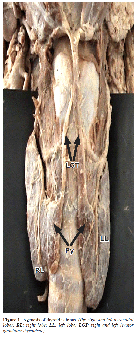
During routine dissection in the Department of Anatomy, 48-year-old male cadaver showed agenesis of isthmus of thyroid gland. There were no scars in the cervical region, suggesting that the patient have not undergone any surgery. The thyroid gland had two separate lobes, with complete agenesis of isthmus. Each lateral lobe had a pyramidal lobe of its own which is connected with two levator glandulae thyroideae. There were no ectopic thyroid tissues present between the root of the tongue to the gland’s position. The two lobes were separate without any tissue intervening between them (Figure 1).
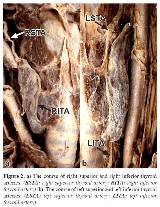
The length of the lobes was 5.8 cm at the right and 6.3 cm at the left lobe; widths were 3.2 and 3.8 cm, respectively. The individual lobes were supplied by branches of superior and inferior thyroid arteries. No accessory thyroid arteries were present. The anterior branch of superior thyroid artery (STA) anastomosed with ascending branch of inferior thyroid artery (ITA) between the pyramidal lobe and lateral lobe on each side. On the posterior border, anastomoses were identified between the posterior and inferior branches of STA & ITA, respectively. But there were no anastomoses between the arteries of right and left side (Figure 2).
Discussion
Agenesis of thyroid isthmus can be explained as an anomaly of embryological development [1,3]. Phylogenetically, the thyroid follicles are structured to acquire a bi-lobe shaped gland. The two lobes were joined together by an isthmus in the upper part of trachea [2,4].
The isthmus may be missing in amphibians, birds and among mammals - Monotremes, certain Marsupials, Cetaceans, Carnivores and Rodents. In rhesus monkey (Macacus rhesus), the thyroid glands are normal in position but there were no isthmus [5].
The morphological difference in the evolutionary origin does not result in any changes in thyroid function. Usually agenesis of isthmus is difficult to determine unless the patients refer for other thyroid diseases.
What is the cause of agenesis of isthmus?
Thyroglossal duct (TGD) arises from the endodermic epithelium of primordial pharynx at the level of 2nd & 3rd pharyngeal arch, when it descends downward, its caudal end bifurcates and gives origin to the thyroid lobes and the isthmus. At the same time the cephalic end of the thyroglossal duct degenerates [6]. This isolates it from the pharyngeal endoderm with the cessation of proliferation of the endodermic cells from which follicular cells of the gland are derived. Rarely, a high separation of thyroglossal duct can engender two independent thyroid lobes and pyramidal lobes with the absence of isthmus [3].
Agenesis of isthmus can be associated with dysorganogenesis related to developmental anomalies of thyroid gland such as absence of either lobe [7] or presence of ectopic thyroid tissue in the nearest cervical region [5].
In our case, the agenesis of thyroid isthmus was not associated with other anomalies of gland, and it may be a congenital anomaly. Interestingly, the glandular branches of STA and ITA did not anastomose in the median plane, which varies from other studies of agenesis of thyroid isthmus. This type of variations should be kept in mind during transthyroid tracheotomy procedures.
Agenesis of isthmus can be diagnosed via scintigraphy, ultrasonography, CT and MRI. When suspected, the individual may be directed for a differential pathological diagnosis such as autonomous thyroid nodule; thyroiditis; primary carcinoma; neoplastic metastases; and infiltrative diseases such as amyloidosis.
References
- Marshall CF. Variation in the form of the thyroid in man. J Anat Physiol. 1895; 29: 234–239.
- Testut L, Latarjet A. Anatomia Humana. Tomo IV. Ed. Barcelona, Salvat. 1978: 402–404. Spanish.
- Sgalitzer KE. Contribution to the study of the morphogenesis of the thyroid gland. J Anat. 1941; 75: 389–405.
- Williams PL, Bannister LK, Berry MM, Collins P, Dyson M, Dussek JE, Ferguson MWJ. Grays Anatomy. 38th Ed., Edinburgh, Churchill Livingstone. 2000; 1891–1897.
- Pastor Vazquez JF, Gil Verona JA, De Paz Fernandez FJ, Barbosa Cachorro M. Agenesis of the thyroid isthmus. Eur J Anatomy, 2006; 10: 83–84.
- Moore KL, Persaud TVN. The developing human, clinical oriented embryology. 6th Ed., Philadelphia, W.B. Saunders Company. 2003; 230–233.
- Burman KD, Adler RA, Wartofsky L. Hemiagenesis of the thyroid gland. Am J Med. 1975; 58: 143–146.
Devi Sankar K*, Sharmila Bhanu P, Susan PJ and Gajendra K
Department of Anatomy, Narayana Medical College, Chinthareddy Palem, Nellore, Andhra Pradesh, India
- *Corresponding Author:
- Devi Sankar K, MSc
Assistant Professor, Department of Anatomy, Narayana Medical College, Chinthareddy Palem, Nellore Andhra Pradesh, 524 002, India
Tel: +91 949 0948006
Fax: +91 861 2317962
E-mail: lesanshar@gmail.com
Date of Received: November 11th, 2008
Date of Accepted: March 3rd, 2009
Published Online: March 5th, 2009
© Int J Anat Var (IJAV). 2009; 2: 29–30.
Abstract
Among endocrine glands, thyroid gland is well known for its developmental anomalies ranging from common to rare. During routine dissection of the male cadaver, agenesis of isthmus of thyroid gland in the midline was noted. It resulted in two lateral lobes with two pyramidal lobe and levator glandulae thyroidae. There is no anastamosis of blood vessels between the right and left lobes as normally seen. The agenesis of isthmus of thyroid gland is extremely rare and very few cases have been reported in literature.
-Keywords
thyroid isthmus, thyroid agenesis, thyroglossal duct anomalies, pyramidal lobe
Introduction
Thyroid gland is the first endocrine gland to start developing in the embryo. It is well known for its developmental anomalies ranging from common to rare. Common anomalies include persistence of pyramidal lobe and thyroglossal duct cyst. Rare anomalies are agenesis or hemi-agenesis of thyroid gland, agenesis of isthmus alone or aberrant thyroid glands [1,2]
In all the higher primates and human, the gland is composed of two lateral lobes joined by an isthmus located in the anterior cervical region, at the level of the second to fourth tracheal rings and protected by the infrahyoid muscles.
It is difficult to determine the incidence of agenesis of the thyroid isthmus because it is usually diagnosed in cohort of individuals presenting with other thyroid diseases.
Case Report
During routine dissection in the Department of Anatomy, 48-year-old male cadaver showed agenesis of isthmus of thyroid gland. There were no scars in the cervical region, suggesting that the patient have not undergone any surgery. The thyroid gland had two separate lobes, with complete agenesis of isthmus. Each lateral lobe had a pyramidal lobe of its own which is connected with two levator glandulae thyroideae. There were no ectopic thyroid tissues present between the root of the tongue to the gland’s position. The two lobes were separate without any tissue intervening between them (Figure 1).
The length of the lobes was 5.8 cm at the right and 6.3 cm at the left lobe; widths were 3.2 and 3.8 cm, respectively. The individual lobes were supplied by branches of superior and inferior thyroid arteries. No accessory thyroid arteries were present. The anterior branch of superior thyroid artery (STA) anastomosed with ascending branch of inferior thyroid artery (ITA) between the pyramidal lobe and lateral lobe on each side. On the posterior border, anastomoses were identified between the posterior and inferior branches of STA & ITA, respectively. But there were no anastomoses between the arteries of right and left side (Figure 2).
Discussion
Agenesis of thyroid isthmus can be explained as an anomaly of embryological development [1,3]. Phylogenetically, the thyroid follicles are structured to acquire a bi-lobe shaped gland. The two lobes were joined together by an isthmus in the upper part of trachea [2,4].
The isthmus may be missing in amphibians, birds and among mammals - Monotremes, certain Marsupials, Cetaceans, Carnivores and Rodents. In rhesus monkey (Macacus rhesus), the thyroid glands are normal in position but there were no isthmus [5].
The morphological difference in the evolutionary origin does not result in any changes in thyroid function. Usually agenesis of isthmus is difficult to determine unless the patients refer for other thyroid diseases.
What is the cause of agenesis of isthmus?
Thyroglossal duct (TGD) arises from the endodermic epithelium of primordial pharynx at the level of 2nd & 3rd pharyngeal arch, when it descends downward, its caudal end bifurcates and gives origin to the thyroid lobes and the isthmus. At the same time the cephalic end of the thyroglossal duct degenerates [6]. This isolates it from the pharyngeal endoderm with the cessation of proliferation of the endodermic cells from which follicular cells of the gland are derived. Rarely, a high separation of thyroglossal duct can engender two independent thyroid lobes and pyramidal lobes with the absence of isthmus [3].
Agenesis of isthmus can be associated with dysorganogenesis related to developmental anomalies of thyroid gland such as absence of either lobe [7] or presence of ectopic thyroid tissue in the nearest cervical region [5].
In our case, the agenesis of thyroid isthmus was not associated with other anomalies of gland, and it may be a congenital anomaly. Interestingly, the glandular branches of STA and ITA did not anastomose in the median plane, which varies from other studies of agenesis of thyroid isthmus. This type of variations should be kept in mind during transthyroid tracheotomy procedures.
Agenesis of isthmus can be diagnosed via scintigraphy, ultrasonography, CT and MRI. When suspected, the individual may be directed for a differential pathological diagnosis such as autonomous thyroid nodule; thyroiditis; primary carcinoma; neoplastic metastases; and infiltrative diseases such as amyloidosis.
References
- Marshall CF. Variation in the form of the thyroid in man. J Anat Physiol. 1895; 29: 234–239.
- Testut L, Latarjet A. Anatomia Humana. Tomo IV. Ed. Barcelona, Salvat. 1978: 402–404. Spanish.
- Sgalitzer KE. Contribution to the study of the morphogenesis of the thyroid gland. J Anat. 1941; 75: 389–405.
- Williams PL, Bannister LK, Berry MM, Collins P, Dyson M, Dussek JE, Ferguson MWJ. Grays Anatomy. 38th Ed., Edinburgh, Churchill Livingstone. 2000; 1891–1897.
- Pastor Vazquez JF, Gil Verona JA, De Paz Fernandez FJ, Barbosa Cachorro M. Agenesis of the thyroid isthmus. Eur J Anatomy, 2006; 10: 83–84.
- Moore KL, Persaud TVN. The developing human, clinical oriented embryology. 6th Ed., Philadelphia, W.B. Saunders Company. 2003; 230–233.
- Burman KD, Adler RA, Wartofsky L. Hemiagenesis of the thyroid gland. Am J Med. 1975; 58: 143–146.


