High up division of brachial artery into laterally placed ulnar artery and medially placed radial artery
Banani Mitra*, Anirban Sadhu, Rudradev Meyur, Satabdi Sarkar
Department of Anatomy, R. G. Kar Medical College, Khudiram Bose Sarani, Kolkata 700 004, India.
- *Corresponding Author:
- Dr. Banani Mitra
Assistant Professor, Department of Anatomy, R. G. Kar Medical College, Khudiram Bose Sarani, Kolkata 700 004, India.
Tel: +91 9433366549
E-mail: dr.bmitra@yahoo.com
Date of Received: March 19th, 2012
Date of Accepted: October 4th, 2012
Published Online: April 21st, 2013
© Int J Anat Var (IJAV). 2013; 6: 64–65.
[ft_below_content] =>Keywords
brachial artery, higher bifurcation, radial artery, ulnar artery
Introduction
The axillary artery begins as the continuation of the brachial artery at the distal border of teres major. It terminates at the level of the neck of the radius by giving rise to the radial and ulnar arteries. However variations in this usual arterial pattern have been frequently seen.
Some observed variations include: Sometimes the artery divides more proximally into two trunks which may reunite. In some cases there is a superficial brachial artery arising from the axillary and continues in the forearm as radial artery [1]. Sometimes bifurcation or trifurcation (radial, ulnar and common interosseous artery) of brachial artery occurs more proximally than usual [2]. In some cases it is also seen that radial artery arises at higher level but follows usual course in forearm [3,4].
So variations of arterial system of upper limb are very common and it can be explained embryologically. Blood vessel formation is regulated by ectodermal-mesenchymal interactions and extracellular matrix components present within the limb bud. Alteration of any factor leads to deviation from the usual way. These are important from clinical and surgical point of view [5].
But in the present case a rare variation of brachial artery was noted. It divides in upper part of arm into two branches of which ulnar artery was lateral and radial artery was medial.
Case Report
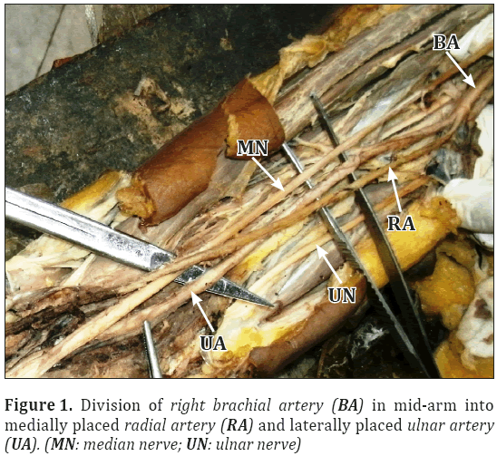
Routine dissection of the right upper extremity for undergraduate teaching in a 69-year-old male cadaver in the Department of Anatomy, R. G. Kar Medical College, revealed the following: the brachial artery divided into radial and ulnar arteries after a short course in the upper half of the arm. The cadaver was donated by relatives after dying of cerebral hemorrhage in this hospital. His medical history included hypertension. The radial artery was located medially and the ulnar artery laterally. In its further course the radial artery, was located laterally after crossing the ulnar artery. Further course of the ulnar artery was as usual. No other variations related to the cords of brachial plexus and their branches were found. Arterial pattern was as usual in the left upper limb.
Discussion
Variations in the upper limb arteries are very common. These variations are reported during the surgical procedures or at the time of angiography. The earliest studies of variations in the arterial system was reported by Senior [6]. Majority of these occur in radial artery followed by ulnar artery [7]. Variations in the division of brachial artery in cubital fossa were reported but mid arm division of brachial artery was rare.
In the present case, brachial artery bifurcated in mid-arm and gave radial and ulnar arteries. But radial artery was present on the medial side and ulnar artery on the lateral side. Then after passing some distance radial artery crosses ulnar artery superficially and followed usual course. This was present only in right side, left side showed no variations.
High origin of radial artery from brachial artery was noted by Keen in 1961. He explained that brachial artery first gives radial artery than ulnar artery. Then new connection developed between radial artery and brachial artery at the level of ulnar artery and previous radial artery was obliterated. If this new connection failed to develop then brachial artery showed high up division [8].
Embryological Explanation
Every variation in the peripheral vascular anatomy is due to obliteration or persistence of any segment of axis artery [9]. The artery to the developing upper limbs is derived from the 7th cervical intersegmental artery. This artery gradually gives rise to other branches to supply upper limb. Distal to the level of teres major it continues as the brachial artery and in the cubital fossa it continues as the interosseous artery. The ectodermal-mesenchymal interaction and extracellular matrix components also control blood vessel formation [10]. The radial and ulnar artery developed late in the forearm from axis artery, subsequently interosseous artery reduces in size and becomes a branch of ulnar artery.
In the present case, early division of brachial artery occured in mid-arm, as a result it became very short. This may be due to alteration of some factors, which may be hemodynamic, that cause early division of brachial artery. In early stage of development there is a capillary plexus which gradually differentiate to form definite blood vessels. Variations in the formation of stages of this capillary plexus may also be responsible for the present case [11].
The knowledge regarding the course of upper limb blood vessels is very important.
It helps surgeons to avoid unnecessary injury at the time of surgery. Radial artery graft is used now-a-days for coronary artery bypass graft surgery [12]. With increasing radial artery harvesting, cardiac catheterization and angiographic procedures, knowledge of usual course as well as the variations of radial artery can be very useful.
Acknowledgements
The authors express their heartfelt gratitude to all the members of the Department of Anatomy, R. G. Kar Medical College, Kolkata for their kind cooperation and generosity in the conduct of this study.
References
- Yang HJ, Gil YC, Jung WS, Lee HY. Variations of the superficial brachial artery in Korean cadavers. J Korean Med Sci. 2008; 23: 884–887.
- Williams PL, Bannister LH, Berry MM, Dyson M, Dussek JE, Ferguson MW, eds. Gray’s Anatomy. 38th Ed., London, Churchill Livingstone. 1999; 319, 1539.
- Okoro IO, Jiburum BC. Rare high origin of the radial artery: A bilateral symmetrical case. Nig J Surg Res 2003; 5: 70–72.
- Singh H, Gupta N, Bargotra RN, Singh NP. Higher bifurcation of brachial artery with superficial course of radial artery in forearm. JK Science. 2010; 12: 39–40.
- Hollinshed WH. Anatomy for Surgeons. Vol. 3. New York, Hoeber and Harper Inc. 1958; 66–73.
- Bhanu PS, Sankar KD, Susan PJ. High origin and superficial course of radial artery. Int J Anat Var (IJAV). 2010; 3: 162–164.
- McCormack LJ, Cauldwell EW, Anson BJ. Brachial and antebrachial arterial patterns; a study of 750 extremities. Surg Gynecol Obstet. 1953; 96: 43–54.
- Satyanarayana N, Sunitha P, Shaik MM, Devi PSV. Brachial artery with high up division with its embryological basis and clinical significance. Int J Anat Var (IJAV). 2010; 3: 56–58.
- Rodriguez-Baeza A, Nebot J, Ferreira B, Reina F, Perez J, Sanudo JR, Roig M. An anatomical study and ontogenic explanation of 23 cases with variations in the main pattern of the human brachio-antebrachial arteries. J Anat. 1995; 187: 473–479.
- Feinbery RN. Vascular development in the embryonic limb bud. In: Feinbery RN, Sherer GK, Auerbach R, eds. The Development of The Vascular System. Basel, Karger (Issues Biomed). 1991; 14: 136–148.
- Larsen WJ. Human Embryology. New York, Churchill-Livingstone. 1993; 222–234.
- Desai ND, Cohen EA, Naylor CD, Fremes SE. Radial Artery Patency Study Investigators. A randomized comparison of radial artery and saphenous-vein coronary bypass grafts. N Engl J Med. 2004; 351: 2302–2309.
Banani Mitra*, Anirban Sadhu, Rudradev Meyur, Satabdi Sarkar
Department of Anatomy, R. G. Kar Medical College, Khudiram Bose Sarani, Kolkata 700 004, India.
- *Corresponding Author:
- Dr. Banani Mitra
Assistant Professor, Department of Anatomy, R. G. Kar Medical College, Khudiram Bose Sarani, Kolkata 700 004, India.
Tel: +91 9433366549
E-mail: dr.bmitra@yahoo.com
Date of Received: March 19th, 2012
Date of Accepted: October 4th, 2012
Published Online: April 21st, 2013
© Int J Anat Var (IJAV). 2013; 6: 64–65.
Abstract
The objective of the present study was to document high up division of brachial artery into radial and ulnar arteries in the middle of the arm, of which ulnar artery lied laterally and radial artery lied medially. The finding was noted during routine dissection of upper limbs of both sides in the department of Anatomy, R. G. Kar Medical College. This variant was noted only in right arm, left arm showed usual arterial distribution pattern. This was the result of unusual developmental condition. Information regarding the variation was important for vascular surgery and angiography.
-Keywords
brachial artery, higher bifurcation, radial artery, ulnar artery
Introduction
The axillary artery begins as the continuation of the brachial artery at the distal border of teres major. It terminates at the level of the neck of the radius by giving rise to the radial and ulnar arteries. However variations in this usual arterial pattern have been frequently seen.
Some observed variations include: Sometimes the artery divides more proximally into two trunks which may reunite. In some cases there is a superficial brachial artery arising from the axillary and continues in the forearm as radial artery [1]. Sometimes bifurcation or trifurcation (radial, ulnar and common interosseous artery) of brachial artery occurs more proximally than usual [2]. In some cases it is also seen that radial artery arises at higher level but follows usual course in forearm [3,4].
So variations of arterial system of upper limb are very common and it can be explained embryologically. Blood vessel formation is regulated by ectodermal-mesenchymal interactions and extracellular matrix components present within the limb bud. Alteration of any factor leads to deviation from the usual way. These are important from clinical and surgical point of view [5].
But in the present case a rare variation of brachial artery was noted. It divides in upper part of arm into two branches of which ulnar artery was lateral and radial artery was medial.
Case Report
Routine dissection of the right upper extremity for undergraduate teaching in a 69-year-old male cadaver in the Department of Anatomy, R. G. Kar Medical College, revealed the following: the brachial artery divided into radial and ulnar arteries after a short course in the upper half of the arm. The cadaver was donated by relatives after dying of cerebral hemorrhage in this hospital. His medical history included hypertension. The radial artery was located medially and the ulnar artery laterally. In its further course the radial artery, was located laterally after crossing the ulnar artery. Further course of the ulnar artery was as usual. No other variations related to the cords of brachial plexus and their branches were found. Arterial pattern was as usual in the left upper limb.
Discussion
Variations in the upper limb arteries are very common. These variations are reported during the surgical procedures or at the time of angiography. The earliest studies of variations in the arterial system was reported by Senior [6]. Majority of these occur in radial artery followed by ulnar artery [7]. Variations in the division of brachial artery in cubital fossa were reported but mid arm division of brachial artery was rare.
In the present case, brachial artery bifurcated in mid-arm and gave radial and ulnar arteries. But radial artery was present on the medial side and ulnar artery on the lateral side. Then after passing some distance radial artery crosses ulnar artery superficially and followed usual course. This was present only in right side, left side showed no variations.
High origin of radial artery from brachial artery was noted by Keen in 1961. He explained that brachial artery first gives radial artery than ulnar artery. Then new connection developed between radial artery and brachial artery at the level of ulnar artery and previous radial artery was obliterated. If this new connection failed to develop then brachial artery showed high up division [8].
Embryological Explanation
Every variation in the peripheral vascular anatomy is due to obliteration or persistence of any segment of axis artery [9]. The artery to the developing upper limbs is derived from the 7th cervical intersegmental artery. This artery gradually gives rise to other branches to supply upper limb. Distal to the level of teres major it continues as the brachial artery and in the cubital fossa it continues as the interosseous artery. The ectodermal-mesenchymal interaction and extracellular matrix components also control blood vessel formation [10]. The radial and ulnar artery developed late in the forearm from axis artery, subsequently interosseous artery reduces in size and becomes a branch of ulnar artery.
In the present case, early division of brachial artery occured in mid-arm, as a result it became very short. This may be due to alteration of some factors, which may be hemodynamic, that cause early division of brachial artery. In early stage of development there is a capillary plexus which gradually differentiate to form definite blood vessels. Variations in the formation of stages of this capillary plexus may also be responsible for the present case [11].
The knowledge regarding the course of upper limb blood vessels is very important.
It helps surgeons to avoid unnecessary injury at the time of surgery. Radial artery graft is used now-a-days for coronary artery bypass graft surgery [12]. With increasing radial artery harvesting, cardiac catheterization and angiographic procedures, knowledge of usual course as well as the variations of radial artery can be very useful.
Acknowledgements
The authors express their heartfelt gratitude to all the members of the Department of Anatomy, R. G. Kar Medical College, Kolkata for their kind cooperation and generosity in the conduct of this study.
References
- Yang HJ, Gil YC, Jung WS, Lee HY. Variations of the superficial brachial artery in Korean cadavers. J Korean Med Sci. 2008; 23: 884–887.
- Williams PL, Bannister LH, Berry MM, Dyson M, Dussek JE, Ferguson MW, eds. Gray’s Anatomy. 38th Ed., London, Churchill Livingstone. 1999; 319, 1539.
- Okoro IO, Jiburum BC. Rare high origin of the radial artery: A bilateral symmetrical case. Nig J Surg Res 2003; 5: 70–72.
- Singh H, Gupta N, Bargotra RN, Singh NP. Higher bifurcation of brachial artery with superficial course of radial artery in forearm. JK Science. 2010; 12: 39–40.
- Hollinshed WH. Anatomy for Surgeons. Vol. 3. New York, Hoeber and Harper Inc. 1958; 66–73.
- Bhanu PS, Sankar KD, Susan PJ. High origin and superficial course of radial artery. Int J Anat Var (IJAV). 2010; 3: 162–164.
- McCormack LJ, Cauldwell EW, Anson BJ. Brachial and antebrachial arterial patterns; a study of 750 extremities. Surg Gynecol Obstet. 1953; 96: 43–54.
- Satyanarayana N, Sunitha P, Shaik MM, Devi PSV. Brachial artery with high up division with its embryological basis and clinical significance. Int J Anat Var (IJAV). 2010; 3: 56–58.
- Rodriguez-Baeza A, Nebot J, Ferreira B, Reina F, Perez J, Sanudo JR, Roig M. An anatomical study and ontogenic explanation of 23 cases with variations in the main pattern of the human brachio-antebrachial arteries. J Anat. 1995; 187: 473–479.
- Feinbery RN. Vascular development in the embryonic limb bud. In: Feinbery RN, Sherer GK, Auerbach R, eds. The Development of The Vascular System. Basel, Karger (Issues Biomed). 1991; 14: 136–148.
- Larsen WJ. Human Embryology. New York, Churchill-Livingstone. 1993; 222–234.
- Desai ND, Cohen EA, Naylor CD, Fremes SE. Radial Artery Patency Study Investigators. A randomized comparison of radial artery and saphenous-vein coronary bypass grafts. N Engl J Med. 2004; 351: 2302–2309.


