Multiple variations of extensor muscles in a single hand
Karin Fischer1*, Tino Breitfeld1, Hans-Georg Damert2, Hermann-Josef Rothkotter1
1Institute of Anatomy, Medical Faculty, Otto-von-Guericke-University, Leipziger Str. 44, 39120 Magdeburg, Germany.
2Department of Plastic, Aesthetic and Hand Surgery, HELIOS Bördeklinik, Kreiskrankenhaus 4, 39387 Oschersleben , Germany.
- *Corresponding Author:
- Karin Fischer
Institut für Anatomie, Otto von Guericke Universität, Leipziger Strasse 44, 39120 Magdeburg, Germany.
Tel: +49 391 6713607
E-mail: karin.fischer@med.ovgu.de
Date of Received: July 28th, 2015
Date of Accepted: August 8th, 2016
Published Online: December 24th, 2016
© Int J Anat Var (IJAV). 2016; 9: 32–34.
[ft_below_content] =>Keywords
Extensor muscles of the hand, extensor tendons, hand, 4th compartment, radial nerve
Introduction
The extensor muscles of the hand showed many variations in shape. Its of peculiar interest for surgeons in the field of tendon repair, transfer or reconstruction. In literature several forms of anatomical variations were described: variations of long muscles, supernumerary tendons and additional muscles [1].
Different classification systems were developed by Kosugi [2], Yoshida [3], Komiyama [4] and Türker [5].
Case Report
In our dissection course we found in the right hand of an 81-year-old male multiple variations of extensor muscles. The left hand did not show any variations.
In the superficial level there were regular extensor carpi radials longus and brevis (ECR) as well as extensor carpi ulnaris (ECU) muscles.
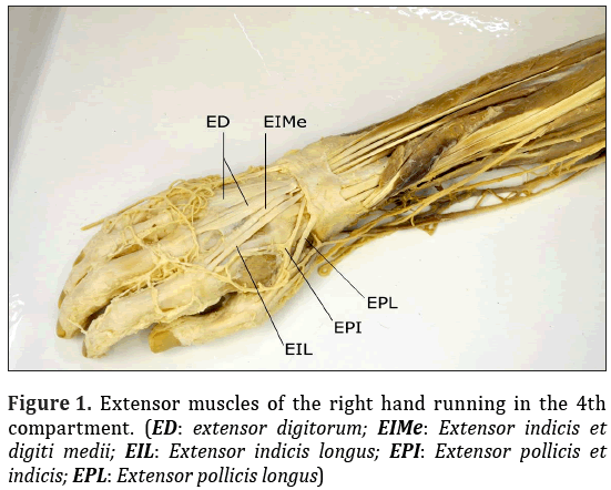
The extensor digitorum muscle (ED) was split into two portions.
One was originated at the lateral epicondyle of the humerus together with ECR brevis and runs on the radial side of the fourth compartment and adds to extensor apparatus of the index finger. This tendon was located radial to the tendon of the extensor indicis and digiti medii muscle (EIMe).
Moreover, there was an interdendinous connection to the tendon of ED of the middle finger. (Figure 1) We called this part extensor indicis longus muscle (EIL).
The second part arose from the ulna and the interosseous membrane. One tendon, separating in the half of the muscle, runs in the superficial level of the fourth compartment to the middle finger. Furthermore, there was a second tendon to the ring finger. Both tendons expanded and add to the intertendinous connection and inserted in the extensor tendon apparatus of the third and fourth finger (Figure 1).
Furthermore, we found a strong intertendineous connection to the tendon of the extensor digiti minimi muscle (EMi).
Each part was innervated by an own direct short branch of deep branch of radial nerve.
Additional we found an EMi with a doubled tendon (Figure 1).
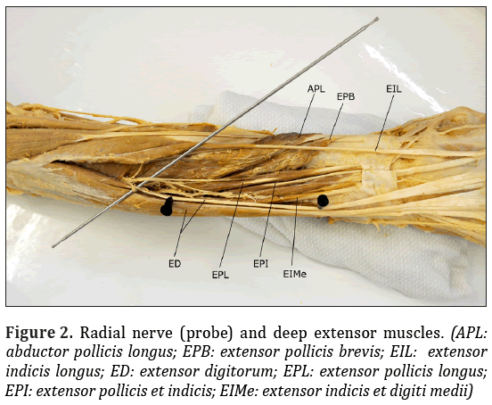
In profound level we found a regular extensor pollicis brevis (EPB), abductor pollicis longus (APL) and extensor pollicis longus muscle (EPL) (Figure 2).
An additional muscle arising from the lower third of the ulna and interosseal membrane was the extensor pollicis and indicis muscle (EPI). Its tendon was separating on the base of the metacarpale bone: one part, leaded to the thumb and the other smaller part to the tendon of the index finger. The part to the thumb meets the tendon of EPL. The tendon of the index finger inserted to the extensor apparatus radial from the tendon of the extensor digitorum (Figure 1).
The muscle was innervated by a long ulnar branch of profound radial nerve.
Distal to the EPI the additional EIMe arose from the ulna and the interosseus membrane. Its belly ended directly in front of the fourth compartment of extensor retinaculum. From this point there were two tendons, one running to the extensor apparatus of the index finger adding deeper, ulnar to the tendon of ED. Some fibers inserted to the capsulate of the carpometacarpal joint. The smaller one leaded to the middle finger. The second tendon was covered from the tendon of ED and becoming expanded till insertion.
This muscle was also innervated by the long ulnar branch of deep branch of the radial nerve.
We classified a superficial and a deep level of tendons in the fourth compartment. From radial to ulnar there were in superficial level the tendons of ED to the index, the middle finger and to the ring finger. In the deep level we found tendons of EPI, EIMe, from radial to ulnar.
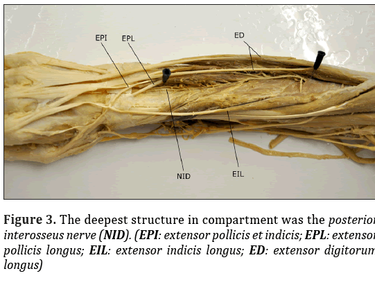
It is remarkable that the tendon of ED to the index intersects the tendon of EPI in the compartment. Both levels were divided by tendon sheaths. The deepest structure in compartment was posterior interosseus nerve (Figure 3).
The dimensions of the variant muscles shown in Table 1.
| Compartment | Belly of the muscle | Tendon | ||||
|---|---|---|---|---|---|---|
| Length (cm) | Width (cm) | Length (cm) | Width (cm) | |||
| M. extensor indicis „longus“ | 4.; radial, superficial | 13,0 | 0.9 | 17,0 | 0,35 | |
| M. extensor digitorum | 4, ulnar , superficial | middle finger 20,0 | 2,5 | middle finger | 11,5 | 0,5 to 2,0 |
| ring finger 10,0 | ring finger | 25,0 | 0,4 to 1,2 | |||
| M. extensor pollicis et indicis | 4.; radial, deep | 5,5 | 0,3 | before splitting | 8,0 | 0,2 |
| thumb | 2,5 | 0,2 | ||||
| index finger | 5,0 | 0,15 | ||||
| M. extensor indicis et digiti medii | 4.; ulnar, deep | 8,5 | 0,4 | index finger | 10,5 | 0,4 |
| middle finger | 10,0 | 0,07 to 0,2 | ||||
| M. extensor digiti minimi | 5. | 15,0 | 0,9 | 14,0 | proximal the compartment 0,3 distal 2 x 0,4 | |
Table 1: Dimensions of the variations of the extensor muscles of the forearm.
In our case extensor EPL, EPI, also EIMe were innervated by a branch of deep branch of radial nerve running ulnar (Figure 2). Exceptional was that the EPL was additionally innervated in the lower third by another branch of deep radial nerve running radial. We could identify this radial branch as posterior interosseous nerve (Figure 3).
Discussion
The muscle variations in the present case were individual described in literature but not combined on one hand.
Metha described an ED which accords to our case. He named this profound muscle extensor indicis [6].
In our case the intertendinous connections in the second intermetacarpal space (between index and middle finger) were in accord to Schroeder [7] case 1; the intertendinous connection in the 3. and 4. intermetacarpal space were described as type 3r.
Remarkable is, that humans in contrast to some vertebrates always have an EPL, EPB and EI when there is a complete EPI [1,3].
Based on classification systems of Yoshida [3], Komiyama [4] and Türker [5] we classified our EPI as Yoshida type 2b, Komiyama type 2b or rather Türker type 1e.
When a classification of the compartment was mentioned, all authors described that the EPI running through the fourth compartment, as well. Merely Gruber [9] found in one out of 408 hands a tendon running through the third compartment. In anatomical studies the frequency of EPI was very different between 0.5 and 5.1% [1].
The EIMe muscle was described quite often in literature. The prevalence of this muscle is between 0.5 and 16% [1]. Yoshida [3] classified this muscle in nine types. Our case equates type IIa which Yoshida [3] found in 3.3 % of his cases. Komiyama [4] classified this case as type 3a. Kosugi [2] described our variation as type IIIb-1.
An EMi with a doubled tendon Kosughi [2] found in 78 % of cases (403 of 516), Mesdagh [9] found only 2 out of 150 extensor digiti minimi muscles with only one tendon and Zilber [10] found doubled tendons in all 50 hands he prepared.
In contrast to the literature we could identify exactly the innervation (Figure 2 and 3).
Conclusion
The described variations of extensor muscles were asymptomatic in most cases. But the knowledge is important for surgical interventions.
References
- Yammine K. The prevalence of the extensor indicis tendon and its variants: a systematic review and metha-analysis. Surg Radiol Anat. 2015; 37: 247-254.
- Kosugi K, Shibata S, Yamashita H. Anatomical study on the variation of extensor muscles of the human forearm 11. The relation between differentiation and variation. Jikeikai Med J. 1989; 36: 93-111.
- Yoshida, Y. Anatomical study on the extensor digitorum profundus muscle in the Japanese. Okajimas Folia Anatomica Japonica. 1990; 71: 339-354.
- Komiyama M, Nwe TM, Toyota N, Shimada Y. Variations of the extensor indicis muscle and tendon. J Hand Surg Br. 1999; 24B: 575-578.
- Türker T, Robertson GA, Thirkannad SM. A classification system for anomalies of the extensor pollicis longus. Hand. 2010; 5: 403-407.
- Mehta V, Arora J, Suri RK, Rath G. An assembly of anomalous extensor tendons of the hand – anatomical description and clinical relevance. ACTA Medica. (Hradec Králové) 2009; 52: 27-30.
- Schroeder von HP; Botte MJ; Gellmann H. Anatomy of the juncturae tendinum of the hand. The Journal of Hand Surgery. 1990; 5A: 4: 595-602.
- Gruber W. Über den constanten Musculus extensor pollicis et indicis gewisser Säugethiere homologen supernumerären Muskel beim Menschen. Virchows Arch path Anat. 1881; 86: 471-491.
- Mestdagh JP. Organisation of the extensor complex of the digits. Anat Ciln. 1985; 7: 49-53.
- Zilber S. Anatomical variation of the extensor tendons to the fingers over the dorsum of the hand: a study of 50 hands and a review in literature. Plast Reconstr Surg. 2004; 113: 214-221.
Karin Fischer1*, Tino Breitfeld1, Hans-Georg Damert2, Hermann-Josef Rothkotter1
1Institute of Anatomy, Medical Faculty, Otto-von-Guericke-University, Leipziger Str. 44, 39120 Magdeburg, Germany.
2Department of Plastic, Aesthetic and Hand Surgery, HELIOS Bördeklinik, Kreiskrankenhaus 4, 39387 Oschersleben , Germany.
- *Corresponding Author:
- Karin Fischer
Institut für Anatomie, Otto von Guericke Universität, Leipziger Strasse 44, 39120 Magdeburg, Germany.
Tel: +49 391 6713607
E-mail: karin.fischer@med.ovgu.de
Date of Received: July 28th, 2015
Date of Accepted: August 8th, 2016
Published Online: December 24th, 2016
© Int J Anat Var (IJAV). 2016; 9: 32–34.
Abstract
Multiple variations of extensor muscles on one hand are very rare. We found in the right hand: 1. M. extensor digitorum was split into two separate muscles; 2. M. extensor pollicis et indicis; 3. M. extensor indicis et digiti medii. All their tendons were in the 4th compartment. The extensor digiti minimi muscle showed a doubled tendon. Besides, we classified the course of tendons in the fourth compartment into a superficial and a profound level. Moreover, we defined the innervation by dissecting the deep branch of the radial nerve having direct branches into the muscles. In our case the extensor pollicis longus muscle was double innervated by a branch of the deep radial nerve and also by a small branch of interosseus antebrachii posterior nerve.
-Keywords
Extensor muscles of the hand, extensor tendons, hand, 4th compartment, radial nerve
Introduction
The extensor muscles of the hand showed many variations in shape. Its of peculiar interest for surgeons in the field of tendon repair, transfer or reconstruction. In literature several forms of anatomical variations were described: variations of long muscles, supernumerary tendons and additional muscles [1].
Different classification systems were developed by Kosugi [2], Yoshida [3], Komiyama [4] and Türker [5].
Case Report
In our dissection course we found in the right hand of an 81-year-old male multiple variations of extensor muscles. The left hand did not show any variations.
In the superficial level there were regular extensor carpi radials longus and brevis (ECR) as well as extensor carpi ulnaris (ECU) muscles.
The extensor digitorum muscle (ED) was split into two portions.
One was originated at the lateral epicondyle of the humerus together with ECR brevis and runs on the radial side of the fourth compartment and adds to extensor apparatus of the index finger. This tendon was located radial to the tendon of the extensor indicis and digiti medii muscle (EIMe).
Moreover, there was an interdendinous connection to the tendon of ED of the middle finger. (Figure 1) We called this part extensor indicis longus muscle (EIL).
The second part arose from the ulna and the interosseous membrane. One tendon, separating in the half of the muscle, runs in the superficial level of the fourth compartment to the middle finger. Furthermore, there was a second tendon to the ring finger. Both tendons expanded and add to the intertendinous connection and inserted in the extensor tendon apparatus of the third and fourth finger (Figure 1).
Furthermore, we found a strong intertendineous connection to the tendon of the extensor digiti minimi muscle (EMi).
Each part was innervated by an own direct short branch of deep branch of radial nerve.
Additional we found an EMi with a doubled tendon (Figure 1).
In profound level we found a regular extensor pollicis brevis (EPB), abductor pollicis longus (APL) and extensor pollicis longus muscle (EPL) (Figure 2).
An additional muscle arising from the lower third of the ulna and interosseal membrane was the extensor pollicis and indicis muscle (EPI). Its tendon was separating on the base of the metacarpale bone: one part, leaded to the thumb and the other smaller part to the tendon of the index finger. The part to the thumb meets the tendon of EPL. The tendon of the index finger inserted to the extensor apparatus radial from the tendon of the extensor digitorum (Figure 1).
The muscle was innervated by a long ulnar branch of profound radial nerve.
Distal to the EPI the additional EIMe arose from the ulna and the interosseus membrane. Its belly ended directly in front of the fourth compartment of extensor retinaculum. From this point there were two tendons, one running to the extensor apparatus of the index finger adding deeper, ulnar to the tendon of ED. Some fibers inserted to the capsulate of the carpometacarpal joint. The smaller one leaded to the middle finger. The second tendon was covered from the tendon of ED and becoming expanded till insertion.
This muscle was also innervated by the long ulnar branch of deep branch of the radial nerve.
We classified a superficial and a deep level of tendons in the fourth compartment. From radial to ulnar there were in superficial level the tendons of ED to the index, the middle finger and to the ring finger. In the deep level we found tendons of EPI, EIMe, from radial to ulnar.
It is remarkable that the tendon of ED to the index intersects the tendon of EPI in the compartment. Both levels were divided by tendon sheaths. The deepest structure in compartment was posterior interosseus nerve (Figure 3).
The dimensions of the variant muscles shown in Table 1.
| Compartment | Belly of the muscle | Tendon | ||||
|---|---|---|---|---|---|---|
| Length (cm) | Width (cm) | Length (cm) | Width (cm) | |||
| M. extensor indicis „longus“ | 4.; radial, superficial | 13,0 | 0.9 | 17,0 | 0,35 | |
| M. extensor digitorum | 4, ulnar , superficial | middle finger 20,0 | 2,5 | middle finger | 11,5 | 0,5 to 2,0 |
| ring finger 10,0 | ring finger | 25,0 | 0,4 to 1,2 | |||
| M. extensor pollicis et indicis | 4.; radial, deep | 5,5 | 0,3 | before splitting | 8,0 | 0,2 |
| thumb | 2,5 | 0,2 | ||||
| index finger | 5,0 | 0,15 | ||||
| M. extensor indicis et digiti medii | 4.; ulnar, deep | 8,5 | 0,4 | index finger | 10,5 | 0,4 |
| middle finger | 10,0 | 0,07 to 0,2 | ||||
| M. extensor digiti minimi | 5. | 15,0 | 0,9 | 14,0 | proximal the compartment 0,3 distal 2 x 0,4 | |
Table 1: Dimensions of the variations of the extensor muscles of the forearm.
In our case extensor EPL, EPI, also EIMe were innervated by a branch of deep branch of radial nerve running ulnar (Figure 2). Exceptional was that the EPL was additionally innervated in the lower third by another branch of deep radial nerve running radial. We could identify this radial branch as posterior interosseous nerve (Figure 3).
Discussion
The muscle variations in the present case were individual described in literature but not combined on one hand.
Metha described an ED which accords to our case. He named this profound muscle extensor indicis [6].
In our case the intertendinous connections in the second intermetacarpal space (between index and middle finger) were in accord to Schroeder [7] case 1; the intertendinous connection in the 3. and 4. intermetacarpal space were described as type 3r.
Remarkable is, that humans in contrast to some vertebrates always have an EPL, EPB and EI when there is a complete EPI [1,3].
Based on classification systems of Yoshida [3], Komiyama [4] and Türker [5] we classified our EPI as Yoshida type 2b, Komiyama type 2b or rather Türker type 1e.
When a classification of the compartment was mentioned, all authors described that the EPI running through the fourth compartment, as well. Merely Gruber [9] found in one out of 408 hands a tendon running through the third compartment. In anatomical studies the frequency of EPI was very different between 0.5 and 5.1% [1].
The EIMe muscle was described quite often in literature. The prevalence of this muscle is between 0.5 and 16% [1]. Yoshida [3] classified this muscle in nine types. Our case equates type IIa which Yoshida [3] found in 3.3 % of his cases. Komiyama [4] classified this case as type 3a. Kosugi [2] described our variation as type IIIb-1.
An EMi with a doubled tendon Kosughi [2] found in 78 % of cases (403 of 516), Mesdagh [9] found only 2 out of 150 extensor digiti minimi muscles with only one tendon and Zilber [10] found doubled tendons in all 50 hands he prepared.
In contrast to the literature we could identify exactly the innervation (Figure 2 and 3).
Conclusion
The described variations of extensor muscles were asymptomatic in most cases. But the knowledge is important for surgical interventions.
References
- Yammine K. The prevalence of the extensor indicis tendon and its variants: a systematic review and metha-analysis. Surg Radiol Anat. 2015; 37: 247-254.
- Kosugi K, Shibata S, Yamashita H. Anatomical study on the variation of extensor muscles of the human forearm 11. The relation between differentiation and variation. Jikeikai Med J. 1989; 36: 93-111.
- Yoshida, Y. Anatomical study on the extensor digitorum profundus muscle in the Japanese. Okajimas Folia Anatomica Japonica. 1990; 71: 339-354.
- Komiyama M, Nwe TM, Toyota N, Shimada Y. Variations of the extensor indicis muscle and tendon. J Hand Surg Br. 1999; 24B: 575-578.
- Türker T, Robertson GA, Thirkannad SM. A classification system for anomalies of the extensor pollicis longus. Hand. 2010; 5: 403-407.
- Mehta V, Arora J, Suri RK, Rath G. An assembly of anomalous extensor tendons of the hand – anatomical description and clinical relevance. ACTA Medica. (Hradec Králové) 2009; 52: 27-30.
- Schroeder von HP; Botte MJ; Gellmann H. Anatomy of the juncturae tendinum of the hand. The Journal of Hand Surgery. 1990; 5A: 4: 595-602.
- Gruber W. Über den constanten Musculus extensor pollicis et indicis gewisser Säugethiere homologen supernumerären Muskel beim Menschen. Virchows Arch path Anat. 1881; 86: 471-491.
- Mestdagh JP. Organisation of the extensor complex of the digits. Anat Ciln. 1985; 7: 49-53.
- Zilber S. Anatomical variation of the extensor tendons to the fingers over the dorsum of the hand: a study of 50 hands and a review in literature. Plast Reconstr Surg. 2004; 113: 214-221.


