Unilateral variant origin of musculocutaneous nerve*
Yogesh A Sontakke*, RR Fulzele, DW Tamgire, Manoj Joshi, Ujwal L Gajbe and Ravindra R Marathe
Department of Anatomy, Jawaharlal Nehru Medical College, Sawangi (Meghe), Wardha, M.S., India
- *Corresponding Author:
- Dr. Yogesh A Sontakke, MBBS, MD
Department of Anatomy, Jawaharlal Nehru Medical College Wardha 442004, M.S., India
Tel: +91 988 1013826
E-mail: dryogeshas@rediffmail.com
Date of Received: August 26th, 2009
Date of Accepted: March 5th, 2010
Published Online: April 23rd, 2010
© Int J Anat Var (IJAV). 2010; 3: 59–60.
[ft_below_content] =>Keywords
anatomical variant, musculocutaneous nerve, median nerve, lateral cutaneous nerve of forearm
Introduction
As per the medical and surgical aspects, nerve supply of arm is very important. The musculocutaneous nerve (MCN) branch out from lateral cord of brachial plexus. It passes through coracobrachialis muscle to innervate it and biceps brachii muscle. Then passes between biceps brachii and brachialis muscles, gives a branch to supply later and continues as the lateral cutaneous nerve of forearm without exhibiting any communication with median nerve (MN) or any other nerve. MN passes through anterior compartment of arm without innervating any muscle [1].
Variations in the formation and branching pattern of brachial plexus and the nerves supplying upper limb are common and they are reported since 19th century [2]. Anatomical variations of peripheral nerves are important to medical staff especially to orthopedic surgeons, neurophysicians, physiotherapist and radiologists. Such comprehension is useful in nerve grafting, neurophysiological evaluation for diagnosing peripheral neuropathies. Present variation was observed during routine dissection at the Department of Anatomy, Jawaharlal Nehru Medical College.
Case Report
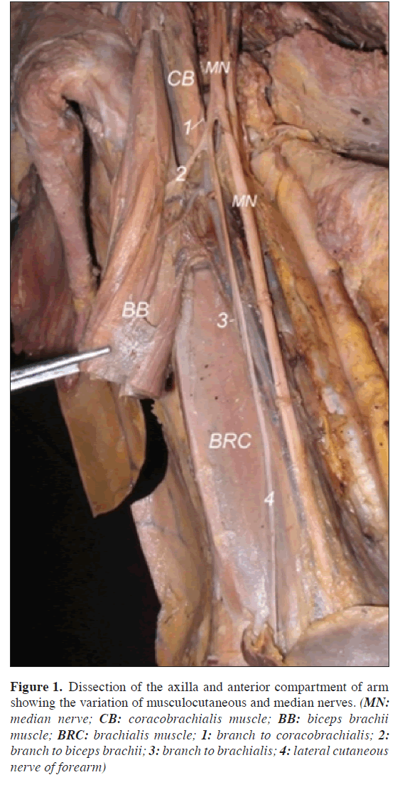
In an adult male cadaver MN was seen to be different from its usual course. In the right upper limb, a branch of MN represents MCN. The motor branches to the muscles of anterior compartment of the right upper arm (i.e. coracobrachialis, biceps brachii, brachialis) found to arise from the branch of the right MN (Figure 1) and the same continued as the lateral cutaneous nerve of forearm. This branch was not passing through coracobrachialis muscle. When traced upwards, the fibers were found to be coming from the lateral root of MN. Left sided structures were as usual.
Figure 1: Dissection of the axilla and anterior compartment of arm showing the variation of musculocutaneous and median nerves. (MN: median nerve; CB: coracobrachialis muscle; BB: biceps brachii muscle; BRC: brachialis muscle; 1: branch to coracobrachialis; 2: branch to biceps brachii; 3: branch to brachialis; 4: lateral cutaneous nerve of forearm)
Discussion
Variants of branching pattern of MCN and MN have been well described by many authors [3,4]. Le Minor (1992) classified these variations in to five types [5]. Type 1: no communication between the MN and MCN; type 2: the fibers of medial root of MN pass through the MCN and join the MN in the middle of the arm; type 3: fibers of the lateral root of the MN pass through the MCN and after some distance leave it to form lateral root of MN; type 4: the MCN fibers join the lateral root of the MN and after some distance the MCN arise from the MN; type 5: The MCN is absent and the entire fibers of MCN pass through lateral root of MN and fibers to the muscles supplied by MCN branch out directly from MN. In this type the MCN does not pierce the coracobrachialis muscle. Present finding indicated presence of Le Minor type V variant. Other classification for variations is suggested by Venieratos and Anangnostopoulou (1998) in relation to coracobrachialis muscle [6]. Type I: communication is proximal to coracobrachialis muscle; type II: communication is distal to muscle; type III: neither the nerve nor the communicating branch pierce the coracobrachialis muscle. The present variation did not coincide with any of Venieratos’s classification.
The existence of this variation described in our case report may be attributed to the random factors influencing the mechanism of formation of limb muscles and the peripheral nerves during embryonic life. In the context that ontogeny recapitulates phylogeny; it is possible that the variation seen in the current study is the result of developmental anomaly. In human being forelimb muscles develops from mesenchyme of paraxial mesoderm in the fifth week of intrauterine life [7]. Regional expression of five Hox D (Hox D 1 to Hox D 5) genes is responsible for upper limb development [8]. The motor axons arrive at the base of limb bud; they mix to form brachial plexus in upper limb. The growth cones of axons continue in the limb bud [7]. The guidance of the developing axons is regulated by the expression of chemo-attractants and chemo-repulsunt in highly coordinated sight specific fission. The tropic substances attract the correct growth cones or support the viability of the growth cones that happen to take the right path. Tropic substances include brain-derived neurotropic growth factor, c-kit ligand, neutrin-1, neutrin-2, etc. [9]. Significant variations in nerve pattern may be result of altered signaling between mesenchymal cells and neuronal growth cones or circulatory factors at the time of fission of brachial plexus cords.
Clinical Significance
Meticulous knowledge of possible variations of MCN and the MN may endow with valuable help in the management of traumatology of shoulder joint and arm as well as in circumventing iatrogenic damage during repair operations of these regions.
References
- Standring S, Borley NR, Collins P, Crossman AR, Gatzoulis MA, Healy JC, Johnson D, Mahadevan V, Newell RLM, Wigley CB. Gray’s Anatomy. 40th Ed. Churchill Livingstone, London. 2008; 791–822.
- Linell EA. The distribution of nerves in the upper limb, with reference to variability and their clinical significance. J Anat. 1921; 55: 79–112.
- Chauhan R, Roy TS. Communication between the Median and musculocutaneous nerve- a case report. J Anat Soc India. 2002; 51: 72–75.
- Choi D, Rodriguez-Niedenfuhr M, Vazquez T, Parkin I, Sanudo JR. Patterns of connections between the musculocutaneous and median nerves in the axilla and in arm. Clin Anat. 2002; 15: 11–17.
- Le Minor JM. A rare variation of the median and musculocutaneous nerves in man. Arch Anat Histol Embryol. 1990; 73: 33–42. (French)
- Venieratos D, Anagnostopoulou S. Classification of communications between the musculocutaneous and median nerves. Clin Anat. 1998; 11: 327–331.
- Moore KL, Persaud TVN. Before we are born. 7th Ed. The musculoskeletal system. Philadelphia, Saunders Elsevier. 2003; 243–244.
- Morgan BA, Tabin C. Hox genes and growth: early and late roles in limb bud morphogenesis. Dev Suppl. 1994: 181–186.
- Larson WJ. Human Embryology. Development of peripheral nervous system. 3rd Ed. Pennsylvania, Churchill Livingstone. 2001; 115–116.
Yogesh A Sontakke*, RR Fulzele, DW Tamgire, Manoj Joshi, Ujwal L Gajbe and Ravindra R Marathe
Department of Anatomy, Jawaharlal Nehru Medical College, Sawangi (Meghe), Wardha, M.S., India
- *Corresponding Author:
- Dr. Yogesh A Sontakke, MBBS, MD
Department of Anatomy, Jawaharlal Nehru Medical College Wardha 442004, M.S., India
Tel: +91 988 1013826
E-mail: dryogeshas@rediffmail.com
Date of Received: August 26th, 2009
Date of Accepted: March 5th, 2010
Published Online: April 23rd, 2010
© Int J Anat Var (IJAV). 2010; 3: 59–60.
Abstract
Musculocutaneous nerve branch out from lateral cord of brachial plexus. It innervates coracobrachialis, biceps brachii and brachialis muscles and continues as the lateral cutaneous nerve of forearm without exhibiting any communication with median nerve or any other nerve. Here, unilateral variant origin of musculocutaneous nerve is reported. In an adult male cadaver, a branch of median nerve represents musculocutaneous nerve which supplies coracobrachialis, biceps brachii and brachialis muscles and continues as lateral cutaneous nerve of forearm. This branch does not pass through coracobrachialis muscle. Such several variations surgeons should keep in mind while performing surgeries of axilla and upper arm.
-Keywords
anatomical variant, musculocutaneous nerve, median nerve, lateral cutaneous nerve of forearm
Introduction
As per the medical and surgical aspects, nerve supply of arm is very important. The musculocutaneous nerve (MCN) branch out from lateral cord of brachial plexus. It passes through coracobrachialis muscle to innervate it and biceps brachii muscle. Then passes between biceps brachii and brachialis muscles, gives a branch to supply later and continues as the lateral cutaneous nerve of forearm without exhibiting any communication with median nerve (MN) or any other nerve. MN passes through anterior compartment of arm without innervating any muscle [1].
Variations in the formation and branching pattern of brachial plexus and the nerves supplying upper limb are common and they are reported since 19th century [2]. Anatomical variations of peripheral nerves are important to medical staff especially to orthopedic surgeons, neurophysicians, physiotherapist and radiologists. Such comprehension is useful in nerve grafting, neurophysiological evaluation for diagnosing peripheral neuropathies. Present variation was observed during routine dissection at the Department of Anatomy, Jawaharlal Nehru Medical College.
Case Report
In an adult male cadaver MN was seen to be different from its usual course. In the right upper limb, a branch of MN represents MCN. The motor branches to the muscles of anterior compartment of the right upper arm (i.e. coracobrachialis, biceps brachii, brachialis) found to arise from the branch of the right MN (Figure 1) and the same continued as the lateral cutaneous nerve of forearm. This branch was not passing through coracobrachialis muscle. When traced upwards, the fibers were found to be coming from the lateral root of MN. Left sided structures were as usual.
Figure 1: Dissection of the axilla and anterior compartment of arm showing the variation of musculocutaneous and median nerves. (MN: median nerve; CB: coracobrachialis muscle; BB: biceps brachii muscle; BRC: brachialis muscle; 1: branch to coracobrachialis; 2: branch to biceps brachii; 3: branch to brachialis; 4: lateral cutaneous nerve of forearm)
Discussion
Variants of branching pattern of MCN and MN have been well described by many authors [3,4]. Le Minor (1992) classified these variations in to five types [5]. Type 1: no communication between the MN and MCN; type 2: the fibers of medial root of MN pass through the MCN and join the MN in the middle of the arm; type 3: fibers of the lateral root of the MN pass through the MCN and after some distance leave it to form lateral root of MN; type 4: the MCN fibers join the lateral root of the MN and after some distance the MCN arise from the MN; type 5: The MCN is absent and the entire fibers of MCN pass through lateral root of MN and fibers to the muscles supplied by MCN branch out directly from MN. In this type the MCN does not pierce the coracobrachialis muscle. Present finding indicated presence of Le Minor type V variant. Other classification for variations is suggested by Venieratos and Anangnostopoulou (1998) in relation to coracobrachialis muscle [6]. Type I: communication is proximal to coracobrachialis muscle; type II: communication is distal to muscle; type III: neither the nerve nor the communicating branch pierce the coracobrachialis muscle. The present variation did not coincide with any of Venieratos’s classification.
The existence of this variation described in our case report may be attributed to the random factors influencing the mechanism of formation of limb muscles and the peripheral nerves during embryonic life. In the context that ontogeny recapitulates phylogeny; it is possible that the variation seen in the current study is the result of developmental anomaly. In human being forelimb muscles develops from mesenchyme of paraxial mesoderm in the fifth week of intrauterine life [7]. Regional expression of five Hox D (Hox D 1 to Hox D 5) genes is responsible for upper limb development [8]. The motor axons arrive at the base of limb bud; they mix to form brachial plexus in upper limb. The growth cones of axons continue in the limb bud [7]. The guidance of the developing axons is regulated by the expression of chemo-attractants and chemo-repulsunt in highly coordinated sight specific fission. The tropic substances attract the correct growth cones or support the viability of the growth cones that happen to take the right path. Tropic substances include brain-derived neurotropic growth factor, c-kit ligand, neutrin-1, neutrin-2, etc. [9]. Significant variations in nerve pattern may be result of altered signaling between mesenchymal cells and neuronal growth cones or circulatory factors at the time of fission of brachial plexus cords.
Clinical Significance
Meticulous knowledge of possible variations of MCN and the MN may endow with valuable help in the management of traumatology of shoulder joint and arm as well as in circumventing iatrogenic damage during repair operations of these regions.
References
- Standring S, Borley NR, Collins P, Crossman AR, Gatzoulis MA, Healy JC, Johnson D, Mahadevan V, Newell RLM, Wigley CB. Gray’s Anatomy. 40th Ed. Churchill Livingstone, London. 2008; 791–822.
- Linell EA. The distribution of nerves in the upper limb, with reference to variability and their clinical significance. J Anat. 1921; 55: 79–112.
- Chauhan R, Roy TS. Communication between the Median and musculocutaneous nerve- a case report. J Anat Soc India. 2002; 51: 72–75.
- Choi D, Rodriguez-Niedenfuhr M, Vazquez T, Parkin I, Sanudo JR. Patterns of connections between the musculocutaneous and median nerves in the axilla and in arm. Clin Anat. 2002; 15: 11–17.
- Le Minor JM. A rare variation of the median and musculocutaneous nerves in man. Arch Anat Histol Embryol. 1990; 73: 33–42. (French)
- Venieratos D, Anagnostopoulou S. Classification of communications between the musculocutaneous and median nerves. Clin Anat. 1998; 11: 327–331.
- Moore KL, Persaud TVN. Before we are born. 7th Ed. The musculoskeletal system. Philadelphia, Saunders Elsevier. 2003; 243–244.
- Morgan BA, Tabin C. Hox genes and growth: early and late roles in limb bud morphogenesis. Dev Suppl. 1994: 181–186.
- Larson WJ. Human Embryology. Development of peripheral nervous system. 3rd Ed. Pennsylvania, Churchill Livingstone. 2001; 115–116.


