Variant arteries at the base of the brain
Satheesha Nayak B.1*, Somayaji SN1 and Soumya KV2
1Department of Anatomy, Melaka Manipal Medical College (Manipal Campus), Madhav Nagar, Manipal, Karnataka State, India
2Department of Mathematics, KMC International Centre, Madhav Nagar, Manipal, Karnataka State, India
- *Corresponding Author:
- Dr.Satheesha Nayak B., MSc, PhD
Associate Professor of Anatomy, Melaka Manipal Medical College (Manipal Campus), International Centre for Health Sciences, Madhav Nagar, Manipal, Udupi District, 576104, Karnataka State, India
Tel: +91 820 2922519
Fax: +91 820 2571905
E-mail: nayaksathish@yahoo.com
Date of Received: January 22th, 2009
Date of Accepted: June 9th, 2009
Published Online: June 10th, 2009
© IJAV. 2009; 2: 60–61.
[ft_below_content] =>Keywords
Circle of Willis, posterior cerebral artery, anterior cerebral artery, anterior communicating artery, posterior communicating artery, vertebral artery, internal carotid artery
Introduction
The base of the brain is the place where the vertebro-basilar system and internal carotid system of vessels anastomose. The anastomosis of these two systems occurs in the interpeduncular cistern and forms an arterial circle called ‘Circle of Willis’. This anastomosis assists to slow down the blood before it reaches the brain and helps in collateral circulation. The pulsations of the arteries also help in drainage of the cerebrospinal fluid in the interpeduncular cistern.
The internal carotid artery terminates by dividing into anterior and middle cerebral arteries. It also gives anterior choroidal, posterior communicating and ophthalmic arteries at the base of the brain. The basilar artery is formed by the union of right and left vertebral arteries at the lower border of the pons. It terminates by dividing into two posterior cerebral arteries at the upper border of the pons.
The anterior part of the circle of Willis is formed by the two anterior cerebral arteries and anterior communicating artery; the lateral part of the circle is formed by the two posterior communicating arteries and the posterior part of the circle is formed by the basilar artery with its two terminal branches.
In the present case, there were two anterior communicating arteries and an abnormally large left posterior communicating artery.
Case Report
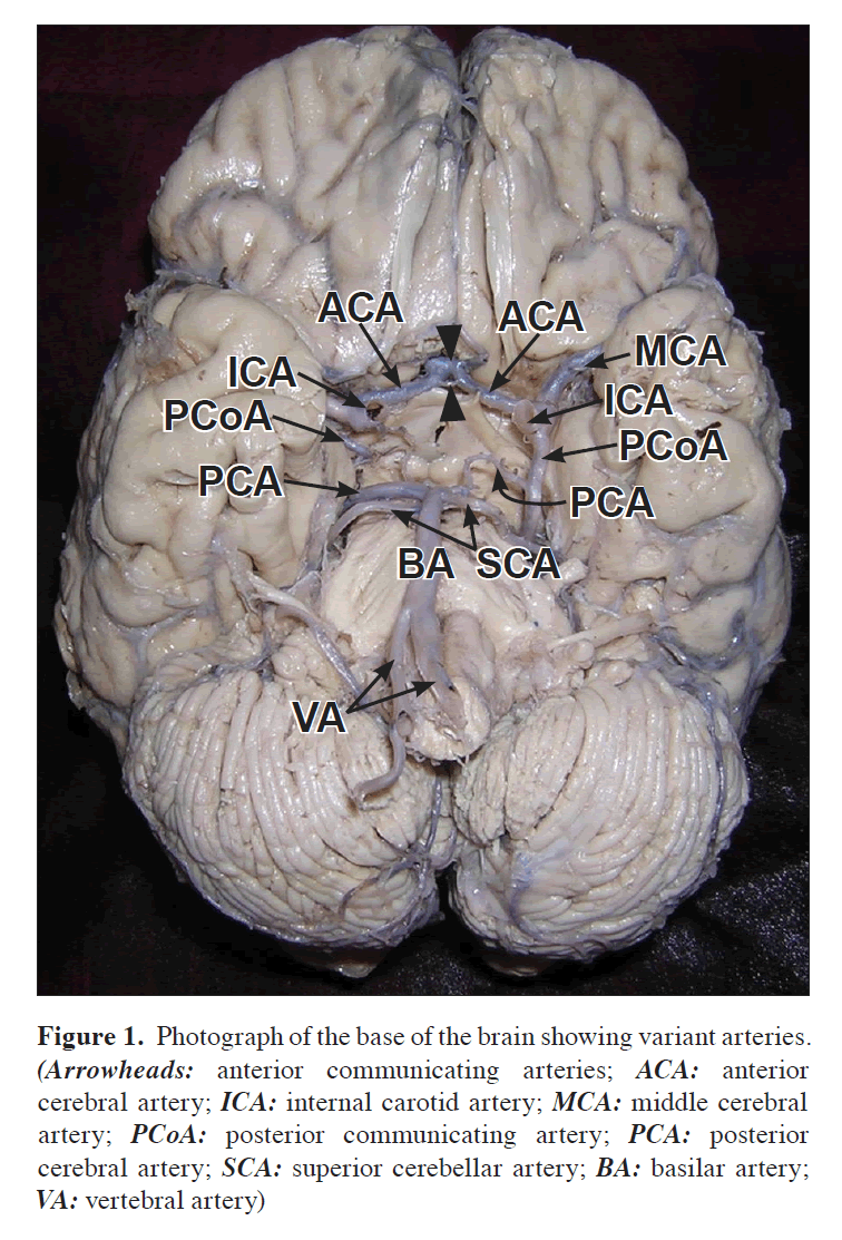
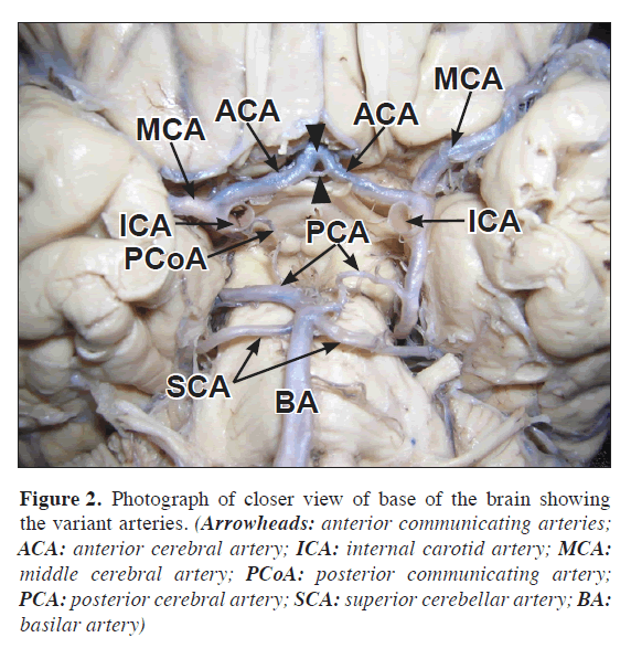
During routine dissection classes for undergraduate medical students, arterial anomalies were noted at the base of the brain, in an approximately 65-year-old male cadaver. The left internal carotid artery was slightly larger compared to the right internal carotid artery. It terminated by dividing into anterior cerebral and middle cerebral arteries. It gave a large posterior communicating artery, which was almost same in size as that of the middle cerebral artery (Figures 1, 2). The basilar artery terminated at the upper border of the pons by dividing into right and left posterior cerebral arteries. The left posterior cerebral artery was smaller than that of the right. The left posterior cerebral artery merged with the left posterior communicating artery and thereafter, the posterior communicating artery continued as the posterior cerebral artery. The two anterior cerebral arteries were connected to one another by two anterior communicating arteries, out of which the proximal one was much smaller than the distal (Figures 1, 2). The left vertebral artery was larger than the right vertebral artery (Figure 1).
Figure 1. Photograph of the base of the brain showing variant arteries. (Arrowheads: anterior communicating arteries; ACA: anterior cerebral artery; ICA: internal carotid artery; MCA: middle cerebral artery; PCoA: posterior communicating artery; PCA: posterior cerebral artery; SCA: superior cerebellar artery; BA: basilar artery; VA: vertebral artery)
Figure 2. Photograph of closer view of base of the brain showing the variant arteries. (Arrowheads: anterior communicating arteries; ACA: anterior cerebral artery; ICA: internal carotid artery; MCA: middle cerebral artery; PCoA: posterior communicating artery; PCA: posterior cerebral artery; SCA: superior cerebellar artery; BA: basilar artery)
Discussion
Variations in the origin, termination and distribution of the arteries at the base of the brain are common. The disappearance of the vessels that normally persist or the persistence of the vessels that normally disappear or formation of new vessels due to hemodynamic factors is the probable reason for the anomalies. In most of the arterial variations the brain function may not be affected due to the collateral circulation and compensation from the arteries of the other side. In a report by Kapoor et al., the circle of Willis showed variations in 54.8% of cases [1]. In the same study, the circle was absent in 3.2% of cases; the anterior cerebral artery was absent in 0.4%; the anterior communicating artery was absent in 1.8%; multiplication of posterior cerebral artery was observed in 2.4% cases while it was hypoplastic in 10.6% brains; posterior communicating artery was absent in 1% of cases. In a study on the Northwest Indian brains, the posterior communicating artery showed aneurysms in 0.92% of cases [2]. The variations of the right posterior communicating artery are more common than the left as evident by a study conducted by Ardakani et al. [3]. Anterior communicating arteries may be duplicated or absent [4]. In a study by Merkkola et al., anterior communicating artery was absent in 22% of cases and left posterior communicating arteries were absent in 46% of cases [5]. In such cases there was defective perfusion of blood into the left hemisphere. In a report by Satheesha Nayak, the A1 segment of left anterior cerebral artery the left posterior communicating artery was absent [6].
Variations of the posterior cerebral artery are rare. In a study by Caruso et al., it showed variations in 3% of cases [7]. The variations they noted include the duplication of its P1 segment, its fenestration and a common trunk for the origin of itself and superior cerebellar artery. Very rarely the proximal segment of the posterior cerebral artery may be reduced in size [8]. In such cases, the distal part of the artery will be replaced or reinforced by the posterior communicating artery, which will be very large as in our case.
Though variations of the arteries at the base of the brain are common, it is important to be aware of all the variations. If there are more than one variations, as presented in this case report, they may breed problems in the blood flow to the brain and they may cause confusions in radiological procedures.
References
- Kapoor K, Singh B, Dewan LI. Variations in the configuration of the circle of Willis. Anat Sci Int. 2008; 83: 96–106.
- Sahni D, Jit I, Lal V. Variations and anomalies of the posterior communicating artery in Northwest Indian brains. Surg Neurol. 2007; 68: 449–453.
- Ardakani SK, Dadmehr M, Nejat F, Ansari S, Eftekhar B, Tajik P, El Khashab M, Yazdani S, Ghodsi M, Mahjoub F, Monajemzadeh M, Nazparvar B, Abdi-Rad A. The cerebral arterial circle (circulus arteriosus cerebri): an anatomical study in fetus and infant samples. Pediatr Neurosurg. 2008; 44: 388–392.
- Gurdal E, Cakmak O, Yalcinkaya M, Uzun I, Cavdar S. Two variations of the anterior communicating artery: a clinical reminder. Neuroanatomy. 2004; 3: 32–34.
- Merkkola P, Tulla H, Ronkainen A, Soppi V, Oksala A, Koivisto T, Hippelainen M. Incomplete circle of Willis and right axillary artery perfusion. Ann Thorac Surg. 2006; 82: 74–79.
- Nayak SB. Anomalous arteries at the base of the brain – a case report. Neuroanatomy. 2008; 7: 45–46.
- Caruso G, Vincentelli F, Rabehanta P, Giudicelli G, Grisoli F. Anomalies of the P1 segment of the posterior cerebral artery: early bifurcation or duplication, fenestration, common trunk with the superior cerebellar artery. Acta Neurochir (Wien). 1991; 109: 66–71.
- Bergman RA, Afifi AK, Miyauchi R. Posterior Cerebral Artery. Illustrated Encyclopedia of Human Anatomic Variation: Opus II: Cardiovascular System: Arteries: Head, Neck, and Thorax. http://www.anatomyatlases.org/AnatomicVariants/Cardiovascular/Text/Arteries/CerebralPosterior.shtml (accessed on 17 January 2009).
Satheesha Nayak B.1*, Somayaji SN1 and Soumya KV2
1Department of Anatomy, Melaka Manipal Medical College (Manipal Campus), Madhav Nagar, Manipal, Karnataka State, India
2Department of Mathematics, KMC International Centre, Madhav Nagar, Manipal, Karnataka State, India
- *Corresponding Author:
- Dr.Satheesha Nayak B., MSc, PhD
Associate Professor of Anatomy, Melaka Manipal Medical College (Manipal Campus), International Centre for Health Sciences, Madhav Nagar, Manipal, Udupi District, 576104, Karnataka State, India
Tel: +91 820 2922519
Fax: +91 820 2571905
E-mail: nayaksathish@yahoo.com
Date of Received: January 22th, 2009
Date of Accepted: June 9th, 2009
Published Online: June 10th, 2009
© IJAV. 2009; 2: 60–61.
Abstract
Abnormalities of the important arteries at the base of brain can lead to serious clinical conditions like stroke or other functional deficits. We report here a variant pattern of arteries seen at the base of the brain. The left internal carotid artery was slightly larger than the right and it terminated by dividing into anterior and middle cerebral arteries. It gave an abnormally large posterior communicating artery, which was almost equal in size to that of the middle cerebral artery. This posterior communicating artery replaced the distal part of the left posterior cerebral artery. The proximal part of the left posterior cerebral artery was very small. There were two anterior communicating arteries. The left vertebral artery was larger than the right vertebral artery. The knowledge of these anomalous arteries may be useful for radiologists, neurosurgeons and clinicians in general.
-Keywords
Circle of Willis, posterior cerebral artery, anterior cerebral artery, anterior communicating artery, posterior communicating artery, vertebral artery, internal carotid artery
Introduction
The base of the brain is the place where the vertebro-basilar system and internal carotid system of vessels anastomose. The anastomosis of these two systems occurs in the interpeduncular cistern and forms an arterial circle called ‘Circle of Willis’. This anastomosis assists to slow down the blood before it reaches the brain and helps in collateral circulation. The pulsations of the arteries also help in drainage of the cerebrospinal fluid in the interpeduncular cistern.
The internal carotid artery terminates by dividing into anterior and middle cerebral arteries. It also gives anterior choroidal, posterior communicating and ophthalmic arteries at the base of the brain. The basilar artery is formed by the union of right and left vertebral arteries at the lower border of the pons. It terminates by dividing into two posterior cerebral arteries at the upper border of the pons.
The anterior part of the circle of Willis is formed by the two anterior cerebral arteries and anterior communicating artery; the lateral part of the circle is formed by the two posterior communicating arteries and the posterior part of the circle is formed by the basilar artery with its two terminal branches.
In the present case, there were two anterior communicating arteries and an abnormally large left posterior communicating artery.
Case Report
During routine dissection classes for undergraduate medical students, arterial anomalies were noted at the base of the brain, in an approximately 65-year-old male cadaver. The left internal carotid artery was slightly larger compared to the right internal carotid artery. It terminated by dividing into anterior cerebral and middle cerebral arteries. It gave a large posterior communicating artery, which was almost same in size as that of the middle cerebral artery (Figures 1, 2). The basilar artery terminated at the upper border of the pons by dividing into right and left posterior cerebral arteries. The left posterior cerebral artery was smaller than that of the right. The left posterior cerebral artery merged with the left posterior communicating artery and thereafter, the posterior communicating artery continued as the posterior cerebral artery. The two anterior cerebral arteries were connected to one another by two anterior communicating arteries, out of which the proximal one was much smaller than the distal (Figures 1, 2). The left vertebral artery was larger than the right vertebral artery (Figure 1).
Figure 1. Photograph of the base of the brain showing variant arteries. (Arrowheads: anterior communicating arteries; ACA: anterior cerebral artery; ICA: internal carotid artery; MCA: middle cerebral artery; PCoA: posterior communicating artery; PCA: posterior cerebral artery; SCA: superior cerebellar artery; BA: basilar artery; VA: vertebral artery)
Figure 2. Photograph of closer view of base of the brain showing the variant arteries. (Arrowheads: anterior communicating arteries; ACA: anterior cerebral artery; ICA: internal carotid artery; MCA: middle cerebral artery; PCoA: posterior communicating artery; PCA: posterior cerebral artery; SCA: superior cerebellar artery; BA: basilar artery)
Discussion
Variations in the origin, termination and distribution of the arteries at the base of the brain are common. The disappearance of the vessels that normally persist or the persistence of the vessels that normally disappear or formation of new vessels due to hemodynamic factors is the probable reason for the anomalies. In most of the arterial variations the brain function may not be affected due to the collateral circulation and compensation from the arteries of the other side. In a report by Kapoor et al., the circle of Willis showed variations in 54.8% of cases [1]. In the same study, the circle was absent in 3.2% of cases; the anterior cerebral artery was absent in 0.4%; the anterior communicating artery was absent in 1.8%; multiplication of posterior cerebral artery was observed in 2.4% cases while it was hypoplastic in 10.6% brains; posterior communicating artery was absent in 1% of cases. In a study on the Northwest Indian brains, the posterior communicating artery showed aneurysms in 0.92% of cases [2]. The variations of the right posterior communicating artery are more common than the left as evident by a study conducted by Ardakani et al. [3]. Anterior communicating arteries may be duplicated or absent [4]. In a study by Merkkola et al., anterior communicating artery was absent in 22% of cases and left posterior communicating arteries were absent in 46% of cases [5]. In such cases there was defective perfusion of blood into the left hemisphere. In a report by Satheesha Nayak, the A1 segment of left anterior cerebral artery the left posterior communicating artery was absent [6].
Variations of the posterior cerebral artery are rare. In a study by Caruso et al., it showed variations in 3% of cases [7]. The variations they noted include the duplication of its P1 segment, its fenestration and a common trunk for the origin of itself and superior cerebellar artery. Very rarely the proximal segment of the posterior cerebral artery may be reduced in size [8]. In such cases, the distal part of the artery will be replaced or reinforced by the posterior communicating artery, which will be very large as in our case.
Though variations of the arteries at the base of the brain are common, it is important to be aware of all the variations. If there are more than one variations, as presented in this case report, they may breed problems in the blood flow to the brain and they may cause confusions in radiological procedures.
References
- Kapoor K, Singh B, Dewan LI. Variations in the configuration of the circle of Willis. Anat Sci Int. 2008; 83: 96–106.
- Sahni D, Jit I, Lal V. Variations and anomalies of the posterior communicating artery in Northwest Indian brains. Surg Neurol. 2007; 68: 449–453.
- Ardakani SK, Dadmehr M, Nejat F, Ansari S, Eftekhar B, Tajik P, El Khashab M, Yazdani S, Ghodsi M, Mahjoub F, Monajemzadeh M, Nazparvar B, Abdi-Rad A. The cerebral arterial circle (circulus arteriosus cerebri): an anatomical study in fetus and infant samples. Pediatr Neurosurg. 2008; 44: 388–392.
- Gurdal E, Cakmak O, Yalcinkaya M, Uzun I, Cavdar S. Two variations of the anterior communicating artery: a clinical reminder. Neuroanatomy. 2004; 3: 32–34.
- Merkkola P, Tulla H, Ronkainen A, Soppi V, Oksala A, Koivisto T, Hippelainen M. Incomplete circle of Willis and right axillary artery perfusion. Ann Thorac Surg. 2006; 82: 74–79.
- Nayak SB. Anomalous arteries at the base of the brain – a case report. Neuroanatomy. 2008; 7: 45–46.
- Caruso G, Vincentelli F, Rabehanta P, Giudicelli G, Grisoli F. Anomalies of the P1 segment of the posterior cerebral artery: early bifurcation or duplication, fenestration, common trunk with the superior cerebellar artery. Acta Neurochir (Wien). 1991; 109: 66–71.
- Bergman RA, Afifi AK, Miyauchi R. Posterior Cerebral Artery. Illustrated Encyclopedia of Human Anatomic Variation: Opus II: Cardiovascular System: Arteries: Head, Neck, and Thorax. http://www.anatomyatlases.org/AnatomicVariants/Cardiovascular/Text/Arteries/CerebralPosterior.shtml (accessed on 17 January 2009).


