Abnormal foramina on the posterior arch of atlas vertebra
Satheesha Nayak B*
Department of Anatomy, Melaka Manipal Medical College (Manipal Campus) Manipal, India.
- *Corresponding Author:
- Satheesha Nayak B., MSc. PhD
Associate Professor of Anatomy, Melaka Manipal Medical College (Manipal Campus), International Centre for Health Sciences, Madhav Nagar, Manipal, Udupi District, Karnataka State, 576 104 India.
Tel: +91 820 2922519
Fax: +91 820 2571905
E-mail: nayaksathish@yahoo.com
Date of Received: July 30th, 2008
Date of Accepted: September 22nd, 2008
Published Online: October 13th, 2008
© IJAV. 2008; 1: 21–22.
[ft_below_content] =>Keywords
atlas vertebra,posterior arch,foramen,cervical vertebra,vertebra
Introduction
Atlas is the first cervical vertebra. It is ring shaped and does not have a body like other cervical vertebrae. It has two arches named anterior and posterior arches. The anterior arch is shorter than the posterior arch and articulates with the dens of the axis vertebra. The posterior arch bears a groove on its superior surface for the vertebral artery and the dorsal ramus of the first cervical spinal nerve. It has two transverse processes, each one of which bears a foramen transversarium. The vertebral artery passes through this foramen. The atlas has two parts called lateral masses. The lateral masses articulate with the occipital condyles to form ellipsoid type of synovial joints. We noted abnormal foramina in the posterior arch of the atlas vertebra.
Case Report
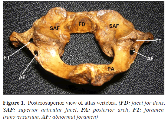
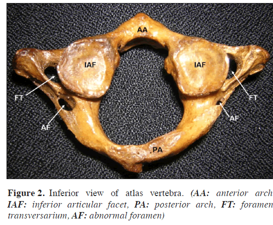
During the osteology demonstration classes for undergraduate medical students, we noticed two abnormal foramina on the posterior arch of the atlas vertebra. The foramina were situated at the lateral part of the posterior arch, one behind each lateral masses (Figures 1 and 2). The foramen on the right side was larger than the left and was almost as large as the foramen transversarium. The foramen on the left was almost half the size of that of the right. There were foramina tranversaria on both transverse processes and their sizes were normal. The posterior arch of the atlas had the groove for the vertebral artery. There were no other abnormalities in the bone.
Discussion
Atlas vertebra shows greatest variability among the cervical vertebrae. The reported variations of atlas include partial or total fusion of atlas vertebra with the occipital bone [1,2]. In a recent study conducted by Wysocki et al., [3], atlas showed the highest variability among the cervical vertebrae. The variations recorded in their study include the split superior articular process (47.8%), split posterior (3%) or anterior (1%) arches, and the presence of some accessory bony arches embracing the vertebral artery.
Absence of foramen transversarium in atlas is a very rare variation. Absence of foramen transversarium unilaterally, on the left side has been reported [4]. In this case however, the groove for vertebral artery was present on the posterior arch. Bilateral absence of foramen transversarium has been reported by Satheesha Nayak [5]. Bergman et al., [6] have reported many variations of atlas vertebra. According to their study, atlas may show incomplete ossification of anterior and posterior arches, anterior arch may be absent, posterior arch may have facets and groove for vertebral artery may get converted into a foramen. Existence of a foramen called “retroarticular foramen” has been reported. In a study on South Africans, retroarticular canal was found in 9.8% of cases [7]. Presence of such a foramen might hinder the flow of blood in the vertebral artery and also make instrumentation in the area difficult. Presence of a “retrotransverse groove/canal” has also been reported [8]. Such a canal was found in 14.7% of cases. The foramen that we have reported here might fall in one of the categories (retroarticular or retrotransverse) reported earlier. Since it was noted in a dry bone, we are not very sure about the structures passing through it. The knowledge of this foramen may be needed for the orthopedic surgeons and neurosurgeons. The radiologists and anthropologists may also benefit from this report.
References
- Nayak S. Asymmetric atlas assimilation and potential danger to the brainstem - a case report. The Internet Journal of Biological Anthropology. 2008;1 (2). http://www.ispub.com/ostia/index.php?xmlFilePath=journals/ijba/vol1n2/atlas.xml (accessed July 2008).
- Nayak S, Vollala VR, Raghunathan D. Total fusion of atlas with occipital bone: a case report. Neuroanatomy. 2005; 4: 39–40.
- Wysocki J, Bubrowski M, Reymond J, Kwiatkowski J. Anatomical variants of the cervical vertebrae and the first thoracic vertebra in man. Folia Morphol. (Warsz). 2003; 62: 357–363.
- Vasudeva N, Kumar R. Absence of foramen transversarium in the human atlas vertebra: a case report. Acta Anat. (Basel). 1995; 152: 230–233.
- Nayak S. Bilateral absence of foramen transversarium in atlas vertebra: a case report. Neuroanatomy. 2007; 6: 28–29.
- Bergman RA, Afifi AK, Miyauchi R. Skeletal systems: Cranium. In: Compendium of human anatomical variations. Baltimore, Urban and Schwarzenberg. 1988; 197–205.
- Mitchell J. The incidence and dimensions of the retroarticular canal of the atlas vertebra. Acta Anat. (Basel). 1998; 163: 113–120.
- Bilodi AK, Gupta SC. Presence of retro transverse groove or canal in atlas vertebrae. J. Anat. Soc. India. 2005; 54: 16–18.
Satheesha Nayak B*
Department of Anatomy, Melaka Manipal Medical College (Manipal Campus) Manipal, India.
- *Corresponding Author:
- Satheesha Nayak B., MSc. PhD
Associate Professor of Anatomy, Melaka Manipal Medical College (Manipal Campus), International Centre for Health Sciences, Madhav Nagar, Manipal, Udupi District, Karnataka State, 576 104 India.
Tel: +91 820 2922519
Fax: +91 820 2571905
E-mail: nayaksathish@yahoo.com
Date of Received: July 30th, 2008
Date of Accepted: September 22nd, 2008
Published Online: October 13th, 2008
© IJAV. 2008; 1: 21–22.
Abstract
Atlas is the first cervical vertebra. It articulates with the occipital bone above and the axis vertebra below. It plays an important role in movement of the skull and the neck. We found a rare variation of the atlas vertebra. The posterior arch of the atlas had one accessory foramen just behind each lateral mass. Foramen on the right side was larger than that of the left. The knowledge of this variation may be of importance to orthopedic surgeons, neurosurgeons, radiologists and anthropologists.
-Keywords
atlas vertebra,posterior arch,foramen,cervical vertebra,vertebra
Introduction
Atlas is the first cervical vertebra. It is ring shaped and does not have a body like other cervical vertebrae. It has two arches named anterior and posterior arches. The anterior arch is shorter than the posterior arch and articulates with the dens of the axis vertebra. The posterior arch bears a groove on its superior surface for the vertebral artery and the dorsal ramus of the first cervical spinal nerve. It has two transverse processes, each one of which bears a foramen transversarium. The vertebral artery passes through this foramen. The atlas has two parts called lateral masses. The lateral masses articulate with the occipital condyles to form ellipsoid type of synovial joints. We noted abnormal foramina in the posterior arch of the atlas vertebra.
Case Report
During the osteology demonstration classes for undergraduate medical students, we noticed two abnormal foramina on the posterior arch of the atlas vertebra. The foramina were situated at the lateral part of the posterior arch, one behind each lateral masses (Figures 1 and 2). The foramen on the right side was larger than the left and was almost as large as the foramen transversarium. The foramen on the left was almost half the size of that of the right. There were foramina tranversaria on both transverse processes and their sizes were normal. The posterior arch of the atlas had the groove for the vertebral artery. There were no other abnormalities in the bone.
Discussion
Atlas vertebra shows greatest variability among the cervical vertebrae. The reported variations of atlas include partial or total fusion of atlas vertebra with the occipital bone [1,2]. In a recent study conducted by Wysocki et al., [3], atlas showed the highest variability among the cervical vertebrae. The variations recorded in their study include the split superior articular process (47.8%), split posterior (3%) or anterior (1%) arches, and the presence of some accessory bony arches embracing the vertebral artery.
Absence of foramen transversarium in atlas is a very rare variation. Absence of foramen transversarium unilaterally, on the left side has been reported [4]. In this case however, the groove for vertebral artery was present on the posterior arch. Bilateral absence of foramen transversarium has been reported by Satheesha Nayak [5]. Bergman et al., [6] have reported many variations of atlas vertebra. According to their study, atlas may show incomplete ossification of anterior and posterior arches, anterior arch may be absent, posterior arch may have facets and groove for vertebral artery may get converted into a foramen. Existence of a foramen called “retroarticular foramen” has been reported. In a study on South Africans, retroarticular canal was found in 9.8% of cases [7]. Presence of such a foramen might hinder the flow of blood in the vertebral artery and also make instrumentation in the area difficult. Presence of a “retrotransverse groove/canal” has also been reported [8]. Such a canal was found in 14.7% of cases. The foramen that we have reported here might fall in one of the categories (retroarticular or retrotransverse) reported earlier. Since it was noted in a dry bone, we are not very sure about the structures passing through it. The knowledge of this foramen may be needed for the orthopedic surgeons and neurosurgeons. The radiologists and anthropologists may also benefit from this report.
References
- Nayak S. Asymmetric atlas assimilation and potential danger to the brainstem - a case report. The Internet Journal of Biological Anthropology. 2008;1 (2). http://www.ispub.com/ostia/index.php?xmlFilePath=journals/ijba/vol1n2/atlas.xml (accessed July 2008).
- Nayak S, Vollala VR, Raghunathan D. Total fusion of atlas with occipital bone: a case report. Neuroanatomy. 2005; 4: 39–40.
- Wysocki J, Bubrowski M, Reymond J, Kwiatkowski J. Anatomical variants of the cervical vertebrae and the first thoracic vertebra in man. Folia Morphol. (Warsz). 2003; 62: 357–363.
- Vasudeva N, Kumar R. Absence of foramen transversarium in the human atlas vertebra: a case report. Acta Anat. (Basel). 1995; 152: 230–233.
- Nayak S. Bilateral absence of foramen transversarium in atlas vertebra: a case report. Neuroanatomy. 2007; 6: 28–29.
- Bergman RA, Afifi AK, Miyauchi R. Skeletal systems: Cranium. In: Compendium of human anatomical variations. Baltimore, Urban and Schwarzenberg. 1988; 197–205.
- Mitchell J. The incidence and dimensions of the retroarticular canal of the atlas vertebra. Acta Anat. (Basel). 1998; 163: 113–120.
- Bilodi AK, Gupta SC. Presence of retro transverse groove or canal in atlas vertebrae. J. Anat. Soc. India. 2005; 54: 16–18.


