Rare anatomical variation of the cystic artery giving off 3 branches to the gallbladder
Received: 27-Mar-2018 Accepted Date: Apr 06, 2018; Published: 19-Apr-2018, DOI: 10.37532/1308-4038.18.11.53
Citation: Indigahawela I, Arachchi A, Briggs C, et al. Rare anatomical variation of the cystic artery giving off 3 branches to the gallbladder. Int J Anat Var. 2018;11(2):53-54.
This open-access article is distributed under the terms of the Creative Commons Attribution Non-Commercial License (CC BY-NC) (http://creativecommons.org/licenses/by-nc/4.0/), which permits reuse, distribution and reproduction of the article, provided that the original work is properly cited and the reuse is restricted to noncommercial purposes. For commercial reuse, contact reprints@pulsus.com
Abstract
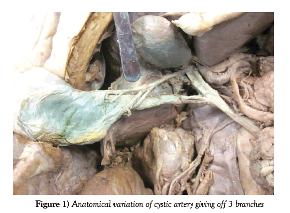
A routine cadaveric dissection demonstrated the cystic artery giving off 3 branches to the gallbladder (also known as Calot’s arteries). The arteries of Calot branched out as the cystic artery traverses along the cystic duct. A thorough literature review could not identify any studies that demonstrated the incidence of this event.
Keywords
Gallbladder; Cystic artery
Introduction
We describe a case of the cystic artery giving off 3 branches to the gallbladder. This was noted during a routine dissection of a cadaver. Variations in the cystic artery and its branches are clinically significant for General and Hepato biliary surgeons who commonly operate on the gallbladder and the hepato biliary tree.
In 80 to 90% of patients, cystic artery arises as a branch of the right hepatic artery, which has its origins from the celiac artery [1-3]. From the celiac trunk, the common hepatic artery is given off, followed by the proper hepatic artery, which splits, into the right and left hepatic arteries, with the former giving off the cystic artery. The cystic artery supplies the proximal portion of the common bile duct as well as the gallbladder itself. It most commonly originates from the cystohepatic triangle of Calot, which is bounded by the cystic duct, common hepatic duct and cystic artery itself [4]. From here, it most commonly traverses posterior to the cystic duct, reaching the neck of the gallbladder where 2-4 minor branches normally arise, also known as Calot’s arteries. Most commonly, the cystic artery branches into the deep and superficial branches at the neck of the gallbladder [1].
However, numerous anatomical variations of the origins of the cystic artery have been described. Despite a single cystic artery branching off the right hepatic artery as the most common anatomical variations, 28% of patients have 2 cystic arteries branching off from the right hepatic artery [1,2]. Less common variants of the cystic artery have them originating from the gastro duodenal artery (7.5%), liver parenchyma (2.5%) or the left hepatic artery (1%) [1,5].
The mean length of the cystic artery is 16.9 mm with ranges between 2 mm and 55 mm, depending on the origin of the cystic artery, and a mean diameter of 1.6 mm, with a range between 1mm and 5 mm [2].
Case Report
As depicted in Figure 1, the cystic artery has its origin from the right hepatic artery at the cystohepatic triangle of Calot. As it approaches the gall bladder, 2 branches are given off just proximal to the neck of the gallbladder while a single branch is given off distal to gallbladder. The distal branch is smaller in diameter than the proximal branches of the cystic artery. Routine cadaveric dissection revealed no further anatomical abnormality.
Discussion
A thorough literature review could not identify any studies that demonstrated the incidence of this anatomical variation. It is well reviewed in literature that variations in the origin and course of the cystic artery are very common. However, although it is known that there are 2 to 4 branches of the cystic artery, the prevalence of each individual occurrence is not well-researched in medical literature. The reason why is not clearly understood, and it can only be assumed that medical practitioners might identify such a study as irrelevant.
Identifying the variations in the cystic artery is important to determine the approach for operations involving the gallbladder and hepatobiliary tree. With poor visualisation intra-operatively, such branches of the cystic artery can be mistaken as the cystic artery itself, and if not correctly identified, can lead to intra-operative complications such as haemorrhage or damage to surrounding structures. Especially in a laparoscopic environment, visualisation and identification of the correct anatomy prior to proceeding is essential to reduce the risk of complications. This is particularly important for General and Hepatobiliary surgeons who commonly operate in this region.
Such anatomical variations are normally detected intra-operatively either during a laparoscopy or laparotomy [1]. Other modalities that can detect anatomical variations of the cystic artery include digital subtraction angiography or cone beam CT [6,7]. However, these are only used in the context of a hepatocellular carcinoma, in which the cystic artery is known to supply the tumour itself.
Conclusion
Although anatomical variations of the cystic artery are well documented, a thorough literature review could not identify studies that determine the prevalence of the number of branches of the cystic artery. Further research is required to determine such data. General and Hepatobiliary surgeons should fully appreciate the different variations of the cystic artery and determine the best route of approach for each operation they partake in that involves the gallbladder and the hepatobiliary tree.
REFERENCES
- Ding YM. New classification of the anatomic variations of cystic artery during laparoscopic cholecystectomy. World J Gastroenterol. 2007;13:5629-34.
- Dandekar U, Dandekar K. Cystic Artery: Morphological Study and Surgical Significance. Anat Res Int. 2016:p. 7201858.
- Hlaing KP, Thwin SS, Shwe N. Unique origin of the cystic artery. Singapore Med J. 2011;52:e262-4.
- Veeramootoo D. Calot's Triangle. A common misconception of basic anatomy. Int J Surg. 2012;10:p. S7.
- Andall RG. The clinical anatomy of cystic artery variations: a review of over 9800 cases. Surg Radiol Anat. 2016;38:529-39.
- Kinoshita M. The usefulness of cone-beam computed tomography during chemoembolization of hepatocellular carcinomas fed exclusively by the cystic artery. Jpn J Radiol. 2016;34:747-53.
- Kang B. The origin of the cystic artery supplying hepatocellular carcinoma on digital subtraction angiography in 311 patients. Cardiovasc Intervent Radiol. 2014;37:1268-82.


