Unusual branching pattern of coeliac trunk – a case report
Satheesha Nayak B*, Naveen Kumar, Anitha Guru and Surekha D Shetty
Department of Anatomy, Melaka Manipal Medical College (Manipal Campus), International Centre for Health Sciences, Manipal University, Madhav Nagar, Manipal, Karnataka, India
- *Corresponding Author:
- Dr. Satheesha Nayak B.
Professor and Head, Department of Anatomy, MMMC Int. Centre for Health Sci. Manipal University, Madhav Nagar, Manipal, Udupi District, Karnataka, 576 104, India
Tel: +91 820 2922519
E-mail: nayaksathish@yahoo.com
Date of Received: January 16th, 2012
Date of Accepted: September 23rd, 2012
Published Online: December 26th, 2012
© Int J Anat Var (IJAV). 2012; 5: 134–136.
[ft_below_content] =>Keywords
coeliac trunk, common hepatic artery, left hepatic artery, right hepatic artery, esophageal branch
Introduction
Coeliac trunk is one of the ventral splanchnic branches of abdominal aorta. It arises from the abdominal aorta opposite the intervertebral disc between T12 and L1 vertebrae. It has a short course of about 1,5 cm, after which it terminates by dividing into three branches; splenic artery, left gastric artery and the common hepatic artery. The common hepatic artery divides into gastroduodenal artery and hepatic artery proper. The gastroduodenal artery terminates by dividing into superior pancreaticoduodenal and right gastroepiploic arteries. The hepatic artery proper terminates by dividing into a right and a left hepatic artery, which supply the right and left lobes of the liver, respectively. The right hepatic artery gives a cystic branch which supplies the gallbladder. The right gastric artery may arise from the common hepatic or proper hepatic arteries. The left gastric artery, apart from supplying the stomach, gives one or two esophageal branches, which supply the terminal part of esophagus. The splenic artery gives pancreatic, left gastroepiploic and short gastric arteries in addition to the branches that supply the spleen.
Here, we report the unusual branches of the coeliac trunk and its branches.
Case Report
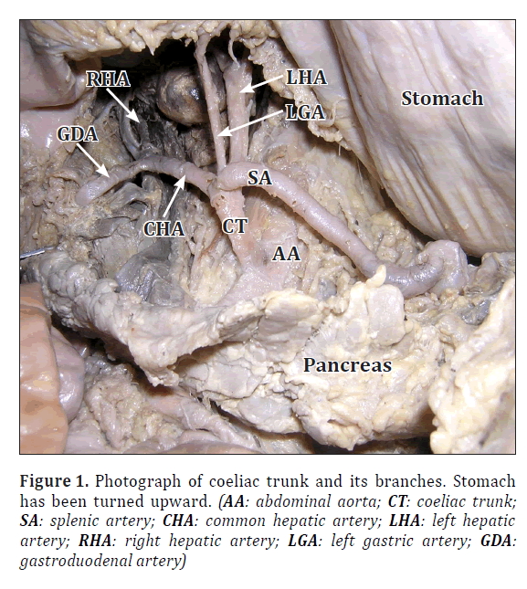
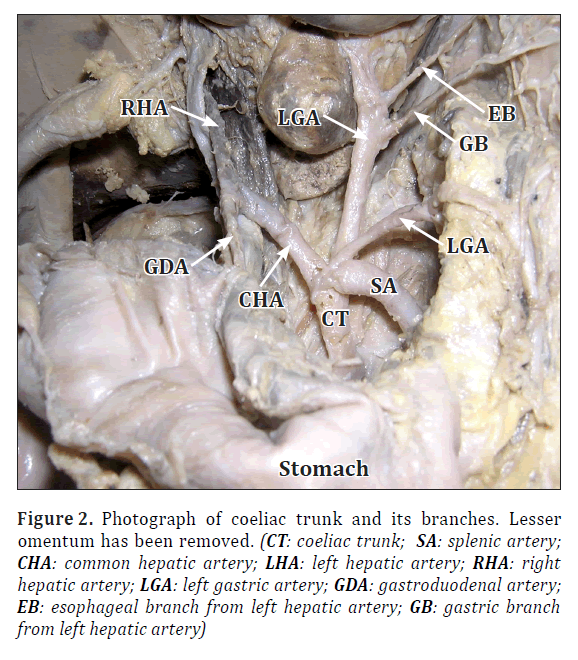
During the dissection classes for medical undergraduate students, we found these variations in a male cadaver aged approximately 65 years. The coeliac trunk was about 5 cm long and terminated by dividing into common hepatic, splenic, left gastric and left hepatic arteries (Figure 1). The common hepatic artery divided into right hepatic and gastroduodenal arteries. The right hepatic artery supplied the gallbladder through a cystic branch. The course and branches of the gastroduodenal artery were normal. The splenic artery also had the usual course and branching pattern. The left gastric artery arose from the coeliac trunk and coursed along the lesser curvature. But it did not give any esophageal branches. The left hepatic artery was larger in size than the right hepatic artery and it originated directly from the coeliac trunk (Figures 1, 2). It coursed upward and to the left and entered the liver through the left end of the porta hepatis. It gave two branches before entering the liver. The upper among the two branches, supplied the terminal part of the esophagus and the lower branch supplied the part of the stomach adjoining esophagus. There were no other significant variations in the abdomen.
Figure 2. Photograph of coeliac trunk and its branches. Lesser omentum has been removed. (CT: coeliac trunk; SA: splenic artery; CHA: common hepatic artery; LHA: left hepatic artery; RHA: right hepatic artery; LGA: left gastric artery; GDA: gastroduodenal artery; EB: esophageal branch from left hepatic artery; GB: gastric branch from left hepatic artery)
Discussion
Coeliac trunk is known to show variation in its branching pattern. It may have more than three branches [1]. Previous studies have classified coeliac trunk into 6 types based on its branching pattern [2]. The six types are as follows; Type 1: usual branching; Type 2: hepatosplenic trunk and left gastric artery coming from aorta; Type 3: hepatosplenomesenteric trunk and left gastric arising from aorta; Type 4: hepatogastric trunk and splenic artery coming from superior mesenteric artery; Type 5: splenogastric type; splenic and left gastric from the coeliac trunk and common hepatic artery from superior mesenteric artery; and Type 6: coeliacomesentric trunk; splenic, left gastric, common hepatic and superior mesenteric arteries originate from a common trunk. Existence of a common coeliaco-mesenterico-phrenic trunk has been reported by Nayak [3]. Deepthinath et al. [4] have reported a variation where the coeliac trunk gave an accessory renal artery, two testicular arteries, middle suprarenal and left inferior phrenic arteries. The coeliac trunk may be absent in 1% of cases and in such cases the common hepatic, splenic and left gastric arteries arise directly from the abdominal aorta [5,6]. In a recent study conducted on 974 cases, a total of 89.8% of cases showed the classical trifurcation of the coeliac trunk [7]. The typical pattern of the coeliac trunk and the hepatic artery was confirmed in 66.6% and 72.4% of the cadavers, respectively. Variations of left hepatic artery were noted in 11% of cases and variations of right hepatic arteries were observed in 4.9% of cases. The variation involving both the left and right hepatic arteries was found in 1.5% cases and presence of common hepatosplenic trunk and a gastrohepatic trunk were seen in 4.4% and 0.3%, respectively.
Though the variations of the common hepatic artery are rare, the right and left hepatic arteries show many variations in their course, branching and distribution. Song et al., [8] did an extensive study on the common hepatic artery and found variations in only 3.71% of cases. In rare cases, the common hepatic artery takes origin from the left gastric artery [9]. Abdullah et al., have reported the variations in the origin of hepatic arteries in 31.9% of cases [10]. In their study involving 932 patients, the variations were divided into three groups having 48 common hepatic artery variations, 236 left or right hepatic artery variations and 13 rare variations including one case of right hepatic artery stemming from the inferior mesenteric artery. Uva et al., have reported the origin of common hepatic artery from the left gastric artery [11]. In a study conducted by Ugurel et al., a hepatosplenomesenteric trunk was seen in 1% and a splenomesenteric trunk was present in 1% patients [5].
In the current case, the common hepatic, splenic, left gastric and left hepatic arteries originated from the coeliac trunk. This kind of quadrifurcation of the coeliac trunk is a rare occurrence. Origin of esophageal and gastric branches from the left hepatic artery has not been reported yet. Knowledge of these variations is important in therapeutic embolization of the left hepatic artery. In cases like what we are reporting here, if the left hepatic artery is embolized, it might result in the infarct of gastroesophageal junction. Hence this variation is of utmost importance for the radiologists and surgeons.
References
- Bergman RA, Thompson SA, Afifi AK, Saadeh FA. Compendium of Human Anatomic Variations. Baltimore, Urban & Schwarzenberg. 1988; 65.
- Michels NA. Blood supply and anatomy of the upper abdominal organs with descriptive atlas. Philadelphia, Lippincott. 1955; 1–581.
- Nayak S. Common celiaco-mesenterico-phrenic trunk and renal vascular variations. Saudi Med J. 2006; 27: 1894–1896.
- Deepthinath R, Satheesha Nayak B, Mehta RB, Bhat S, Rodrigues V, Samuel VP, Venkataramana V, Prasad AM. Multiple variations in the paired arteries of the abdominal aorta. Clin Anat. 2006; 19: 566–568.
- Ugurel MS, Battal B, Bozlar U, Nural MS, Tasar M, Ors F, Saglam M, Karademir I. Anatomical variations of hepatic arterial system, coeliac trunk and renal arteries: an analysis with multidetector CT angiography. Br J Radiol. 2010; 83: 661–667.
- Yi SQ, Terayama H, Naito M, Hirai S, Alimujang S, Yi N, Tanaka S, Itoh M. Absence of the celiac trunk: case report and review of the literature. Clin Anat. 2008; 21: 283–286.
- Chen H, Yano R, Emura S, Shoumura S. Anatomic variation of the celiac trunk with special reference to hepatic artery patterns. Ann Anat. 2009; 191: 399–407.
- Song SY, Chung JW, Yin YH, Jae HJ, Kim HC, Jeon UB, Cho BH, So YH, Park JH. Celiac axis and common hepatic artery variations in 5002 patients: systematic analysis with spiral CT and DSA. Radiology. 2010; 255: 278–288.
- Okada Y, Nishi N, Matsuo Y, Watadani T, Kimura F. The common hepatic artery arising from the left gastric artery. Surg Radiol Anat. 2010; 32: 703–705.
- Abdullah SS, Mabrut JY, Garbit V, De La Roche E, Olagne E, Rode A, Morin A, Berthezene Y, Baulieux J, Ducerf C. Anatomical variations of the hepatic artery: study of 932 cases in liver transplantation. Surg Radiol Anat. 2006; 28: 468–473.
- Uva P, Arvelakis A, Rodriguez-Laiz G, Lerner S, Emre S, Gondolesi G. Common hepatic artery arising from the left gastric artery: a rare anatomic variation identified on a cadaveric liver donor. Surg Radiol Anat. 2007; 29: 93–95.
Satheesha Nayak B*, Naveen Kumar, Anitha Guru and Surekha D Shetty
Department of Anatomy, Melaka Manipal Medical College (Manipal Campus), International Centre for Health Sciences, Manipal University, Madhav Nagar, Manipal, Karnataka, India
- *Corresponding Author:
- Dr. Satheesha Nayak B.
Professor and Head, Department of Anatomy, MMMC Int. Centre for Health Sci. Manipal University, Madhav Nagar, Manipal, Udupi District, Karnataka, 576 104, India
Tel: +91 820 2922519
E-mail: nayaksathish@yahoo.com
Date of Received: January 16th, 2012
Date of Accepted: September 23rd, 2012
Published Online: December 26th, 2012
© Int J Anat Var (IJAV). 2012; 5: 134–136.
Abstract
Variations in the branching pattern of the coeliac trunk are common. We found a relatively rare type of variation in the branching pattern of this vessel. The coeliac trunk was longer than usual and terminated by dividing into common hepatic, splenic, left hepatic and left gastric arteries. The common hepatic artery divided into right hepatic and gastroduodenal arteries. Left hepatic artery gave an esophageal branch and a gastric branch apart from supplying the liver. Knowledge of these variations may be of importance to the surgeons and radiologists. Therapeutic embolization of the left hepatic artery might result in ischemia of the gastroesophageal junction when the esophageal and gastric branches arise from it.
-Keywords
coeliac trunk, common hepatic artery, left hepatic artery, right hepatic artery, esophageal branch
Introduction
Coeliac trunk is one of the ventral splanchnic branches of abdominal aorta. It arises from the abdominal aorta opposite the intervertebral disc between T12 and L1 vertebrae. It has a short course of about 1,5 cm, after which it terminates by dividing into three branches; splenic artery, left gastric artery and the common hepatic artery. The common hepatic artery divides into gastroduodenal artery and hepatic artery proper. The gastroduodenal artery terminates by dividing into superior pancreaticoduodenal and right gastroepiploic arteries. The hepatic artery proper terminates by dividing into a right and a left hepatic artery, which supply the right and left lobes of the liver, respectively. The right hepatic artery gives a cystic branch which supplies the gallbladder. The right gastric artery may arise from the common hepatic or proper hepatic arteries. The left gastric artery, apart from supplying the stomach, gives one or two esophageal branches, which supply the terminal part of esophagus. The splenic artery gives pancreatic, left gastroepiploic and short gastric arteries in addition to the branches that supply the spleen.
Here, we report the unusual branches of the coeliac trunk and its branches.
Case Report
During the dissection classes for medical undergraduate students, we found these variations in a male cadaver aged approximately 65 years. The coeliac trunk was about 5 cm long and terminated by dividing into common hepatic, splenic, left gastric and left hepatic arteries (Figure 1). The common hepatic artery divided into right hepatic and gastroduodenal arteries. The right hepatic artery supplied the gallbladder through a cystic branch. The course and branches of the gastroduodenal artery were normal. The splenic artery also had the usual course and branching pattern. The left gastric artery arose from the coeliac trunk and coursed along the lesser curvature. But it did not give any esophageal branches. The left hepatic artery was larger in size than the right hepatic artery and it originated directly from the coeliac trunk (Figures 1, 2). It coursed upward and to the left and entered the liver through the left end of the porta hepatis. It gave two branches before entering the liver. The upper among the two branches, supplied the terminal part of the esophagus and the lower branch supplied the part of the stomach adjoining esophagus. There were no other significant variations in the abdomen.
Figure 2. Photograph of coeliac trunk and its branches. Lesser omentum has been removed. (CT: coeliac trunk; SA: splenic artery; CHA: common hepatic artery; LHA: left hepatic artery; RHA: right hepatic artery; LGA: left gastric artery; GDA: gastroduodenal artery; EB: esophageal branch from left hepatic artery; GB: gastric branch from left hepatic artery)
Discussion
Coeliac trunk is known to show variation in its branching pattern. It may have more than three branches [1]. Previous studies have classified coeliac trunk into 6 types based on its branching pattern [2]. The six types are as follows; Type 1: usual branching; Type 2: hepatosplenic trunk and left gastric artery coming from aorta; Type 3: hepatosplenomesenteric trunk and left gastric arising from aorta; Type 4: hepatogastric trunk and splenic artery coming from superior mesenteric artery; Type 5: splenogastric type; splenic and left gastric from the coeliac trunk and common hepatic artery from superior mesenteric artery; and Type 6: coeliacomesentric trunk; splenic, left gastric, common hepatic and superior mesenteric arteries originate from a common trunk. Existence of a common coeliaco-mesenterico-phrenic trunk has been reported by Nayak [3]. Deepthinath et al. [4] have reported a variation where the coeliac trunk gave an accessory renal artery, two testicular arteries, middle suprarenal and left inferior phrenic arteries. The coeliac trunk may be absent in 1% of cases and in such cases the common hepatic, splenic and left gastric arteries arise directly from the abdominal aorta [5,6]. In a recent study conducted on 974 cases, a total of 89.8% of cases showed the classical trifurcation of the coeliac trunk [7]. The typical pattern of the coeliac trunk and the hepatic artery was confirmed in 66.6% and 72.4% of the cadavers, respectively. Variations of left hepatic artery were noted in 11% of cases and variations of right hepatic arteries were observed in 4.9% of cases. The variation involving both the left and right hepatic arteries was found in 1.5% cases and presence of common hepatosplenic trunk and a gastrohepatic trunk were seen in 4.4% and 0.3%, respectively.
Though the variations of the common hepatic artery are rare, the right and left hepatic arteries show many variations in their course, branching and distribution. Song et al., [8] did an extensive study on the common hepatic artery and found variations in only 3.71% of cases. In rare cases, the common hepatic artery takes origin from the left gastric artery [9]. Abdullah et al., have reported the variations in the origin of hepatic arteries in 31.9% of cases [10]. In their study involving 932 patients, the variations were divided into three groups having 48 common hepatic artery variations, 236 left or right hepatic artery variations and 13 rare variations including one case of right hepatic artery stemming from the inferior mesenteric artery. Uva et al., have reported the origin of common hepatic artery from the left gastric artery [11]. In a study conducted by Ugurel et al., a hepatosplenomesenteric trunk was seen in 1% and a splenomesenteric trunk was present in 1% patients [5].
In the current case, the common hepatic, splenic, left gastric and left hepatic arteries originated from the coeliac trunk. This kind of quadrifurcation of the coeliac trunk is a rare occurrence. Origin of esophageal and gastric branches from the left hepatic artery has not been reported yet. Knowledge of these variations is important in therapeutic embolization of the left hepatic artery. In cases like what we are reporting here, if the left hepatic artery is embolized, it might result in the infarct of gastroesophageal junction. Hence this variation is of utmost importance for the radiologists and surgeons.
References
- Bergman RA, Thompson SA, Afifi AK, Saadeh FA. Compendium of Human Anatomic Variations. Baltimore, Urban & Schwarzenberg. 1988; 65.
- Michels NA. Blood supply and anatomy of the upper abdominal organs with descriptive atlas. Philadelphia, Lippincott. 1955; 1–581.
- Nayak S. Common celiaco-mesenterico-phrenic trunk and renal vascular variations. Saudi Med J. 2006; 27: 1894–1896.
- Deepthinath R, Satheesha Nayak B, Mehta RB, Bhat S, Rodrigues V, Samuel VP, Venkataramana V, Prasad AM. Multiple variations in the paired arteries of the abdominal aorta. Clin Anat. 2006; 19: 566–568.
- Ugurel MS, Battal B, Bozlar U, Nural MS, Tasar M, Ors F, Saglam M, Karademir I. Anatomical variations of hepatic arterial system, coeliac trunk and renal arteries: an analysis with multidetector CT angiography. Br J Radiol. 2010; 83: 661–667.
- Yi SQ, Terayama H, Naito M, Hirai S, Alimujang S, Yi N, Tanaka S, Itoh M. Absence of the celiac trunk: case report and review of the literature. Clin Anat. 2008; 21: 283–286.
- Chen H, Yano R, Emura S, Shoumura S. Anatomic variation of the celiac trunk with special reference to hepatic artery patterns. Ann Anat. 2009; 191: 399–407.
- Song SY, Chung JW, Yin YH, Jae HJ, Kim HC, Jeon UB, Cho BH, So YH, Park JH. Celiac axis and common hepatic artery variations in 5002 patients: systematic analysis with spiral CT and DSA. Radiology. 2010; 255: 278–288.
- Okada Y, Nishi N, Matsuo Y, Watadani T, Kimura F. The common hepatic artery arising from the left gastric artery. Surg Radiol Anat. 2010; 32: 703–705.
- Abdullah SS, Mabrut JY, Garbit V, De La Roche E, Olagne E, Rode A, Morin A, Berthezene Y, Baulieux J, Ducerf C. Anatomical variations of the hepatic artery: study of 932 cases in liver transplantation. Surg Radiol Anat. 2006; 28: 468–473.
- Uva P, Arvelakis A, Rodriguez-Laiz G, Lerner S, Emre S, Gondolesi G. Common hepatic artery arising from the left gastric artery: a rare anatomic variation identified on a cadaveric liver donor. Surg Radiol Anat. 2007; 29: 93–95.


