Variations in Vasculatures of Kidney - A Case Report
2 Department of Biomedical Sciences, MLB Medical College, Jhansi, UP, India, Email: Amrita@gmail.com
3 Department of Biomedical Sciences, UPUMS, Saifai, Etawah, India
Received: 16-Mar-2021 Accepted Date: Apr 23, 2021; Published: 30-Apr-2021
Citation: Deka D, Nidhi A, Yadav N. Variations in vasculatures of kidney – A case. Int J Anat Var. 2021;14(4):84-85.
This open-access article is distributed under the terms of the Creative Commons Attribution Non-Commercial License (CC BY-NC) (http://creativecommons.org/licenses/by-nc/4.0/), which permits reuse, distribution and reproduction of the article, provided that the original work is properly cited and the reuse is restricted to noncommercial purposes. For commercial reuse, contact reprints@pulsus.com
Abstract
Anatomy- Kidneys are two retroperitoneal paired organs situated at the level of L1, L2,L3 position. Superior aspect of kidney is within lower thorasic cage at the level of tenth rib. Right kidney is lower than the left kidney due to presence of liver. The adult kidney weights about 150 gm. Right kidney is broad and short and left kidney is narrow and long. Right renal artery is longer than the left renal artery and right renal vein is shorter than the left renal vein. Renal arteries are end arteries while veins anastomose freely. Left gonadal vein and left suprarenal vein (adrenal vein) drain into the left renal vein. Right suprarenal vein and gonadal vein drain into inferior vena cava. Both the ureters are draining from the kidneys behind the renal artery. From anterior to posterior we get renal vein, renal artery and ureter.
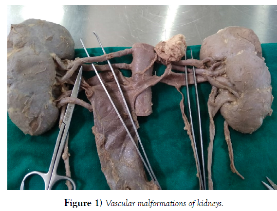
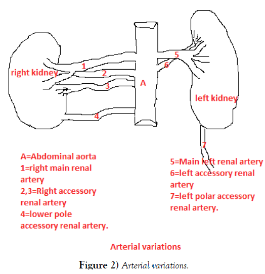
In this case report we found pair of kidneys with renal vein and renal artery. Right renal artery is longer than the left renal artery and left renal vein is longer than the right renal vein. We can see the posterior aspect of inferior vena cava.Left gonadal vein and left suprarenal vein (adrenal vein) are draining into the left renal vein. Right suprarenal vein and gonadal vein are draining into inferior vena cava. Both the ureters are draining from lower endbehind the renal artery. From anterior to posterior we get renal artery, renal vein and ureter. Here we are getting duplication of ureter in the left side. On right side, there three accessory renal arteries, one to the inferior pole and two to the hilum behind the main renal artery of the kidney. On the left side,there are two renal arteries one going to the hilum behind the main renal artery and one going to the inferior pole of the kidney. On right side ureter is draining behind the renal vein. On left side there is duplex ureter draining behind the renal vein. Here the left suprarenal vein is twisted and the gland is also showed lower down as the venacava is rotated down showing its posterior aspect and we are not getting any venous tributaries draining from it.
Keywords
kidney, Artery, Vein, Ureter, Pole
Introduction
In duplication of renal artery it is usual for each renal segment to have its own arterial supply. Regarding ureter, most common congenital anomaly is duplication of renal ureter. It is autosomal dominant in inheritance. It is mostly found in females and is often bilateral. There are two types of duplication of ureter- complete and incomplete. In incomplete type, both ureters join together and a single ureteric opening is found.This is called “Yo – Yo ”effect. In complete type,both ureters open separately. a) Weigert- Meyer’s rule: both upper pole and lower pole ureters rotate along their long axis so that upper pole ureter drain medially and caudally than the lower pole ureter. Lower pole ureter opens laterally and have short intravesicle course leading to vesicoureteral reflux(VUR). Lower down in the urinary bladder. In Females, the upper pole ureter is ectopic and obstructed. It drainsmainly into the distal part of the external urethral sphincter and sometimes may be outside the urogenital tract. Classic symptoms isincontinence of urine with normal voiding pattern. In Males, ectopic opening is always proximal to the external urethral sphincter and so incontinence does not occur. Other positions of ectopic ureteric orifices are – Males: prostate or posterior urethra or lateral urinary bladder. Females: Anterior urethra, vestibule or vagina. The intraluminal diameter of ureter is 1.5-2.5 mm and its length is around 30cm.volume of renal pelvis is around 7ml. It is partly intrarenal and partly extrarenal. Urine formed in kidney drain into the collecting system which again drains into the renal pelvis and from there to the urinary bladder via peristaltic movement. Pacemakers for urinary peristalsis is found in the collecting system. Along the course of ureter in the retroperitonium, the ureteric orifice is narrower than normal at the level of renal pelvis, pelvic brim when it is crossing the iliac vessels, in the intravesicle course and inside broad ligament. In patients passing a stone These are common sites of impaction of stone.
A single renal artery arises bilaterally from the lateral portion of the abdominal aorta immediately caudal to the origin of the superior mesenteric artery. The right renal artery passes behind the inferior vena cava (IVC) and is typically posterior and superior to the left and right renal veins. The left renal artery is higher than the right renal artery. In relation to the renal pelvis, the renal artery forms an anterior division and a posterior division. These divisions are most often formed outside the renal hilum. Along the lateral border of the kidney, between the arterial divisions, lies the avascular plane (Brödel’s line), which is located in the axis of the posterior. This avascular plane is not in the exact mid-lateral portion of the kidney, but is located slightly posteriorly. Brödel’s line can be used for avascular access for anatrophic nephrolithotomy and for endophytic tumors. The right renal vein drains directly into the IVC. There are usually no tributaries; rarely, the right gonadal vein may drain into the right renal vein. The left renal vein is approximately two to three times longer than the right renal vein, enters the IVC anterior to the aorta. Left renal vein tributaries include the gonadal vein, adrenal vein, inferior phrenic veins, the first or second lumbar veins, and paravertebral veins in one-third of cases [1]. There are three main types of anatomical variants of renal veins: multiple renal veins, in which are identifable two or more renal veins, either uni or bilaterally; retroaortic left renal vein (RLRV), in which the renal vein has a retro aortic course before entering the inferior vena cava; and circumaortic left renal vein (CLRV), in which there are two or more renal veins forming a ring around the aorta [2]. Different origins of the renal arteries and its frequent variations are explained in various literatures owing to the development of mesonephric arteries. These mesonephric arteries extend from C6 to L3 during the development. Most cranial vessels disappear while the caudal arteries form a network, the rete arteriosum urogenitale that supplies in future the metanephros. The metanephros in future develops into adult kidney deriving its blood supply from the lowest suprarenal artery which gives out a permanent renal artery [3,4]. The term abberent has been applied equally to an additional artery in the renal pedicle, or to a vessel entering the kidney at either pole, whether derived from the main renal artery, from the aorta or from a branch of the aorta [5].
Accessory renal artery usually arises from the aorta above or below the main renal artery. The variation in the number of arteriesis because of persistence of lateral splanchnic arteries or due to the persistence of blood supply from lower level than normal [6].
Case Report
In this case report (Figure 1) we found pair of kidneys with renal vein and renal artery. Right renal artery is longer than the left renal artery and left renal vein is longer than the right renal vein. We can see the posterior aspect of inferior vena cava. Left gonadal vein and left suprarenal vein (adrenal vein) are drainning into the left renal vein. Right suprarenal vein and gonadal vein are draining into inferior vena cava. Both the ureters are draining from lower end behind the renal artery. From anterior to posterior we get renal artery, renal vein and ureter. Here we are getting duplication of ureter in the left side. On right side, there three accessory renal arteries one to the inferior pole and two to the hilum behind the main renal artery of the kidney. On the left side,there are two accessory renal arteries one going to the hilum and one going to the inferior pole of the kidney other than the main left renal artery (Figure 2). On right side ureter is draining behind the renal vein. On left side there is duplex ureter draining behind nearer to the renal artery. Here the left suprarenal vein is twisted and the gland is also showed lower down as the venacava is rotated down showing its posterior aspect and we are not getting any venous tributaries draining from it.
Discussion
Daniel Emilio, Dalledone Siqueira and Ana Terezinha Guillaumon (2019) mentioned in their publication that Accessory renal arteries may be present in one or in both kidneys. Most accessory renal arteries supply the inferior pole of the kidney, arising from the suprarenal aorta to iliac arteries [3]. This is as we found in our case.
Petru Bordei, Sapte Elena, Dan Eliescu found triple renal artery in their study on kidney. They found double renal artery on the left side and triple renal artery on the right side [7]. This coincides with our finding.
Turan Pestemalci, Ayfer Mavi, Yusuf Zeki Yildiz et al. bilateral triple renal arteries were noticed. On the right side, the main renal artery arose from the abdominal aorta; the first additional renal artery (ARA), supplying the upper pole of the kidney, arose from the abdominal aorta and the second ARA, supplying the lower pole of the kidney, arose from the ipsilateral common iliac artery. On the left side, the main renal artery and the aARAs (to upper and lower poles of the kidney) arose from the abdominal aorta [8]. This is according to our findings.
Ning Xiao, Bo Ge, jianfeng Wang, huasheng Zhao, mentioned in their article that ureteric bud divides prematurely before penetrating into metanephric blastema will result in duplex ureter.
There are 2 types - complete and incomplete duplex ureter. The incidence of incomplete duplex ureter, including 3 subtypes, such as proximal, middle, and distal, depending on the location that bifid ureters join a single unit, is 3 times more than the complete [9-11]. This is according to our finding.
Conclusion
In any angiographic interventions and urosurgery, inadvertent injury to accessory renal artery as well as ureter can be prevented after getting awarded about this kind of renovascular anomalies.
REFERENCES
- Klattea T, Ficarrab V, Gratzkec C, et al. A literature review of renal surgical anatomy and surgical strategies for partial nephrectomy. Eur Urol. 2015;68:980-92.
- Hectic S, Rusu M, Negoi L, et al. Anatomical variants of renal veins: a meta-analysis of prevalence. Scien Reports. 2019;9.
- Https://www.intechopen.com/books/angiography/angiography-forrenal- artery-diseases
- Rao TR, Rachana. Aberrant renal arteries and its clinical significance: a case report. IJAV. 2011;4:37-9.
- FT Graves. The aberrant renal artery. J Anat. 1956;90:553-8.
- 6. Mir NS, Hassan A, Rangrez R, et al. Bilateral Duplication of Renal Vessels: Anatomical, Medical and Surgical perspective. IJHS. 2008;2.
- Bordei P, Elena S, Eliescu D. Double renal arteries originating from the aorta. Surg Radiol Anat. 2005;26:474-9.
- Pestemalci T, Mavi A, Yildiz YZ, et al. Bilateral Triple Renal Arteries. Saudi J Kidney Dis Transpl. 2009;20:468-70.
- Xiao M,Gee B, Wang J, et al. Obstruction of bifid ureter by two calculi - A case report. Medicine. 2018;97:30.
- Hyung L, Belldegrun KA, Brunicardi FC, et al. Pollock Schwartz’s Principles Of Surgery / Specific Considerations. Eighth edition.Mc Graw Hill Medical Publishing Division.United States of America. 2005;39:1520.
- Tanagho EA, Mc Aninch JW. Smith’s General urology.Seventeenth edition. Tata Mc Graw Hills Education Private Limited. New York. 2009;4.


