


 5 / 5
5 / 5
Page 16
November 06-07, 2019 | Tokyo, Japan
Volume 02
Journal of Clinical Genetics and Genomics
STEM CELLS AND REGENERATIVE MEDICINE
PEDIATRICS AND CHILD CARE
International Conference on
2
nd
World Congress on
&
J Clin Gen Genomics, Volume 02
Stem Cells 2019 & Pediatrics Congress 2019
November 06-07, 2019
Fabrication and characterization of electrospun PCL nanofibrous scaffolds for tissue
engineering: Biomechanics and cells behavior
Statement of the Problem
: Tissue engineering is a promising solution for the problem of organ or tissue shortage. A main
requirement is the use of biologically functional scaffolds to deliver cells to the implant site and/or provide a structure for cell
attachment to regenerate lost or damaged extracellular matrix (ECM). The natural ECM is structured in the nanoscale range, a
characteristic that should be incorporated into scaffold design for tissue engineering. Scaffolds produced by the electrospinning
process have several unique advantages. In this research, we survey the potential of poly(ε-caprolactone) (PCL) for the synthesis
of electrospun nanofibrous scaffolds and investigate their biomechanics and cell's interaction for successful tissue engineering
applications.
Methodology & Theoretical Orientation
: PCL pellets were dissolved in acetic acid (20% wt.). Electrospinning was
implemented to manufacture the microporous nanofibrous scaffolds. Morphological characterization was observed by SEM.
Mechanical tensile testing and
in vitro
degradation of the scaffolds were also performed. The MTT assay was used to determine
viability of hCMEC/D3 cell line following exposure to electrospun PCL scaffolds surface.
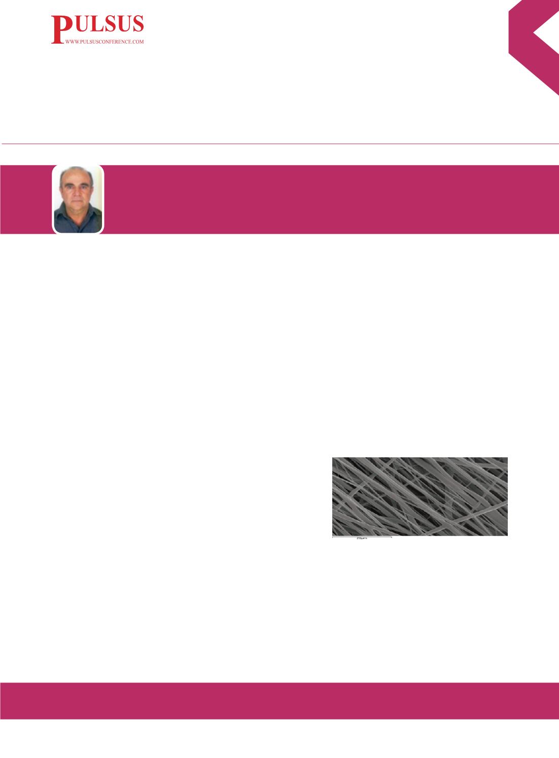
Findings
: Results showed a scaffold morphology consisting of parallel, aligned and homogeneous PCL microfibers with
diameter 1.16 ± 0.45 um, pore size 17.7 ± 5.37 um (Figure 1) and measured elastic modulus 18.3 ± 0.23 MPa, in the fibers
direction. Gravimetric weight loss of the PCL scaffolds immersed to PBS (37°C) was measured weekly over 15 weeks (4-
10% weight loss). Capability of cell infiltration verified by MTT assay where
cytotoxicity was not observed, exhibiting high cell viability (85.64 ± 3.12%).
Conclusion & Significance
: Utilizing the electrospinning process we were
able to produce laminate micro fibrous PCL scaffolds. Their structural
organization and biomechanics mimic natural tissue ECM structure and bio-
functionality, as well as they are capable of hosting cells. This material (PCL)
appears to be a promising candidate for tissue engineering applications.
Biography
Dimosthenis Mavrilas is a professor of Biomedical Engineering in the Laboratory of Biomechanics and Biomedical Engineering, Department of
Mechanical Engineering &Aer/tics, University of Patras, Greece. He is an expert in biomechanics of biomaterials and biomedical engineering
of cardiovascular system. Last decade his research targets in tissue engineering, producing scaffolds either of biological origin (decellularized
animal tissues) or from synthetic polymers. His research team achieved the production of either random or parallel fibrous orientation
of synthetic biodegradable polymeric nanofibers, capable for the structure of multi laminate biomimetic scaffolds, suitable especially for
cardiovascular tissue engineering.
dmauril@upatras.grDimosthenis Mavrilas
University of Patras, Greece
Figure 1. SEM image of PCL electrospun nanofibrous
membrane