A extremely unusual testicular tumour case report
Received: 27-May-2023, Manuscript No. PULCMR-23-6472; Editor assigned: 30-May-2023, Pre QC No. PULCMR-23-6472 (PQ); Reviewed: 14-Jun-2023 QC No. PULCMR-23-6472; Revised: 27-Jul-2023, Manuscript No. PULCMR-23-6472 (R); Published: 04-Aug-2023
Citation: Hilary J. A extremely unusual testicular tumour case report. J Cancer Metastasis Res 2023;5(5):1-2.
This open-access article is distributed under the terms of the Creative Commons Attribution Non-Commercial License (CC BY-NC) (http://creativecommons.org/licenses/by-nc/4.0/), which permits reuse, distribution and reproduction of the article, provided that the original work is properly cited and the reuse is restricted to noncommercial purposes. For commercial reuse, contact reprints@pulsus.com
Abstract
Myoid testicular tumour is an uncommon neoplasm that develops from smooth muscle cells in the tunica albuginea. It is normally painless and appears as a testicular lump. Histopathological testing confirms the diagnosis. The preferred treatment is radical orchiectomy. Recurrence is uncommon, and long-term monitoring is advised. Based on current literature, we examine the clinical appearance, diagnosis, therapy, and prognosis of myoid tumour of the testis in this abstract.
A myoid tumor of the testis, also known as a testicular leiomyoma or leiomyosarcoma, is a rare kind of tumour that arises in the testicular smooth muscle cells. These tumours are usually benign, which means they do not migrate to other regions of the body, but in some circumstances they can be malignant, which means they can metastasis and infect surrounding tissues.
Keywords
Tumour; Environment leiomysarcoma; Testicular; Etiology; Hormonal imbalances
Introduction
Myoid testicular tumours can occur in males of any age, however they are most usually detected in men in their forties or older. They frequently manifest as painless testicular lumps that can be discovered via physical examination or imaging investigations such as ultrasound. Myoid tumours of the testis have no established an etiology; however some experts believe they may be caused by hormonal imbalances or exposure to environmental contaminants [1]. The tumour is normally removed surgically, and in some situations, radiation therapy or chemotherapy may be used to prevent recurrence.
Myoid tumours of the testis have no known cause; however some specialists believe they may be caused by hormonal imbalances or environmental pollutants [2]. The tumour is usually surgically removed, although in some cases, radiation therapy or chemotherapy may be used to prevent recurrence [3].
Case Presentation
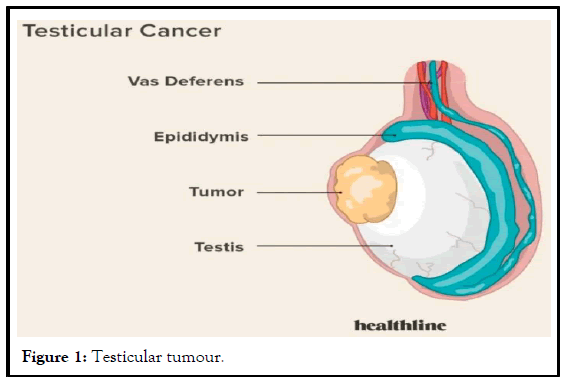
It is about a young patient, 27 years old, married, with no special pathological history, who visited for the appearance of a left testicular mass without other related indicators since 4 months, his physical appearance was normal, with no dysmorphism or signs of feminization (Figure 1).
The clinical examination revealed a left testicular mass that was firm and not too painful to palpate, and the lymph nodes were clear. The hormonal balance (LDH, FP, and HCG) was normal. Doppler ultrasonography revealed a hypoechoic, heterogeneous, well-limited left testicular mass measuring 40/35 mm, a contralateral testicle of normal size with no abnormalities, and a CT scan as an extension assessment revealed no subsequent lesions. The patient benefited from an inguinal complete orchiectomy, and the postoperative complications were minor [4]. The patient left the hospital the same day. Myoid tumours of the testis, also known as leydig cell tumours, are rare testicular neoplasms that grow from the testis interstitial leydig cells. These tumours are often unilateral, solitary masses that account for less than 1% of all testicular tumours. While myoid tumours of the testis are usually benign, they can develop cancerous and spread to other organs. Due to their rarity, there is little knowledge about the best way to treat these tumours, and more study is needed to better understand their pathophysiology and clinical behaviour.
Leydig cell tumours are thought to develop from the leydig cells, which are responsible for testosterone production in the testes. These tumours are frequently asymptomatic and are identified by chance during a normal checkup [5]. Leydig cell tumours are thought to develop from the leydig cells, which are responsible for testosterone production in the testes. These tumours are frequently asymptomatic and are identified by chance during a normal physical examination or ultrasound. They can, however, cause symptoms such as pain, swelling, or a tumour in the testicle. Myoid testicular tumours are normally diagnosed using a combination of physical examination, imaging techniques, and histological analysis of a biopsy or excision specimen [6].
Results and Discussion
Given the rarity of these tumours and the lack of rigorous data on their clinical behaviour, the appropriate management of myoid tumours of the testis remains a matter of discussion. Current standards, however, propose surgical removal of the afflicted testis. Myoid tumours of the testis, according to Ceballos-Quintal et al., can show as a large testicular mass with infiltrative signs and may be linked with reactive lymphoid follicles. The tumours have fascicles of spindle cells with eosinophilic cytoplasm and elongated nuclei organised in a whorled or storiform pattern on histology. Positive immunohistochemistry for smooth muscle markers such as desmin and smooth muscle actin is required to confirm the diagnosis [7].
Myoid tumours of the testis are considered low-grade cancers, with a usually favourable prognosis. However, there have been a few cases of metastasis, such as the one documented by Patel et al., who recorded a case of a myoid tumour of the test is that metastasized to the lungs. As a result, long-term monitoring using imaging is advised. Orchidectomy, which is frequently curative for benign tumours, is the treatment for myoid tumours of the testis. However, for malignant tumours, adjuvant therapy, such as chemotherapy or radiotherapy, may be required. Kim et al. found that 10 individuals with malignant myoid tumours of the testis had a higher risk of recurrence and metastasis, and adjuvant therapy was associated with a better prognosis.
Conclusion
Myoid testicular tumours are treated with orchidectomy, which is typically curative for benign tumours. Adjuvant therapy, such as chemotherapy or radiotherapy, may be required for malignant tumours. Kim et al. discovered that 10 people with malignant myoid testicular tumours had a higher chance of recurrence and metastasis, and that adjuvant therapy was associated with a better prognosis.
References
- Grove J, Komforti MK, Craig-Owens L, et al. A Collision Tumor In The Breast Consisting Of Invasive Ductal Carcinoma And Malignant Melanoma. Cureus. 2022;14(3):23588.
[Crossref] [Google Scholar] [PubMed]
- Donley GM, Liu WT, Pfeiffer RM, et al. Reproductive factors, exogenous hormone use and incidence of melanoma among women in the United States. British J Cancer. 2019;120(7):754-60.
[Crossref] [Google Scholar] [PubMed]
- Farrow NE, Leddy M, Landa K, et al. Injectable therapies for regional melanoma. Surg Oncol Clin. 2020;29(3):433-44.
[Google Scholar] [PubMed]
- Garfield K, LaGrange CA. Renal cell cancer. Stat Pearls International 2022, Treasure Island, Florida. 2022.
- Alzubaidi AN, Sekoulopoulos S, Pham J, et al. Incidence and distribution of new renal cell carcinoma cases: 27-Year trends from a Statewide Cancer Registry. J Kidney Can VHL. 2022;9(2):7.
[Crossref] [Google Scholar] [PubMed]
- Lobo J, Ohashi R. WHO 2022 landscape of papillary and chromophobe renal cell carcinoma. Histopathology. 2022; 81(4):426-38.
[Crossref] [Google Scholar] [PubMed]
- Kuroda N, Sugawara E, Kusano H, et al. A review of ALK- rearranged renal cell carcinomas with a focus on clinical and pathobiological aspects. P J Patho. 2018;69(2):109-13.
[Crossref] [Google Scholar] [PubMed]


