Applicability of histomorphometry analysis for evaluating alveolar bone regeneration after gene transfer
Received: 21-Oct-2017 Accepted Date: Oct 27, 2017; Published: 01-Nov-2017
Citation: Kawai M, Ohura K. Applicability of histomorphometry analysis for evaluating alveolar bone regeneration after gene transfer. J Histol Histopathol Res 2017;1(1):21-22.
This open-access article is distributed under the terms of the Creative Commons Attribution Non-Commercial License (CC BY-NC) (http://creativecommons.org/licenses/by-nc/4.0/), which permits reuse, distribution and reproduction of the article, provided that the original work is properly cited and the reuse is restricted to noncommercial purposes. For commercial reuse, contact reprints@pulsus.com
Abstract
It can be difficult for researchers and clinicians to evaluate bone tissues after some regenerative bone treatments because bone is always undergoing the process of remodeling. Moreover, hard tissues such as bone or teeth are typically very difficult to handle to prepare fresh and proper materials for clinical use. Therefore, regenerative bone tissues must be evaluated by adequate methods. In our previous studies, we developed a bone regeneration system using a combination of a non-viral bone morphogenetic protein gene-expression plasmid vector and in vivo electroporation. Using a model of ectopic bone formation in the skeletal muscles of rats made it easy to evaluate the bone-inductive potential of our developed system. In this study, we used bone morphometry analysis to evaluate the alveolar bone with our gene-transfer system. Upon labeling alveolar bone with calcein and tetracycline in rats and analyzing double-staining, we concluded that histomorphometry analysis is an efficient method for evaluating alveolar bone regeneration induced by a combination of non-viral plasmid vector and in vivo electroporation.
Keywords
Alveolar bone; Histomorphometry analysis; Bone labeling; Gene therapyBone regeneration therapy is pivotal for many patients with bone defects caused by trauma, tumor or malformation [1], and alveolar bone regeneration therapy is important for the maintenance of teeth [2]. Therefore, many researchers are endeavoring to identify efficient and safe therapies for bone regeneration [3]. In these studies, it is sometimes difficult to evaluate the bone regenerated by their developed methods because bone is always undergoing the process of remodeling [4]. Moreover, hard tissues such as bone or teeth are difficult to handle to yield fresh and proper materials [5]. For these reasons, we typically used ectopic bone formation models in the skeletal muscles of rats to evaluate bone regeneration methods [6,7]. However, it is also necessary to evaluate regenerated bone within target bone defect areas for clinical applications.
In previous studies, we developed a gene-transfer system for bone regeneration therapy by combining a non-viral gene expression plasmid vector and in vivo electroporation [8,9]. The ultimate goal is to apply this method for alveolar bone regeneration [10]. Therefore, we require an adequate evaluation method for regenerated alveolar bone after gene therapy treatment. In this study, we used histomorphometry analysis to evaluate alveolar bone regeneration induced by our gene-transfer system.
Materials and Methods
Gene transfer
Nine-week-old male Wistar rats (n=3 per group) were anesthetized via intraperitoneal injection of pentobarbital sodium (5.0 mg/100 g of body weight). The lacZ non-viral gene expression vector detailed in our previous study [11] was diluted to 0.5 μg/μL in phosphate-buffered saline, and a total volume of 50 μL was injected into the palatal region of periodontal tissues in the first molar of the right maxilla using a syringe with 31-gauge needle. Electroporation was then performed immediately, with 32 electrical pulses at 50 V for 50 msec [12].
Double-staining of bone
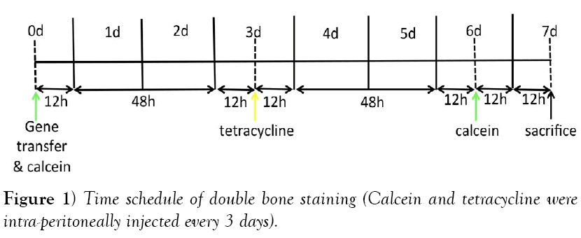
Rats (n=3 per group) were intraperitoneally injected with calcein (10 mg/ kg) on the day of initiation of gene transfer. Three days later, tetracycline hydrochloride (30 mg/kg) was intraperitoneally injected. Six days after gene transfer, rats were again injected with calcein. Seven days after gene transfer, rats were sacrificed with an overdose of pentobarbital sodium. The schedule of bone labeling is described in (Figure 1). The maxillary regions of rats were dissected and fixed with 70% ethanol for 8 days, followed by staining with Villanueva bone stain for 10 days, dehydration with increasing concentrations of ethanol, and embedding in methyl methacrylate without decalcification. After polymerization, frontal 10-μm sections were obtained from the mesiolingual center of the upper first molars and second molars. Sections were observed by fluorescence microscopy under UV irradiation for tetracycline (364 nm) and calcein (477 nm) labeling. The width of areas labeled with calcein and tetracycline was measured vertically at 10 points within the region in which gene transfer had been performed, using a Histometry RT Camera (System Supply, Tokyo, Japan). Statistical analyses were performed by unpaired t test.
Results
Villanueva bone staining
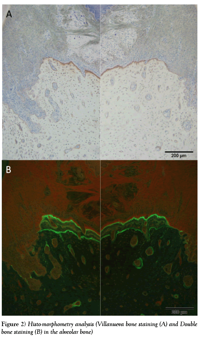
Villanueva bone staining was visualized as follows: transparent green to jade green or homogeneous red represented osteoid, red represented lowdensity bone, greenish-blue to dark purple represented nuclei of osteoblasts or osteoblasts, and green or light green represented cytoplasm. After gene transfer, no significant differences in osteoid or low-density bone were observed between control and gene-transfer sites (Figure 2A).
Bone labeling and MAR
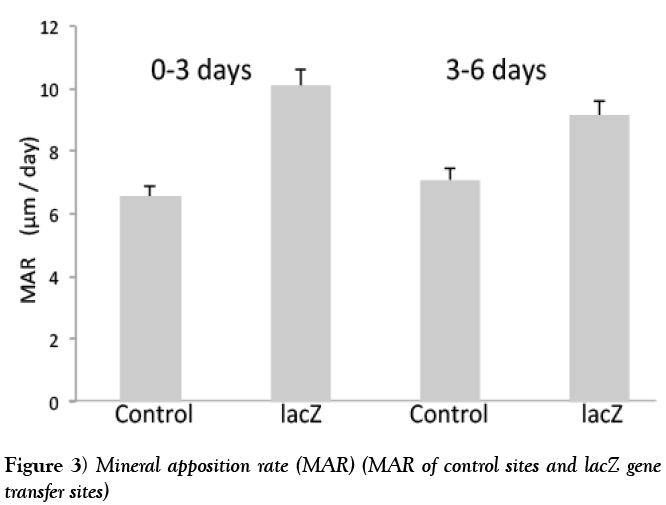
We found three labels in the alveolar bone of both control and gene-transfer sites (Figure 2B). Moreover, we did not observe significant differences in the width of each label between control and gene-transfer sites. Results of MAR from 0–3 days and 3–6 days after gene transfer were not significantly different from the control sites (Figure 3).
Discussion
After transferring a lacZ non-viral vector into the periodontal tissues of rats with electroporation, we revealed that lacZ gene transfer with electroporation did not influence alveolar bone formation and remodeling using histomorphometry analyses. In our developed gene-transfer system, electrical pulses are needed to induce the efficiency of gene transfection [8]. However, some reports have described the influence of electronic pulses on bone formation [13,14]. In this study, histomorphometry analyses of alveolar bone after lacZ gene transfer with electroporation revealed that electrical stimulation did not influence alveolar bone regeneration.
Preparing histomorphometry samples is sometimes difficult and requires a lot of time because it is not possible to accelerate decalcification [5].
However, histomorphometry analyses can reveal the time-variant continuous conditions of bones [15]. In contrast, histological staining of sections with hematoxylin and eosin demonstrates only a snapshot or the fragmental changes of bone. This is an important distinction, as bone is always remodeling and changing [15].
Conclusion
We concluded that histomorphometry analysis of alveolar bone with genetransfer treatment is effective for evaluating regenerated bone. In the future, we plan to analyze alveolar bone treated with electroporation transfer of osteogenic cytokine genes, such as bone morphogenetic protein, by histomorphometry analysis.
Acknowledgement
This study was supported by Grants‐in‐Aid for Scientific Research from the Japan Society for the Promotion of Science (Basic Research B Number 24300182) and the Daiwa Health Sciences Foundation.
REFERENCES
- Dimitriou R, Jones E, McGonagle D, et al. Bone regeneration: current concepts and future directions. BMC Med 2011;31:9-66.
- Intini G, Katsuragi Y, Kirkwiid KL, et al. Alveolar bone loss: mechanisms, potential therapeutic targets, and intervenetions. Adv Dent Res 2014;26(1):38-46.
- Mardas N, Dereka X, Donos N, et al. Experimental model for bone regeneration in oral and cranio-maxillo-facial surgery. J Invest Surg 2014;27(1):32-49.
- Peric M, Dumic-Cule L, Grcevic D, et al. The rational use of animal models in the evaluation of noble bone regenerative therapies. Bone 2014;70:73-86.
- Mashiba T. Morphological analysis of bone dynamics and metabolic bone disease. Histological findings in animal fracture model—effests of osteoporosis treatment drugs on fracture healing process. Clin Calcium. 2011;21(4): 551-8.
- Kaihara S, Bessho K, Ohkubo Y, et al. Simple and effective osteoinductive gene therapy by local injection of a bone morphogenetic protein-2-expressing recombinant adenoviral vector and FK506 mixture in rats. Gene Ther 2004;11: 439-47.
- Bessho K, Carnes DL, Cabin R, et al. Experimental studies on bone induction using low molecular weight poly (DL-lactide-co-glycolide) as a carrier for recombinant human bone morphogenetic protein-2. J Biomed Mater Res 2002;61:61-5.
- Kawai M, Bessho K, Kaihara S, et al. Ectopic bone formation by human bone morphogenetic protein-2 gene transfer to skeletal muscle using transcutaneous electroporation. Hum Gene Ther. 2003;14:1547-56.
- Kawai M, Maruyama H, Bessho K, et al. Simple strategy for bone regeneration with a BMP-2/7 gene expression cassette vector. Biochem Biophys Res Commun 2009;390:1012-17.
- Kawai M, Ohura K. Gene therapy using non-viral gene expression vector and in vivo electroporation for bone regeneration: Challenge to gene transfer into the periodontal tissues. JBEB 2016;3:18-21.
- Tsuchida M, Koike T, Takekubo M, et al. Electroporation-mediated transfer of plasmid DNA encoding IL-10 attenuates orthotopic tracheal allograft stenosis in rats. Transpl Immunorol 2008;19:173-77.
- Yamamoto H, Kawai M, Shiotsu N, et al. BMP-2 gene transfer under various conditions with in vivo electroporation and bone induction. A J Oral Maxillo Surg 2012;24: 49-53.
- Ciombor DM, Aaron RK. Influence of electromagnetic fields on endochondral bone formation. J Cell Biochem 1993;52(1)37-41.
- Ciombor DM, Aaron RK. The role of electrical stimulation in bone repair. Foot Ankle Clin 2005;10(4):579-93.
- Xiao W, Wang Y, Pacios S, et al. Cellular and molecular aspects of bone remodeling. Front Oral Biol 2016;18:9-16.


