Minisrew-assisted management midline deviation caused by impacted canine and replacement of 2 mandibular first molars by protraction of second molars and impacted third molars in Class II malocclusion
2 Chongqing Municipal Key Laboratory of Oral Biomedical Engineering of Higher Education, College of Stomatology, Chongqing Medical University, Chongqing, China, Email: dentist123@gmail.com
3 Department of Orthodontics, College of Stomatology, Chongqing Medical University, P.R. China, Email: wuxiaomian898@163.com
4 Department of Oral and Maxillofacial Surgery, Stomatological Hospital, Chongqing Medical University, Chongqing, China, Email: dentist123@gmail.com
Dr. Xiaomian Wu, Department of Orthodontics, College of Stomatology, Chongqing Medical University, P.R. China, Email: wuxiaomian898@163.com
Received: 08-Aug-2017 Accepted Date: Jan 15, 2018; Published: 22-Jan-2018
Citation: Wu X, Chen J and Ji P. Minisrew-assisted management midline deviation caused by impacted canine and replacement of 2 mandibular first molars by protraction of second molars and impacted third molars in Class II malocclusion. Dentist Case Rep 2018;2(1):18-22.
This open-access article is distributed under the terms of the Creative Commons Attribution Non-Commercial License (CC BY-NC) (http://creativecommons.org/licenses/by-nc/4.0/), which permits reuse, distribution and reproduction of the article, provided that the original work is properly cited and the reuse is restricted to noncommercial purposes. For commercial reuse, contact reprints@pulsus.com
Abstract
Impacted molar is the most common impacted teeth followed by impacted canine. In this case with the assistance of minisrews, a 20-year-old college student, who had maxillary left impacted canine and mandibular impacted third molars, damaged mandibular first molars with residual roots and maxillary midline deviated to the left, was successfully treated by extraction of 2 maxillary first premolars and replacement the mandibular first molars by protraction of mandibular second molars. Posttreatment, the patient accepted the result and her personality became sparky and talkable.
Keywords
Impacted canine; Miniscrew; Impacted third molar; Class II; Molar replacementTeeth impaction is the failed eruption of teeth with a completely developed root (1). It was caused by the interaction of environmental and genetic factors, such as hard tissue obstructions, soft tissue lesions and anomalies of neighboring teeth and so on (2,3). Different teeth has different prevalence rate. The most frequently impacted teeth are the third molars and the second one is the maxillary canine. Impacted canine in the mandible ranged from 0.92 to 5.1, and 89 percent of them were surgically extracted while 20-32 percent of them were treated by orthodontic traction (1,4).
Facial aesthetics, especially the anterior tooth aesthetics, has strong influences on psychological development and self-esteem from early childhood to adulthood (5,6). Many factors related to the anterior tooth aesthetics, such as migration of anterior teeth, anterior tooth crowding, lip profile and soon (6). And the influences of anterior teeth aesthetics on psychological development have attracted attention of many clinicians and researchers (5,7).
The disaster result for impacted teeth tracing case is that the teeth tracing fail but other healthy teeth has been extracted to offer space for alignment. So, well design and analyses is vital and CBCT (cone-beam computed tomography) is quiet helpful for diagnosis and treatment plan design (8). While maxillary impacted canine has a high prevalence rate, many studies in literature have shown the appliance of CBCT in evaluation the alveolar bone dimensions, maxillary transverse dimension in canine (9,10), and the diagnosis of impacted canine in orthodontic design (4,11). If analyses are necessary for the mandibular molar, CBCT is also powerful.
Material and Methods
Diagnosis and etiology
A young girl, 20-year-old, was transferred to Orthodontics department, the Affiliated Hospital of Stomatology, Chongqing Medical University, from general dentist after root canal treatment of her upper right first molar, with the chief complaint of tooth aesthetics such as maxillary dental midline deviation to the left, and clinical absence of the lower first molars and maxillary left canine. She was in good general health, but she was shy, silent and disliked to open her mouth or smile to show her tooth, because she felt unsatisfied with her anterior teeth aesthetics. Orthodontic documentation was requested before treatment.
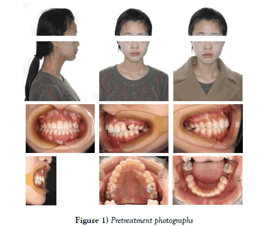
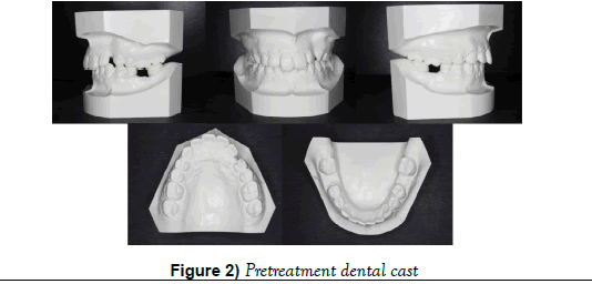
Frontal analysis showed her chin asymmetry to the right side due to unilateral and habitual mastication (Figures 1 and 2.). The profile analysis showed a Class II aspect, with the protrusion of upper lips and the retrusion of mandibular. Intraoral examination, impaction of maxillary left canine, 3-mm maxillary midline deviation to the left, residual roots of mandibular left and right permanent first molar with 7 mm space in each side, anterior tipped mandibular second molars, extensive carious lesion in first and second molars (some of which have been treated), and server abrasion in maxillary right canine and premolars were found.
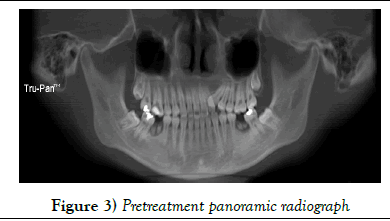
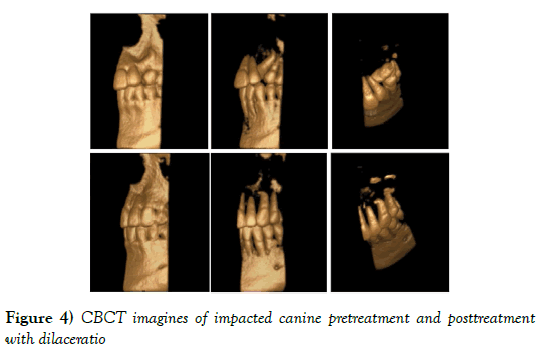
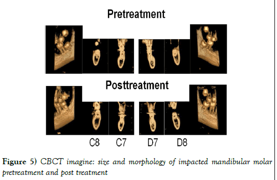
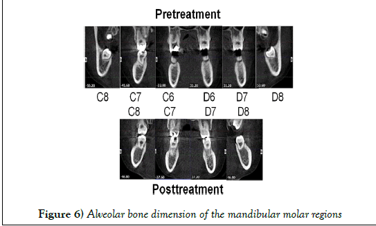
Radiography, it was observed that maxillary left canine was impacted between maxillary left lateral incisor and left first premolar (Figure 3). The size and morphology of canine and impacted mandibular third molars were well as CBCT imagine shown (Figures 4 and 5), but the dilacerations of impacted maxillary left canine was detected in CBCT image (Figure 4). And the alveolar bone dimension around the residual roots was fine in CBCT evaluation (Figure 6). There wasn’t obvious morphological asymmetry of TMJ. No obvious lateral shift of the mandibular incisor and no limited or asymmetric movement could be found during the maximum open-close excise.
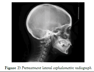
In comparison with Chinese norms, the cephalometric analysis exhibited a skeletal Class I jaw relationship (ANB angle 2.1°) and a normal mandibular plane angle (FMA 24.4°) (Figure 7). The maxillary incisors were labial inclined (U1-Maxillary Plane 116.1°) with an over jet of 5 mm, while the mandibular incisors were normal (IMPA 90.7°).
Retreatment objectives
The patient had maxillary midline deviation, impacted canine, residual roots of mandibular first molars and protrusion of maxillary incisors and upper lips, with a Class II division 1 malocclusion. Treatment objectives for this patient were to (1) managed the midline deviation by serial upper right first premolar extraction, correction of maxillary midline, traction of maxillary left impacted canine, and then extraction of upper left first premolar, Becker ACS (2) replaced space of the missing mandibular first molars by protraction of mandibular second molars bilaterally, Aktan et al. (3) improved soft tissue profile, D’Amico et al. (4) reduced increased over jet, Perillo et al. (5) maintained the mandibular incisors in the appropriate position, Xie et al. (6) modulated molars to a simulate Class II relationship and canines to a Class I relationship.
Treatment alternatives
The first alternative treatment plan was that extraction of maxillary right first premolar and the impacted left canine, which avoided the risk of traction failure of impacted canine after extraction of left first premolar. But extraction of canine could cause Bolton tooth-size discrepancy and occlusion problem.
The second alternative treatment plant was extraction of mandibular third molar, distal movement of mandibular second molar and opening enough space for implants or placing fixed partial dentures for the missing mandibular first molar. But the implant would increase much more cost for the patient, and fixed partial denture might compromise the longevity of prepared adjacent tooth. The patient couldn’t accept this plan.
The third option was that extraction of two maxillary premolar and the residual root of mandibular first molar and protracts the second molar to replace the first molar. After a carefully examination and evaluation of the alveolar dimension of edentulous sites by CBCT, it exhibited that the possibility to replace mandibular first molar by protraction of second molars bilaterally was quiet high. It also showed that obstruction of impacted canine would be the maxillary left lateral incisor and first premolar. While the dilacerations of canine could be found, the impacted canine traction should be successful after opening enough space. And after having a talk in detail with the patient and her parents, they choose this plan after a carefully discussion.
Treatment progress
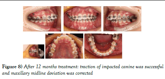
The maxillary right first premolar and the residual roots of mandibular first molar were extracted followed by placement of fixed appliance Mini Uni- Twin Bracket (3M Unitek) 0.018” slot up to the second molars, which was considered as a low friction appliance. The treatment was initiated by 0.012- in nickel-titanium alloy(Ni-Ti) archwires and leveled and aligned from 0.014- in 0.016-in to 0.018-in Ni-Ti archwires. Then 0.018“Australian arch wire” was applied. For the maxillary, Open Coil Spring was set between the left lateral incisor and first premolar and laceback was used on the right side. For the mandibular, an omega loop was set in the mesial of second molar and a tip back bend were utilized bilaterally, to upright the second molars. After 12 months treatment, traction of impacted canine was successful and maxillary midline deviation was corrected (Figure 8).
After the maxillary midline deviation was treated, maxillary first premolar was extracted. And for the mandibular, ligation from the left second premolar to right second premolar was applied to reinforce the anchorage, and “lace back” was set between the anterior teeth and the second molar bilaterally to protract the second molar. After the second molar protracted forward, it offered enough space for the eruption and anterior movement of third molars.
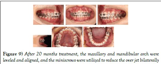
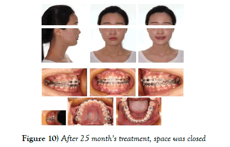
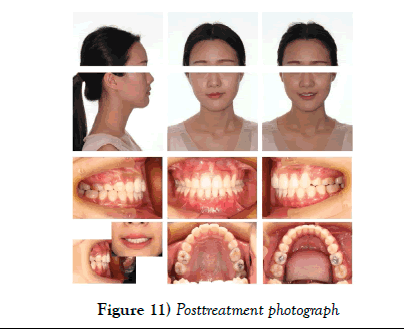
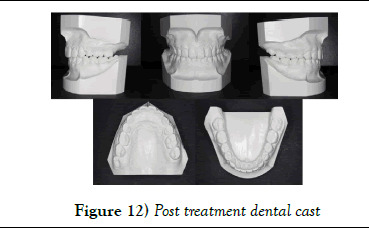
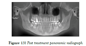
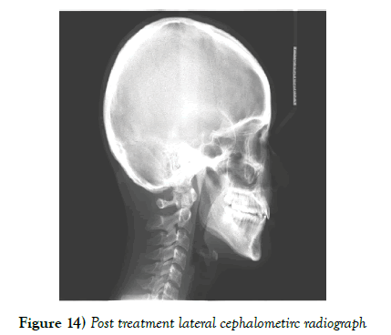
And after the maxillary left canine successfully entered the arch, the upper and lower arch were further leveled and aligned from 0.016 NiTi archwires followed by 0.018 NiTi, 0.016 × 0.022 NiTi, 0.018 × 0.025 NiTi and 0.018 × 0.025 stainless steel arch wires. While bilateral spaces in the maxillary is different, two 11 mm miniscrews were planted between the roots of maxillary second premolar and first molar bilaterally to retract the protrusion of upper incisors. Elastics chains were placed from the implant to the hook on the maxillary arch wire and from the second molar to the hook on mandibular arch wire, applying approximately 60 cN of force (Figure 9). The patient was recalled 4 weeks until the space was closed (Figure 10). The total active treatment period was 27 months (Figures 11-14).
While there were no opposing teeth in the mandibular arch, the patient was referred for extraction of upper third molars. For both maxillary and mandibular, an invisible retainer for daytime and a Hawley retainer for nighttime were given.
Results
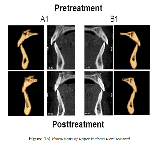
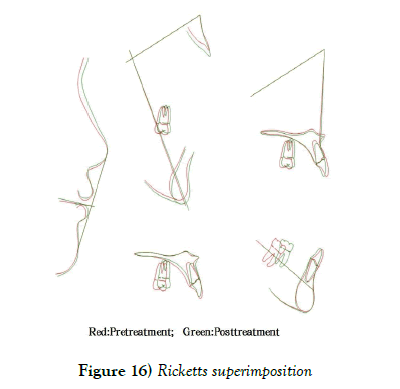
After the active treatment, the deviation of maxillary midline was corrected, the traction of impacted canine was successful (Figures 4 and 11), the protraction of mandibular second molar success, the third molar successfully erupted to the place of second molar (Figure 5), the protrusion of upper incisors was reduced (Figure 15 and 16) and the lip and soft profile were improved (Figure 11). The patient had a Class II molar relationship and a Class I canine relationship. While we continued advising the patient to give up the unilateral and habitual mastication and chin asymmetry to the right side was improved, but the asymmetry still existed.
Lateral cephalograms were taken at pretreatment (Figure 7) and posttreatment (Figure 13). It showed that the skeletal pattern was maintained with no significant change of SNA, SNB, or ANB angles (Table 1). The increased Interincisal angle (U1-L1), reduced U1- Maxillary Plane and overjet indicated that the maxillary incisors proclination was reduced, but the mandibular incisors were kept in an acceptable position as the IMPA and L1-MP(LADH) shown. And the Ricketts superimposition exhibited the improvement of upper and lower lips relationship as the Lower Lip to E-Plane and Upper Lip to E-Plane shown.
| Variables | Norm ± SD | Pretreatment | Posttreatment |
|---|---|---|---|
| FMIA (°) | 64.8 ± 8.5 | 64.9 | 68.2 |
| FMA (°) | 23.9 ± 4.5 | 24.4 | 27.5 |
| IMPA (°) | 95.0 ± 7.0 | 90.7 | 84.3 |
| SNA (°) | 82.0 ± 3.5 | 80 | 81.4 |
| SNB (°) | 80.9 ± 3.4 | 77.9 | 79.4 |
| ANB (°) | 1.6 ± 1.5 | 2.1 | 2 |
| Intervocalic Angle (U1-L1) (°) | 130.0 ± 6.0 | 126.3 | 140 |
| L1 - MP (LADH) (mm) | 40.0 ± 2.0 | 38.2 | 38.5 |
| U1 - Maxillary Plane (°) | 110.0 ± 5.0 | 116.1 | 108.8 |
| Occ Plane to SN (°) | 6.8 ± 5.0 | 14.3 | 14.3 |
| Anterior Cranial Base (SN) (mm) | 75.3 ± 3.0 | 61.7 | 61.7 |
| LPFH (Go-MxP) (mm) | 43.0 ± 4.0 | 36.1 | 35.8 |
| LAFH (Me-MxP) (mm) | 66.0 ± 3.5 | 58.8 | 53.5 |
| Lower Lip to E-Plane (mm) | -2.0 ± 2.0 | -1.8 | -1.3 |
| Upper Lip to E-Plane (mm) | -6.0 ± 2.0 | -1.8 | -2 |
| Chin Thickness (Pg-Pg') (mm) | 13.9 ± 3.5 | 11.9 | 10.4 |
| Overjet (mm) | 2.5 ± 2.5 | 5 | 3.7 |
| Overbite (mm) | 2.5 ± 2 | 1.5 | 1.3 |
Table 1: Cephalometric measurements
Discussion
Orthodontic treatment plane should base on the patient’s facial profile, functional occlusion, dental condition, long term stable and health, skeletal pattern, treatment time and so on. So, in detail examination and proper diagnosis must been done before the decision of orthodontic extraction was made. The extraction of impact canine will cause functional occlusion and aesthetics problems. Extraction of impacted mandibular third molars and opening space for implant or placing fixed partial dentures for first molar would increase the cost of the patient or have influence on the long term health of adjacent prepared tooth (12).
CBCT is a powerful device to evaluate the impact teeth (13,14). In orthodontic clinical daily work, it was quiet often utilized to exam impacted canine, while the prevalence rate of impact maxillary canine is lower than impact molar (9,10). After CBCT analysis, the obstructions of the impacted canine and impacted third molar was obvious and after removing the obstructions, the success of impacted tooth traction should be achieved, though dilacerations of canine could be found (15,16). There were two challenges in the treatment of this case. The first one was to correct the maxillary midline, tract the impacted canine and reduce the increased proincline upper incisor in the same time, when the space was different between the left and right side. In order to deal with this problem, we serial extracted right first premolar, corrected maxillary midline, performed traction on the maxillary left impacted canine, extracted of left first premolar, and then applied miniscrew to assist reducing the increased labial incline of upper incisors (17,18). And the second challenge was to protract the second and third molar to replace the mandibular first molar and kept the mandibular incisors in the appropriate position. To overcome this challenge, Mini Uni-Twin Bracket (3M Unitek) 0.018” slot with Roth prescription appliance, ligation from the left second premolar to right second premolar, “lace back”, and Australian arch wire with omega loop combined molar tip back bend mechanism system were utilized. It was reported that compared to standard double width brackets, Mini Uni-Twin Bracket had a significantly different amount of proclination of the lower anteriors after de-crowding (19,20), and this effect also offered some contribution for the anchorage control of lower incisors. Anterior teeth and soft tissue aesthetics is quiet important for patients and it could have negative influence on the psychological development. After the active orthodontics treatment, the patient accepted this result and her personalities became sparky and talkable. And she was referred again to give up unilateral and habitual mastication (21).
Conclusion
With the assistance of minisrews, a 20-year-old patient with maxillary midline deviation, maxillary impacted canine and mandibular impacted third molar and damage mandibular first molars was treated by extraction of 2 upper first premolars and the residual roots of mandibular first molars. CBCT is a powerful device to evaluate the impact teeth and alveolar diameter before orthodontic plan was made. Minisrews and serial teeth extraction was a useful couple to treat midline midline deviation. In order to pursue perfect result, while replacing the missing mandibular first molars with second molars, we should pay enough attention to the anchorage control of the lower incisor.
Acknowledgement
This study was supported by the National Natural Science Foundation of China (31400808) and the Science and Technology Research Project of Chongqing Municipal Education Commission of China (KJ1500227) to Dr. Xiaomian Wu. For this case, Dr Xiaomian Wu was awarded The Top 60 Youth Orthodontists the Case Contest of 2017 CIOC&COS.
REFERENCES
- Dalessandri DPS, Rubiano R, Gallone D, et al. Impacted and transmigrant mandibular canines incidence, aetiology, and treatment: a systematic review. Eur J Orthod. 2017, 39(2):161-9.
- Becker ACS: Etiology of maxillary canine impaction: A review. Am J Orthod Dentofacial Orthop. 2015;148:557-67.
- Aktan AMKS, Akgünlü F, Malkoç S. The incidence of canine transmigration and tooth impaction in a Turkish subpopulation. Eur J Orthod. 2010;32:575-81.
- D'Amico RMBK, Kurol J, Falahat B. Long-term results of orthodontic treatment of impacted maxillary canines. Angle Orthod. 2003;73(3):231-8.
- Perillo LEM, Caprioglio A, Attanasio S, et al. Orthodontic treatment need for adolescents in the Campania region: the malocclusion impact on self-concept. Patient Prefer Adherence 2014;19:353-9.
- Xie YZQ, Tan Z, Yang S. Orthodontic treatment in a periodontal patient with pathologic migration of anterior teeth. Am J Orthod Dentofacial Orthop. 2014;145:685-93.
- Meazzini MCMF, Gabriele C, Bozzetti A. Mandibular distraction osteogenesis in hemifacial microsomia: Long-term follow-up. J Craniomaxillofac Surg. 2005;33:370-6.
- Edwards RAN, Heo G, Flores-Mir C. Agreement among orthodontists experienced with cone-beam computed tomography on the need for follow-up and the clinical impact of craniofacial findings from multiplanar and 3-dimensional reconstructed views. Am J Orthod Dentofacial Orthop. 2015;148:264-73.
- Tadinada AMM, Vishwanath M, Allareddy V, et al. Evaluation of alveolar bone dimensions in unilateral palatally impacted canine: a cone-beam computed tomographic analyses. Eur J Orthod. 2015;37:596-602.
- Hong WH RR, Chung CH. Relationship between the maxillary transverse dimension and palatally displaced canines: A cone-beam computed tomographic study. Angle Orthod. 2015;85:440-5.
- Alqerban AJR, van Keirsbilck PJ, Aly M, et al. The effect of using CBCT in the diagnosis of canine impaction and its impact on the orthodontic treatment outcome. J Orthod Sci. 2014;3:34-40.
- Tomonari HYT, Kuninori T, Ikemori T, et al. Replacement of a first molar and 3 second molars by the mesial inclination of 4 impacted third molars in an adult with a Class II Division 1 malocclusion. Am J Orthod Dentofacial Orthop. 2015;147:755-65.
- Demiriz L HBE, İçen M, Durmuşlar MC. Evaluation of the accuracy of cone beam computed tomography on measuring impacted supernumerary teeth. Scanning 2016;38:579-84.
- Wang DHX, Wang Y, Li Z, et al. External root resorption of the second molar associated with mesially and horizontally impacted mandibular third molar: evidence from cone beam computed tomography. Clin Oral Investig. 2017;21:1335-42.
- Motamedi MHTF, Navi F, Shafeie HA, et al. Assessment of radiographic factors affecting surgical exposure and orthodontic alignment of impacted canines of the palate: a 15-year retrospective study. Oral Surg Oral Med Oral Pathol Oral Radiol Endod. 2009;107:772-5.
- Bayram MOM, Sener I. Maxillary canine impactions related to impacted central incisors: two case reports. J Contemp Dent Pract. 2007;8:72-81.
- Sh Y. Midline correction with mini-screw anchorage and lingual appliances. J Clin Orthod. 2006;40:314-22.
- Chung KRKS, Kook YA, Kang YG, et al. Dental midline correction using two component C-orthodontic mini-implant. Prog Orthod 2009;10:76-86.
- Krishna NK FA, Issar R, Subramanian S, et al. Clinical Evaluation of Proclination of Lower Anterior Teeth during Alignment using a Single Width Bracket-A Pilot Study. J Clin Diagn Res. 2016;10:113-5.
- Husain NKA. Frictional resistance between orthodontic brackets and archwire: an in vitro study. J Contemp Dent Pract. 2011;1;12:91-9.
- Pasinato FOA, Santos-Couto-Paz CC, Zeredo JL, et al. Study of the kinematic variables of unilateral and habitual mastication of healthy individuals. Codas. 2017;29:e20160074.


