A cadaveric report of unilateral fibularis tertius in absentia: a clinical perspective
Alok Saxena1*, Biswabina Ray2, Snigdha Mishra2 and Vivekanandan Perumal3
1Department of Anatomy, Veer Chandra Singh Garhwali Government Medical Science and Research Institute, Srinagar, Uttarakhand, India
2Department of Anatomy, MMMC, Manipal University, Karnataka, India
3Department of Anatomy, University of Otago, New Zealand
- *Corresponding Author:
- Alok Saxena
Senior Demonstrator Department of Anatomy Veer Chandra Singh Garhwali Government Medical Science and Research Institute Srinagar, Uttarakhand, India
Tel: +91 989 7699599
E-mail: drsan_99@rediffmail.com
Date of Received: November 10th, 2011
Date of Accepted: September 8th, 2012
Published Online: February 5th, 2013
© Int J Anat Var (IJAV). 2013; 6: 20–21.
[ft_below_content] =>Keywords
dorsiflexion, eversion, extensor digitorum longus, fibularis tertius
Introduction
Fibularis tertius (FT), formerly known as peroneus tertius is a unique muscle in human being, often considered as a fifth tendon of extensor digitorum longus. The muscle originates from the distal one third or medial fibular surface, anterior surface of the interosseous membrane and anterior crural intermuscular septum. Thereafter, the tendon runs beneath the superior extensor retinaculum, inferior extensor retinaculum and gets inserted into dorsal surface of fifth metatarsal. Fibularis tertius is supplied by deep peroneal nerve and assists in dorsiflexion of foot in swing phase and evertion of foot [1]. Vertullo et al. considered its insertion site as an important factor in case of Jones fracture [2]. A study conducted by Joshi et al. concluded that muscle might remain rudimentary or even absent in 4.4-10% cases in humans [3]. Another similar study carried out on Caucasian population showed its absence in 5-17% of cases studied [4].
Case Report
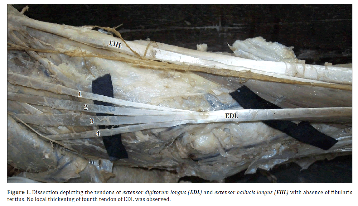
During routine dissection curriculum for undergraduate teaching, we have come across a rare finding of absence of FT unilaterally in formalin fixed male cadaver. Dissection was carried out on both right and left lower limbs using Cunningham’s manual of practical anatomy. Both the limbs were carefully observed and examined to differentiate the absence of FT muscle. There was no mark of external injury or operational scar on the limbs. No relevant sign of existence of the muscle was noticed on left limb whereas right limb was showing its presence. Relationship of extensor digitorum longus tendon (EDL) was noticed with neighboring structures on extensor compartment which revealed following relations medial to lateral: tibialis anterior, extensor hallucis longus, anterior tibial artery, deep peroneal nerve and extensor digitorum longus. Photographic record of the dissected limbs was documented (Figure 1).
Discussion
FT occupies the extensor compartment of leg and assists in dorsiflexion of foot in swing phase and eversion of foot [1]. Study conducted by Soames et al. revealed minimal functional importance of FT since its action is superimposed by actions of other muscles. It has significant influence on neuromuscular control and in prevention of talofibular ligament injuries [5]. Jungers et al. suggested that presence of the FT muscle is in close relation with the development of EDL muscle [6]. Local thickening of fourth tendon of EDL may be expected in case of absent FT [7]. A gender based comparative study was performed to describe its prevalence stating that the rate of its existence was higher in men [5].
We did not observe any sign of presence of FT muscle in left foot and any local thickening of fourth tendon of EDL during dissection. Presence of muscle plays an important role in Jones fracture of fifth metatarsal as suggested by Vertullo et al. [2]. Interestingly, subjects with FT in absentia are less prone to this fracture [5]. As described by Das et al., FT provides proper aid to the lateral border of sole and its absence may weaken the support given by the muscle. The same study also focused on the utilization of its tendon for transplant surgeries in foot drop conditions [8]. Absence of this muscle is a rare incidence in human being, which is actually a favorable aspect in terms of reducing the danger of Jones fracture. Awareness of its absence may be important for anatomists, orthopedic surgeons, physiotherapists and anthropologists.
References
- Standring S, ed. Gray?s Anatomy. London, Churchill Livingstone. 2008; 1419.
- Vertullo CJ, Glisson RR, Nunley JA. Torsional strains in the proximal fifth metatarsal: implications for Jones and stress fracture management. Foot Ankle Int. 2004; 25: 650?656.
- Joshi SD, Joshi SS, Athavale SA. Morphology of peroneus tertius muscle. Clin Anat. 2006; 19: 611?614.
- Witvrouw E, Borre KV, Willems TM, Huysmans J, Broos E, De Clercq D. The significance of peroneus tertius muscle in ankle injuries: a prospective study. Am J Sports Med. 2006; 34: 1159?1163.
- Jungers WL, Meldrum DJ, Stern JT. The functional and evolutionary significance of the human peroneus tertius muscle. J Hum Evol. 1993; 25: 377?386.
- Iyer PB. Bilateral absence of fibularis tertius: clinical implications and phylogeny. Int J Anat Var (IJAV). 2010; 3: 170?172.
- Ramirez D, Gajardo C, Caballero P, Zavando D, Cantin M, Galdames IS. Clinical evaluation of fibularis tertius muscle prevalence. Int J Morphol. 2010; 28: 759?764.
- Das S, Haji Suhaimi F, Abd Latiff A, Pa Pa Hlaing K, Abd Ghafar N, Othman F. Absence of the peroneus tertius muscle: cadaveric study with clinical considerations. Rom J Morphol Embryol. 2009; 50: 509?511.
Alok Saxena1*, Biswabina Ray2, Snigdha Mishra2 and Vivekanandan Perumal3
1Department of Anatomy, Veer Chandra Singh Garhwali Government Medical Science and Research Institute, Srinagar, Uttarakhand, India
2Department of Anatomy, MMMC, Manipal University, Karnataka, India
3Department of Anatomy, University of Otago, New Zealand
- *Corresponding Author:
- Alok Saxena
Senior Demonstrator Department of Anatomy Veer Chandra Singh Garhwali Government Medical Science and Research Institute Srinagar, Uttarakhand, India
Tel: +91 989 7699599
E-mail: drsan_99@rediffmail.com
Date of Received: November 10th, 2011
Date of Accepted: September 8th, 2012
Published Online: February 5th, 2013
© Int J Anat Var (IJAV). 2013; 6: 20–21.
Abstract
Fibularis tertius is a muscle of anterior compartment of leg, often considered as a part of extensor digitorum longus. This muscle contributes to dorsiflexion and eversion of foot. Existence of this muscle in many primates and in humans is still a topic of discussion. Previous reports show many variations on its existence. During routine dissection in the Department of Anatomy we came across an incidental finding of absence of fibularis tertius unilaterally. Clinical importance and functional significance of absence of fibularis tertius is discussed herein.
-Keywords
dorsiflexion, eversion, extensor digitorum longus, fibularis tertius
Introduction
Fibularis tertius (FT), formerly known as peroneus tertius is a unique muscle in human being, often considered as a fifth tendon of extensor digitorum longus. The muscle originates from the distal one third or medial fibular surface, anterior surface of the interosseous membrane and anterior crural intermuscular septum. Thereafter, the tendon runs beneath the superior extensor retinaculum, inferior extensor retinaculum and gets inserted into dorsal surface of fifth metatarsal. Fibularis tertius is supplied by deep peroneal nerve and assists in dorsiflexion of foot in swing phase and evertion of foot [1]. Vertullo et al. considered its insertion site as an important factor in case of Jones fracture [2]. A study conducted by Joshi et al. concluded that muscle might remain rudimentary or even absent in 4.4-10% cases in humans [3]. Another similar study carried out on Caucasian population showed its absence in 5-17% of cases studied [4].
Case Report
During routine dissection curriculum for undergraduate teaching, we have come across a rare finding of absence of FT unilaterally in formalin fixed male cadaver. Dissection was carried out on both right and left lower limbs using Cunningham’s manual of practical anatomy. Both the limbs were carefully observed and examined to differentiate the absence of FT muscle. There was no mark of external injury or operational scar on the limbs. No relevant sign of existence of the muscle was noticed on left limb whereas right limb was showing its presence. Relationship of extensor digitorum longus tendon (EDL) was noticed with neighboring structures on extensor compartment which revealed following relations medial to lateral: tibialis anterior, extensor hallucis longus, anterior tibial artery, deep peroneal nerve and extensor digitorum longus. Photographic record of the dissected limbs was documented (Figure 1).
Discussion
FT occupies the extensor compartment of leg and assists in dorsiflexion of foot in swing phase and eversion of foot [1]. Study conducted by Soames et al. revealed minimal functional importance of FT since its action is superimposed by actions of other muscles. It has significant influence on neuromuscular control and in prevention of talofibular ligament injuries [5]. Jungers et al. suggested that presence of the FT muscle is in close relation with the development of EDL muscle [6]. Local thickening of fourth tendon of EDL may be expected in case of absent FT [7]. A gender based comparative study was performed to describe its prevalence stating that the rate of its existence was higher in men [5].
We did not observe any sign of presence of FT muscle in left foot and any local thickening of fourth tendon of EDL during dissection. Presence of muscle plays an important role in Jones fracture of fifth metatarsal as suggested by Vertullo et al. [2]. Interestingly, subjects with FT in absentia are less prone to this fracture [5]. As described by Das et al., FT provides proper aid to the lateral border of sole and its absence may weaken the support given by the muscle. The same study also focused on the utilization of its tendon for transplant surgeries in foot drop conditions [8]. Absence of this muscle is a rare incidence in human being, which is actually a favorable aspect in terms of reducing the danger of Jones fracture. Awareness of its absence may be important for anatomists, orthopedic surgeons, physiotherapists and anthropologists.
References
- Standring S, ed. Gray?s Anatomy. London, Churchill Livingstone. 2008; 1419.
- Vertullo CJ, Glisson RR, Nunley JA. Torsional strains in the proximal fifth metatarsal: implications for Jones and stress fracture management. Foot Ankle Int. 2004; 25: 650?656.
- Joshi SD, Joshi SS, Athavale SA. Morphology of peroneus tertius muscle. Clin Anat. 2006; 19: 611?614.
- Witvrouw E, Borre KV, Willems TM, Huysmans J, Broos E, De Clercq D. The significance of peroneus tertius muscle in ankle injuries: a prospective study. Am J Sports Med. 2006; 34: 1159?1163.
- Jungers WL, Meldrum DJ, Stern JT. The functional and evolutionary significance of the human peroneus tertius muscle. J Hum Evol. 1993; 25: 377?386.
- Iyer PB. Bilateral absence of fibularis tertius: clinical implications and phylogeny. Int J Anat Var (IJAV). 2010; 3: 170?172.
- Ramirez D, Gajardo C, Caballero P, Zavando D, Cantin M, Galdames IS. Clinical evaluation of fibularis tertius muscle prevalence. Int J Morphol. 2010; 28: 759?764.
- Das S, Haji Suhaimi F, Abd Latiff A, Pa Pa Hlaing K, Abd Ghafar N, Othman F. Absence of the peroneus tertius muscle: cadaveric study with clinical considerations. Rom J Morphol Embryol. 2009; 50: 509?511.


