A third head of the biceps brachii and coexisting fused higher origin of brachioradialis
Anita S.Fating* and Vishal M.Salve
Dr. Pinnamaneni Siddhartha Institute of Medical Sciences & Research Foundation, Chinnaoutpalli, Gannavaram Mandal, Krishna District (AP), India.
- *Corresponding Author:
- Dr. Anita S. Fating, MBBS, MS (Anatomy)
Assistant Professor, Department of Anatomy, Dr. Pinnamaneni Siddhartha Institute of Medical, Sciences & Research Foundation, Chinnaoutpalli, Gannavaram Mandal, Krishna District (AP), 521286, India.
Tel: +91 998 9084307
E-mail: rashmi_vishal2005@yahoo.in
Date of Received: January 3rd, 2010
Date of Accepted: October 25th, 2010
Published Online: February 22nd, 2011
© IJAV. 2011; 4: 31–33.
[ft_below_content] =>Keywords
biceps brachii,brachioradialis,third head of biceps brachii,variation
Introduction
The biceps brachii is a large fusiform muscle in the flexor compartment of the arm. It is the only flexor of the arm crossing the shoulder joint as well as the elbow joint. Thus it acts on the both joints. Among the two classical heads, the long head runs in the intracapsular course over the humeral head and attached to the supraglenoid tubercle and adjacent portion of glenoid labrum. The short head arises from the tip of the coracoid process of scapula. The two heads soon fuse in the upper half of the arm to form the bulk of the biceps brachii muscle. The flattened tendon of biceps brachii crosses the elbow ventrally at the lower end, turns backwards and laterally to get inserted into the posterior rough part of radial tuberosity. Bicipital apponeurosis gets merged with deep fascia of forearm [1,2].
Brachioradialis is the most superficial muscle along the radial side of the forearm. It forms the lateral border of the cubital fossa. It arises from proximal 2/3 of the lateral supracondylar crest of humerus and from the anterior surface of the lateral intermuscular septum. The muscle fibers end above midforearm level in a flat tendon which inserts on the lateral side of the distal end of the radius, just proximal to the styloid process [1,2].
It has been reported that in 10% cases the third head of biceps brachii may arise from the supero-medial part of the brachialis and is attached to the bicipetal apponeurosis and medial side of tendon insertion. The presence of the third head and fused higher origin of brachioradialis is important for academic and clinical purpose [1,2].
Here we describe a case of third head of biceps brachii muscle and fused higher origin of brachioradialis muscle.
Case Report
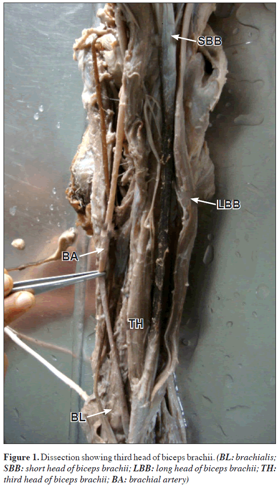
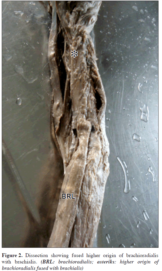
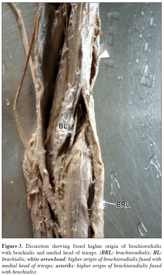
During routine dissection (MBBS Batch 2009-2010) of a middle aged male cadaver at the Dr. Pinnamaneni Siddhartha Institute of Medical Sciences & Research Foundation, Gannavaram, Krishna Dist. AP (INDIA); third head of biceps brachii and fused higher origin of brachioradialis were found in the left upper limb. The third head of biceps brachii arose from superomedial part of brachialis just below the insertion of coracobrachialis (Figure 1). The long head of biceps brachii arose from the supraglenoid tubercle and adjacent portion of glenoid labrum. The short head of biceps brachii arose from the tip of the coracoid process of scapula. The tendon of third head was attached to the lower part of muscle belly. It didn’t get attached to the bicipital apponeurosis. This third head was supplied by a branch from musculocutaneous nerve. The tendon of biceps brachii got inserted into the posterior rough part of radial tuberosity. Bicipital apponeurosis was merged with the deep fascia of forearm. Brachioradialis had higher additional origin beside its usual origin from proximal 2/3 of the lateral supracondylar crest of humerus and from the anterior surface of the lateral intermuscular septum. This higher origin of brachioradialis has got fused with brachialis and medial head of triceps muscles (Figures 2, 3). The tendon of brachioradialis got inserted on the lateral side of the distal end of the radius, just proximal to its styloid process. There were no such variations in the right upper limb.
Figure 3: Dissection showing fused higher origin of brachioradialis with brachialis and medial head of triceps. (BRL: brachioradialis; BL: brachialis; white arrowhead: higher origin of brachioradialis fused with medial head of triceps; asteriks: higher origin of brachioradialis fused with brachialis)
Discussion
Biceps brachii muscles present wide range of variations. They can manifest as a cluster of accessory fascicles arising from coracoid process, pectoralis minor tendon or articular capsule of humerus [3]. The most common variation is the muscle arising from proximal humerus. This variation is also known as the humeral head or third head of the biceps brachii muscle. According to present studies, this variation varies in different population, Chinese 8%, European White 10%, African Black 12%, Japanese 18%, South African blacks 20.55%, South African whites 8.35% and Colombian 37.5% [4].
Kumar et. al. studied 48 cadavers (n=96) at Vardhaman Medical College, Delhi. The third head of biceps was observed on both sides of a one male cadaver (3.3%). None of the rest 47 cadavers (n=94) had any variant third head of the biceps brachii muscle. The variant third head originated from the anterior limb of the “V” shaped insertion of the deltoid muscle on the humerus. This additional head was supplied by a branch from musculocutaneous nerve [5].
Poudel and Bhattarani studied 16 cadavers (n=32) at Manipal College of Medical Sciences, Pokhra, Nepal. Three-headed biceps brachii muscle was observed unilaterally on the right arm of one adult male cadaver (6.2 %). Four-headed biceps brachii muscle was also observed unilaterally on the right arm of one adult male cadaver (6.2%). On the left arm of above mentioned cadavers no third head of biceps brachii muscle was observed. None of the rest 14 cadavers (n=28) had any variant third head of the biceps brachii muscle [6].
Brachioradialis muscle is often fused proximally with brachialis. Its tendon may divide into two or three separately attached slips. In rare instances it is double or absent. Its radial attachment may be much more proximal than the base of styloid process [1].
George and Nayak found a few fleshy fibers from brachialis merged with brachioradialis and other superficial flexor of the forearm after an oblique course. Some of the fibers were inserted on the medial aspects of olecranon process of ulna. The additional slip of brachialis may mechanically stabilize the ulnohumeral joint. It can cause compression neuropathy of median nerve and vascular compression symptoms due to entrapment of brachial artery [7].
Our observation of the third head of biceps brachii muscle is similar to that of described in standard text books of anatomy. The fused higher origin of brachioradialis with brachialis and median head of triceps differ from previous reports. The third head of biceps brachii observed in our case may increase the power of flexion and supination component. The fused higher origin of brachioradialis may cause compression neuropathy of median nerve and vascular compression symptoms due to entrapment of brachial artery.
A variation in the heads of the biceps brachii muscle has already been reported to cause compression of surrounding neurovascular structures. It leads to erroneous interpretation during routine surgeries [8]. Presence of third head of biceps brachii muscle might increase its kinematics. This third head of biceps brachii muscle may increase the power of flexion and the supination component [5].
References
- Williams PL, ed. Gray’s Anatomy (The Anatomical Basis of Medicine and Surgery). 38th Ed., Edinburgh, Churchill Livingstone. 1995; 843, 849.
- Moore KL, Dalley AF. Clinically oriented Anatomy. 4th Ed., Philadelphia, Lippincott Williams & Wilkins. 1999; 784–787.
- Sargon MF, Tuncali D, Celik HH. An unusual origin for the accessory head of biceps brachii muscle. Clin Anat., 1996; 9: 160–162.
- Asvat R, Candler P, Sarmiento EE. High incidence of the third head of biceps brachii in South African populations. J Anat, 1993; 182: 101–104.
- Kumar H, Das S, Rath G. An anatomical insight into the third head of biceps brachii muscle. Bratisl Lek Listy. 2008; 109: 76–78.
- Poudel PP, Bhattarani C. Study on the supernumary heads of biceps brachii muscle in Nepalese. Nepal Med Col J, 2009; 11: 96–98.
- George BM, Nayak SB. Median nerve and brachial artery entrapment in the abnormal brachalis muscle – a case report. Neuroanatomy. 2008; 7: 41–42.
- Warner JJ, Paletta GA, Warren RF. Accessory head of the biceps brachii. Case report demonstrating clinical relevance. Clin Orthop Relat Res. 1992; 280: 179–181.
Anita S.Fating* and Vishal M.Salve
Dr. Pinnamaneni Siddhartha Institute of Medical Sciences & Research Foundation, Chinnaoutpalli, Gannavaram Mandal, Krishna District (AP), India.
- *Corresponding Author:
- Dr. Anita S. Fating, MBBS, MS (Anatomy)
Assistant Professor, Department of Anatomy, Dr. Pinnamaneni Siddhartha Institute of Medical, Sciences & Research Foundation, Chinnaoutpalli, Gannavaram Mandal, Krishna District (AP), 521286, India.
Tel: +91 998 9084307
E-mail: rashmi_vishal2005@yahoo.in
Date of Received: January 3rd, 2010
Date of Accepted: October 25th, 2010
Published Online: February 22nd, 2011
© IJAV. 2011; 4: 31–33.
Abstract
The biceps brachii is a large fusiform muscle in the flexor compartment of the arm. Brachioradialis is the most superficial muscle of the forearm. It has been reported that in 10% cases the third head of biceps brachii may arise from the supero-medial part of the brachialis and is attached to the bicipital apponeurosis. The presence of the third head is important for academic and clinical purpose. During routine dissection of a middle aged male cadaver at the Dr. PSIMS & RF, Gannavaram (INDIA); third head of biceps brachii and fused higher origin of brachioradialis were found in the left upper limb. The third head of biceps brachii arose from superomedial part of brachialis. Brachioradialis had higher additional origin beside its usual origin from proximal 2/3 of the lateral supracondylar crest of humerus. A variation in the heads of the biceps brachii muscle has already been reported to cause compression of surrounding neurovascular structures.
-Keywords
biceps brachii,brachioradialis,third head of biceps brachii,variation
Introduction
The biceps brachii is a large fusiform muscle in the flexor compartment of the arm. It is the only flexor of the arm crossing the shoulder joint as well as the elbow joint. Thus it acts on the both joints. Among the two classical heads, the long head runs in the intracapsular course over the humeral head and attached to the supraglenoid tubercle and adjacent portion of glenoid labrum. The short head arises from the tip of the coracoid process of scapula. The two heads soon fuse in the upper half of the arm to form the bulk of the biceps brachii muscle. The flattened tendon of biceps brachii crosses the elbow ventrally at the lower end, turns backwards and laterally to get inserted into the posterior rough part of radial tuberosity. Bicipital apponeurosis gets merged with deep fascia of forearm [1,2].
Brachioradialis is the most superficial muscle along the radial side of the forearm. It forms the lateral border of the cubital fossa. It arises from proximal 2/3 of the lateral supracondylar crest of humerus and from the anterior surface of the lateral intermuscular septum. The muscle fibers end above midforearm level in a flat tendon which inserts on the lateral side of the distal end of the radius, just proximal to the styloid process [1,2].
It has been reported that in 10% cases the third head of biceps brachii may arise from the supero-medial part of the brachialis and is attached to the bicipetal apponeurosis and medial side of tendon insertion. The presence of the third head and fused higher origin of brachioradialis is important for academic and clinical purpose [1,2].
Here we describe a case of third head of biceps brachii muscle and fused higher origin of brachioradialis muscle.
Case Report
During routine dissection (MBBS Batch 2009-2010) of a middle aged male cadaver at the Dr. Pinnamaneni Siddhartha Institute of Medical Sciences & Research Foundation, Gannavaram, Krishna Dist. AP (INDIA); third head of biceps brachii and fused higher origin of brachioradialis were found in the left upper limb. The third head of biceps brachii arose from superomedial part of brachialis just below the insertion of coracobrachialis (Figure 1). The long head of biceps brachii arose from the supraglenoid tubercle and adjacent portion of glenoid labrum. The short head of biceps brachii arose from the tip of the coracoid process of scapula. The tendon of third head was attached to the lower part of muscle belly. It didn’t get attached to the bicipital apponeurosis. This third head was supplied by a branch from musculocutaneous nerve. The tendon of biceps brachii got inserted into the posterior rough part of radial tuberosity. Bicipital apponeurosis was merged with the deep fascia of forearm. Brachioradialis had higher additional origin beside its usual origin from proximal 2/3 of the lateral supracondylar crest of humerus and from the anterior surface of the lateral intermuscular septum. This higher origin of brachioradialis has got fused with brachialis and medial head of triceps muscles (Figures 2, 3). The tendon of brachioradialis got inserted on the lateral side of the distal end of the radius, just proximal to its styloid process. There were no such variations in the right upper limb.
Figure 3: Dissection showing fused higher origin of brachioradialis with brachialis and medial head of triceps. (BRL: brachioradialis; BL: brachialis; white arrowhead: higher origin of brachioradialis fused with medial head of triceps; asteriks: higher origin of brachioradialis fused with brachialis)
Discussion
Biceps brachii muscles present wide range of variations. They can manifest as a cluster of accessory fascicles arising from coracoid process, pectoralis minor tendon or articular capsule of humerus [3]. The most common variation is the muscle arising from proximal humerus. This variation is also known as the humeral head or third head of the biceps brachii muscle. According to present studies, this variation varies in different population, Chinese 8%, European White 10%, African Black 12%, Japanese 18%, South African blacks 20.55%, South African whites 8.35% and Colombian 37.5% [4].
Kumar et. al. studied 48 cadavers (n=96) at Vardhaman Medical College, Delhi. The third head of biceps was observed on both sides of a one male cadaver (3.3%). None of the rest 47 cadavers (n=94) had any variant third head of the biceps brachii muscle. The variant third head originated from the anterior limb of the “V” shaped insertion of the deltoid muscle on the humerus. This additional head was supplied by a branch from musculocutaneous nerve [5].
Poudel and Bhattarani studied 16 cadavers (n=32) at Manipal College of Medical Sciences, Pokhra, Nepal. Three-headed biceps brachii muscle was observed unilaterally on the right arm of one adult male cadaver (6.2 %). Four-headed biceps brachii muscle was also observed unilaterally on the right arm of one adult male cadaver (6.2%). On the left arm of above mentioned cadavers no third head of biceps brachii muscle was observed. None of the rest 14 cadavers (n=28) had any variant third head of the biceps brachii muscle [6].
Brachioradialis muscle is often fused proximally with brachialis. Its tendon may divide into two or three separately attached slips. In rare instances it is double or absent. Its radial attachment may be much more proximal than the base of styloid process [1].
George and Nayak found a few fleshy fibers from brachialis merged with brachioradialis and other superficial flexor of the forearm after an oblique course. Some of the fibers were inserted on the medial aspects of olecranon process of ulna. The additional slip of brachialis may mechanically stabilize the ulnohumeral joint. It can cause compression neuropathy of median nerve and vascular compression symptoms due to entrapment of brachial artery [7].
Our observation of the third head of biceps brachii muscle is similar to that of described in standard text books of anatomy. The fused higher origin of brachioradialis with brachialis and median head of triceps differ from previous reports. The third head of biceps brachii observed in our case may increase the power of flexion and supination component. The fused higher origin of brachioradialis may cause compression neuropathy of median nerve and vascular compression symptoms due to entrapment of brachial artery.
A variation in the heads of the biceps brachii muscle has already been reported to cause compression of surrounding neurovascular structures. It leads to erroneous interpretation during routine surgeries [8]. Presence of third head of biceps brachii muscle might increase its kinematics. This third head of biceps brachii muscle may increase the power of flexion and the supination component [5].
References
- Williams PL, ed. Gray’s Anatomy (The Anatomical Basis of Medicine and Surgery). 38th Ed., Edinburgh, Churchill Livingstone. 1995; 843, 849.
- Moore KL, Dalley AF. Clinically oriented Anatomy. 4th Ed., Philadelphia, Lippincott Williams & Wilkins. 1999; 784–787.
- Sargon MF, Tuncali D, Celik HH. An unusual origin for the accessory head of biceps brachii muscle. Clin Anat., 1996; 9: 160–162.
- Asvat R, Candler P, Sarmiento EE. High incidence of the third head of biceps brachii in South African populations. J Anat, 1993; 182: 101–104.
- Kumar H, Das S, Rath G. An anatomical insight into the third head of biceps brachii muscle. Bratisl Lek Listy. 2008; 109: 76–78.
- Poudel PP, Bhattarani C. Study on the supernumary heads of biceps brachii muscle in Nepalese. Nepal Med Col J, 2009; 11: 96–98.
- George BM, Nayak SB. Median nerve and brachial artery entrapment in the abnormal brachalis muscle – a case report. Neuroanatomy. 2008; 7: 41–42.
- Warner JJ, Paletta GA, Warren RF. Accessory head of the biceps brachii. Case report demonstrating clinical relevance. Clin Orthop Relat Res. 1992; 280: 179–181.


