Bilateral absence of common iliac artery – a cadaveric observation
Shailaja Shetty*, Lakshmi Kantha and Chowdapurkar Sheshgiri
Department of Anatomy, M.S. Ramaiah Medical College, Bangalore, India
- *Corresponding Author:
- Dr. Shailaja Shetty
Professor of Anatomy M. S. Ramaiah Medical College, MSR Nagar Bangalore, 560054 Karnataka, India
Tel: +91 (944) 8713013
E-mail: drshailajashetty@rediffmail.com
Date of Received: November 6th, 2011
Date of Accepted: September 8th, 2012
Published Online: January 13th, 2013
© Int J Anat Var (IJAV). 2013; 6: 7–8.
[ft_below_content] =>Keywords
abdominal aorta, common iliac artery, aneurysm
Introduction
Vascular malformations involving the iliac and femoral vessels are far more rare than those involving the thoracic and abdominal aorta [1].
The abdominal aorta bifurcates into the right and left common iliac arteries at the level of fourth lumbar vertebra which in turn terminates into external and internal iliac arteries [2].
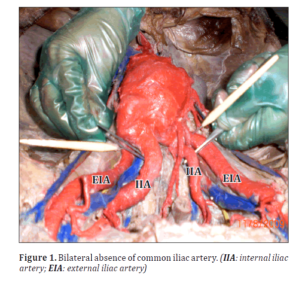
The present case showed multiple variations namely –bilateral absence of common iliac arteries, aneurysm of abdominal aorta.
Vascular malformations involving the iliac and femoral vessels are usually discovered incidentally or during the work-up for lower extremity ischemia. The exact prevalence of iliofemoral variations is unknown, but Greeb identified no more than 6 cases by angiography in a series of 8000 symptomatic patients [1].
Case Report
During the routine dissection teaching of the abdominopelvic region to MBBS students at M.S. Ramaiah Medical College, Bangalore, in an adult male cadaver of about 60 years, a variation was observed in the termination of the abdominal aorta. There was bilateral absence of the common iliac artery with both of its terminal branches (external and internal iliac arteries) arising directly from abdominal aorta. The further course of both iliac arteries and all its branches were traced on both sides and photographs were taken of the associated arterial variations.
The abdominal aorta was directly branching into external and internal iliac arteries on both sides at the level of the intervertebral disc between 4th and 5th lumbar vertebra. There was bilateral absence of common iliac arteries (Figure 1).
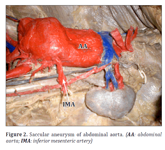
The abdominal aorta also showed saccular dilatation closer to its termination slightly below the level of the origin of renal arteries. Hence inferior mesenteric artery was noticed to be arising from the lateral aspect of abdominal aorta due to the saccular aneurysm of abdominal aorta (Figure 2). Rest of the branches of the aorta were as usual.
Discussion
Complete absence of the common iliac artery specifically is an extremely rare variation. Mansfield and Howard showed the autopsy specimen of a patient with congenital bilateral absence of the common iliac arteries. In their case study, the aorta divided directly into two internal iliac arteries and two external iliac arteries [3].
Dumanian et al. presented a case report of a 44-year-old man with long standing bilateral intermittent claudication secondary to congenital absence of the left common iliac, both external iliac and femoral arteries [4].
More recently, Llauger et al. described congenital absence of right common iliac artery in an asymptomatic patient in whom the blood supply to the right pelvis and the right lower extremity derived from a variant artery originating from the left hypogastric artery [5].
Dabydeen et al. reported a case of complete absence of the right common iliac artery in a 21-year-old woman, incidentally discovered by CT during the investigative procedure for acute abdominal pain. A network of prominent collateral arteries reconstituted the distal portion of the right external iliac artery and the common femoral artery, forming the arterial supply of the right lower limb [1].
The initial segment of the umbilical artery becomes the common iliac artery. It is likely that the absence of common iliac artery in this case is due to the disappearance of the initial segment of the umbilical artery [6].
Leriche and Morel described a syndrome that includes fatigue, pain throughout the lower extremities appearing after exercise, pallor of the legs on elevation, absent or weak pulse in the lower extremities and impotence. The primary etiological agent for this syndrome is atherosclerotic involvement of the aortic bifurcation. Although both occlusive disease of the terminal aorta and congenital absence of the iliac arteries can present with claudication, there are still some differences [7].
Some authors have argued that while congenital absence of the iliac arteries is rare, it should be included in the differential diagnosis for intermittent claudication of the legs [6]. So knowing this variation is very important for surgeons, gynecologists and radiologists before planning for abdominal or vascular surgery in order to avoid complications.
Conclusion
The present case is an observation where there was bilateral absence of common iliac artery due to the disappearance of the initial segment of the umbilical artery. It was associated with aneurysm of abdominal aorta.
Congenital absence of the iliac arteries is rare; it should be included in the differential diagnosis for intermittent claudication of the lower limbs because there is a high prevalence of aneurysm and atherosclerosis.
References
- Dabydeen D, Shabashov A, Shaffer K. Congenital absence of the right common iliac artery. Radiology Case Reports. 2008; 3. DOI: 10.2484/rcr.2008v3i1.47 URL: http://radiology.casereports.net/index.php/rcr/article/view/47/388
- Standring S, ed. Gray’s Anatomy. 39th Ed., Edinburgh, Elsevier Churchill Livingstone. 2005; 1360.
- Mansfield AO, Howard JM. Absence of both common iliac arteries. A Case report. Anat Rec. 1964; 150: 363–364.
- Dumanian AV, Frahm CJ, Benchik FA, Wooden TF. Intermittent claudication secondary to congenital absence of iliac arteries. Arch Surg. 1965; 91: 604–606.
- Llauger J, Sabate JM, Guardia E, Escudero J. Congenital absence of the right common iliac artery: CT and angiographic demonstration. Eur J Radiol. 1995; 21: 128–130.
- Senior HD. The development of the arteries of the human lower extremities. Am J Anat .1919; 25: 54–95.
- Leriche R, Morel A. Syndrome of thrombotic obliteration of aortic bifurcation. Ann Surg. 1948; 127: 193–206.
Shailaja Shetty*, Lakshmi Kantha and Chowdapurkar Sheshgiri
Department of Anatomy, M.S. Ramaiah Medical College, Bangalore, India
- *Corresponding Author:
- Dr. Shailaja Shetty
Professor of Anatomy M. S. Ramaiah Medical College, MSR Nagar Bangalore, 560054 Karnataka, India
Tel: +91 (944) 8713013
E-mail: drshailajashetty@rediffmail.com
Date of Received: November 6th, 2011
Date of Accepted: September 8th, 2012
Published Online: January 13th, 2013
© Int J Anat Var (IJAV). 2013; 6: 7–8.
Abstract
The vascular malformations involving the abdominal aorta, common iliac artery and its branches are very rare. Normally the abdominal aorta bifurcates into the right and left common iliac arteries anterolateral to the fourth lumbar vertebra. In the present case, there was bilateral absence of common iliac arteries which are the terminal branches of the abdominal aorta associated with aneurysm of abdominal aorta. The reason for the absence of common iliac artery is attributed to the disappearance of the initial segment of the umbilical artery. Proper identification of anatomical variations is essential for surgical and radiological interventions in order to prevent inadvertent complications during open surgical procedure or percutaneous intervention.
-Keywords
abdominal aorta, common iliac artery, aneurysm
Introduction
Vascular malformations involving the iliac and femoral vessels are far more rare than those involving the thoracic and abdominal aorta [1].
The abdominal aorta bifurcates into the right and left common iliac arteries at the level of fourth lumbar vertebra which in turn terminates into external and internal iliac arteries [2].
The present case showed multiple variations namely –bilateral absence of common iliac arteries, aneurysm of abdominal aorta.
Vascular malformations involving the iliac and femoral vessels are usually discovered incidentally or during the work-up for lower extremity ischemia. The exact prevalence of iliofemoral variations is unknown, but Greeb identified no more than 6 cases by angiography in a series of 8000 symptomatic patients [1].
Case Report
During the routine dissection teaching of the abdominopelvic region to MBBS students at M.S. Ramaiah Medical College, Bangalore, in an adult male cadaver of about 60 years, a variation was observed in the termination of the abdominal aorta. There was bilateral absence of the common iliac artery with both of its terminal branches (external and internal iliac arteries) arising directly from abdominal aorta. The further course of both iliac arteries and all its branches were traced on both sides and photographs were taken of the associated arterial variations.
The abdominal aorta was directly branching into external and internal iliac arteries on both sides at the level of the intervertebral disc between 4th and 5th lumbar vertebra. There was bilateral absence of common iliac arteries (Figure 1).
The abdominal aorta also showed saccular dilatation closer to its termination slightly below the level of the origin of renal arteries. Hence inferior mesenteric artery was noticed to be arising from the lateral aspect of abdominal aorta due to the saccular aneurysm of abdominal aorta (Figure 2). Rest of the branches of the aorta were as usual.
Discussion
Complete absence of the common iliac artery specifically is an extremely rare variation. Mansfield and Howard showed the autopsy specimen of a patient with congenital bilateral absence of the common iliac arteries. In their case study, the aorta divided directly into two internal iliac arteries and two external iliac arteries [3].
Dumanian et al. presented a case report of a 44-year-old man with long standing bilateral intermittent claudication secondary to congenital absence of the left common iliac, both external iliac and femoral arteries [4].
More recently, Llauger et al. described congenital absence of right common iliac artery in an asymptomatic patient in whom the blood supply to the right pelvis and the right lower extremity derived from a variant artery originating from the left hypogastric artery [5].
Dabydeen et al. reported a case of complete absence of the right common iliac artery in a 21-year-old woman, incidentally discovered by CT during the investigative procedure for acute abdominal pain. A network of prominent collateral arteries reconstituted the distal portion of the right external iliac artery and the common femoral artery, forming the arterial supply of the right lower limb [1].
The initial segment of the umbilical artery becomes the common iliac artery. It is likely that the absence of common iliac artery in this case is due to the disappearance of the initial segment of the umbilical artery [6].
Leriche and Morel described a syndrome that includes fatigue, pain throughout the lower extremities appearing after exercise, pallor of the legs on elevation, absent or weak pulse in the lower extremities and impotence. The primary etiological agent for this syndrome is atherosclerotic involvement of the aortic bifurcation. Although both occlusive disease of the terminal aorta and congenital absence of the iliac arteries can present with claudication, there are still some differences [7].
Some authors have argued that while congenital absence of the iliac arteries is rare, it should be included in the differential diagnosis for intermittent claudication of the legs [6]. So knowing this variation is very important for surgeons, gynecologists and radiologists before planning for abdominal or vascular surgery in order to avoid complications.
Conclusion
The present case is an observation where there was bilateral absence of common iliac artery due to the disappearance of the initial segment of the umbilical artery. It was associated with aneurysm of abdominal aorta.
Congenital absence of the iliac arteries is rare; it should be included in the differential diagnosis for intermittent claudication of the lower limbs because there is a high prevalence of aneurysm and atherosclerosis.
References
- Dabydeen D, Shabashov A, Shaffer K. Congenital absence of the right common iliac artery. Radiology Case Reports. 2008; 3. DOI: 10.2484/rcr.2008v3i1.47 URL: http://radiology.casereports.net/index.php/rcr/article/view/47/388
- Standring S, ed. Gray’s Anatomy. 39th Ed., Edinburgh, Elsevier Churchill Livingstone. 2005; 1360.
- Mansfield AO, Howard JM. Absence of both common iliac arteries. A Case report. Anat Rec. 1964; 150: 363–364.
- Dumanian AV, Frahm CJ, Benchik FA, Wooden TF. Intermittent claudication secondary to congenital absence of iliac arteries. Arch Surg. 1965; 91: 604–606.
- Llauger J, Sabate JM, Guardia E, Escudero J. Congenital absence of the right common iliac artery: CT and angiographic demonstration. Eur J Radiol. 1995; 21: 128–130.
- Senior HD. The development of the arteries of the human lower extremities. Am J Anat .1919; 25: 54–95.
- Leriche R, Morel A. Syndrome of thrombotic obliteration of aortic bifurcation. Ann Surg. 1948; 127: 193–206.


