Sign up for email alert when new content gets added: Sign up
Evaluation of the local scaphoid fracture pathway
Webinar on Osteoporosis, Arthritis and Musculoskeletal Disorders
June 15, 2022 | Webinar
Abdirahman Osman
Lister Hospital, East and North Hertfordshire Trust, UK
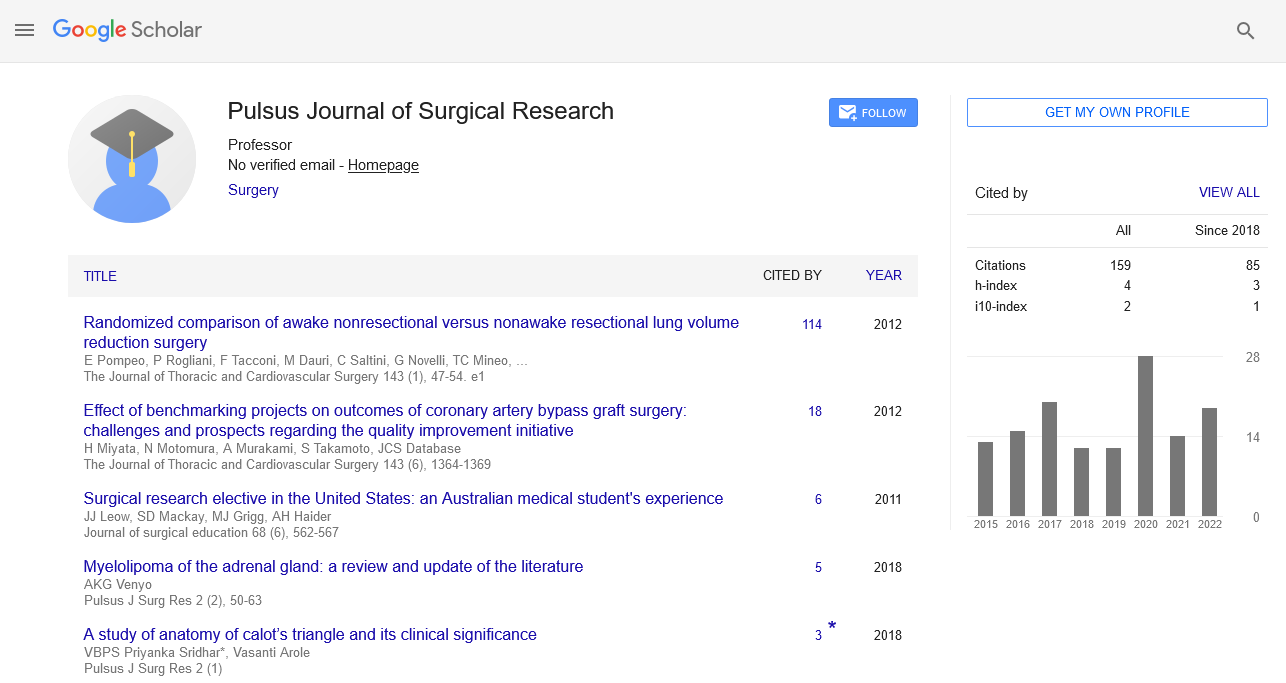
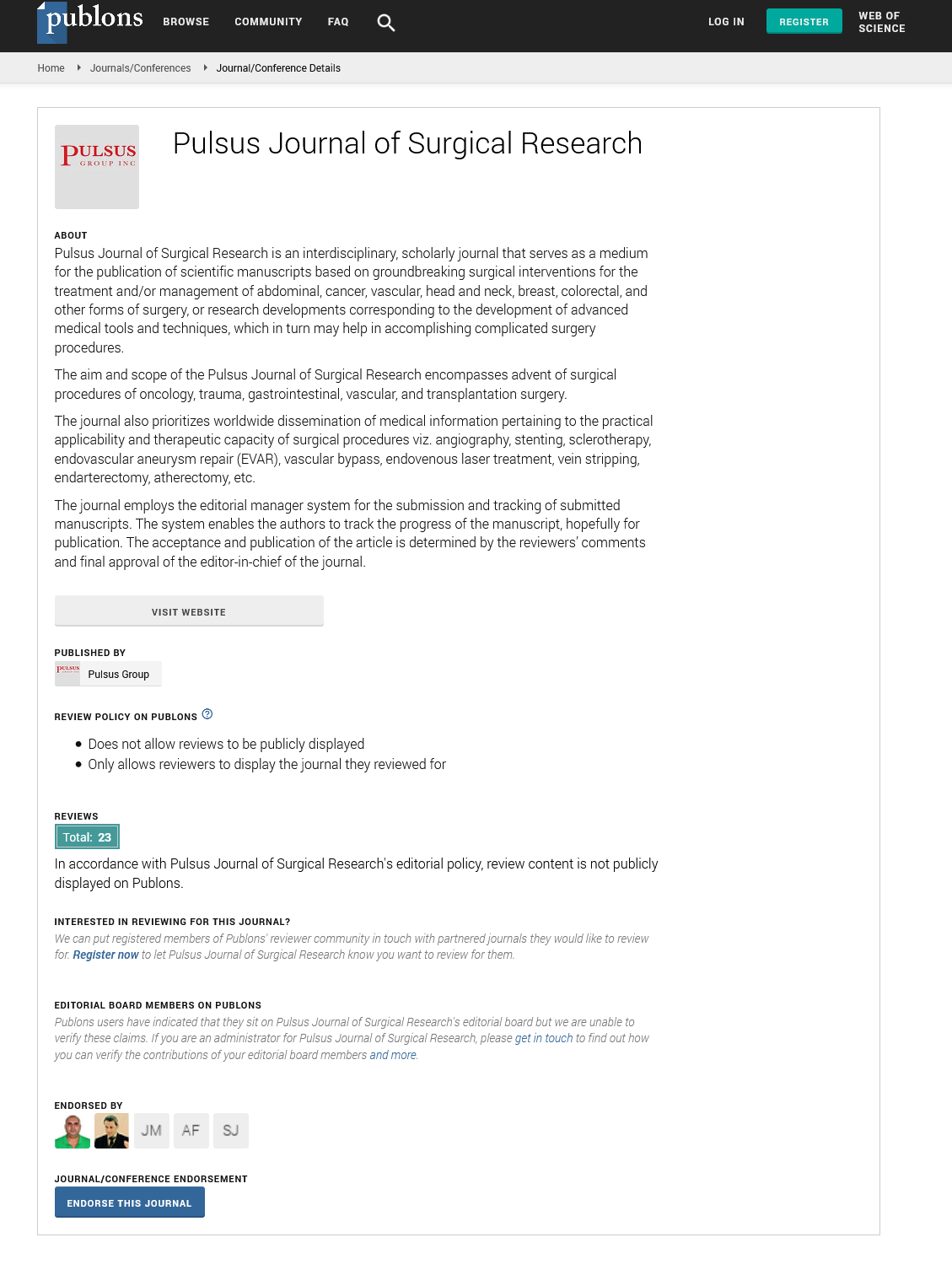
Posters & Accepted Abstracts: Pulsus J Surg Res
Abstract :
Aims: Identify clinical examination findings, initial imaging findings and stabilisation method in the emergency department for suspected scaphoid fractures. Review the follow-up, repeat imaging and further investigations for these patients. Identify the waiting time for further investigations / imaging. Introduction: Scaphoid fractures are the most common carpal fracture and account for 2% to 7% of all fractures1. These fractures are commonly missed through clinical and radiographic examination; it has been reported that up to 40% of scaphoid fractures are missed on initial presentation. There are three clinical findings that can indicate a potential scaphoid fracture; anatomical snuffbox tenderness (AST), scaphoid tubercule tenderness (STT) and telescoping tenderness (TT). ASB, STT and TT all have 100% sensitivity but, in one study, specificities were 9%, 30% and 48% respectively. However, when these tests are combined, multiple studies have illustrated that the specificity does increase2-3. Current National Institute for Health and Clinical Excellence (NICE) guidance advises that MRI directly from the emergency department should be considered for suspected scaphoid fractures. Studies have shown that a minority of trauma centres currently offer further imaging from the emergency department4. Misdiagnosis can increase patient morbidity; non-union, arthritis, deformity and instability. Early definitive diagnosis will not only prevent a missed scaphoid injury but can avert overtreatment for those without a scaphoid fracture and subjection to extended immobilization. A report by the NHSLA has highlighted the litigation cost of negligent scaphoid fracture management in the UK; 0.01% of all orthopaedic-related litigation were attributed to mismanagement of scaphoid fractures and the largest costs ascribed to a combination of failed diagnosis and delay in initiating appropriate management5. Methods: This is a retrospective study of all patients identified on eTrauma (clinical platform for centralised orthopaedic trauma coordination) as referrals for a suspected scaphoid fracture from 01/04/21 – 01/08/21 at the Lister hospital. The following data was collected: - Clinical presentation (anatomical snuffbox tenderness, scaphoid tubercle tenderness and telescoping tenderness) - Initial plain film radiograph, method of immobilization and total time immobilised - Follow-up and repeat plain film radiograph - Further imaging modality and time from initial referral to further imaging. Current local practice and pathway Results and discussion: I. 131 patients identified in this study II. Total number of clinical findings on physical examination: 62% had 1 finding, 28% had 2 findings, 1.5% had 3 findings and 8.5% had none III. Number of patient injuries immobilised, length of time and method of immobilisation: 115 (88%) patients had an immobilisation method. 81 in Futuro splint, 31 in thumb spina splint and 3 in scaphoid POP; more than 90% had a stabilisation method in place for at least two weeks IV. Scaphoid fracture on initial radiograph at presentation: 6% confirmatory / high suspicion, 88% negative and 6% identified other bony injuries V. Scaphoid fracture on repeat radiograph at two weeks: 68 patients had a repeat radiograph at two weeks; 5 (7%) confirmed a scaphoid fracture, 63 (93%) negative for scaphoid fracture VI. Further imaging modalities and waiting time: 28 patients had further imaging requested; 16 for CT and 12 for MRI. The average waiting time from fracture clinic referral to CT and MRI were 5 weeks and 8 weeks respectively Conclusion: 1) The most common reason for referral was from 1 clinical sign 2) 11% of patients had a scaphoid fracture identified on radiograph (on presentation AND at two weeks) 3) 21% of patients in had further imaging modalities requested (CT / MRI) The gold standard investigation tool for identifying scaphoid fractures is MRI and, ideally, all patients with a query diagnosis of scaphoid fracture should have this imaging modality. However, the question remains; are the fracture clinic referrals appropriate with the low efficacy of reduced clinical findings? A multi-pronged approach will be needed to decrease inappropriate referrals, increase the number of patients having further imaging and to reduce the time from presentation to CT / MRI: 1) Diagnostic algorithm education for emergency department 2) Review current pathway to incorporate MRI within 3-5 days from Presentation 6 3) Alternative dedicated imaging (cone-beam computed tomography)7 Further research is needed to fully investigate the facilitators and barriers to the implementation of NICE guidance
Biography :
Abdirahman Osman is currently working at Lister Hospital, Trauma and Orthopaedic Department, East and North Hertfordshire Trust, Stevenage, United Kingdom. His research interests include Trauma and Orthopaedic, Surgery.