A bony canal in the basilar part of occipital bone
Navneet Kumar Chauhan*, Jyoti Chopra, Anita Rani, Archana Rani and Ajay Kumar Srivastava
Department of Anatomy, Chhatrapati Shahuji Maharaj Medical University, Lucknow, India
- *Corresponding Author:
- Dr. Navneet Kumar Chauhan
Associate Professor, Department of Anatomy, Chhatrapati Shahuji Maharaj Medical University (Upgraded King George’s Medical College) Lucknow, 226003, U.P, India
Tel: +91 941 5083580
E-mail: navneetchauhan@yahoo.com
Date of Received: December 19th, 2009
Date of Accepted: July 11th, 2010
Published Online: August 9th, 2010
© Int J Anat Var (IJAV). 2010; 3: 112–113.
[ft_below_content] =>Keywords
clivus, clival canal, occipital bone, notochord remnant
Introduction
The clivus (Latin: slope) is a curved sloppy surface between the dorsum sellae of sphenoid and foramen magnum of occipital bone in human skull. In the present study, a bony canal was observed in the clivus. There are very few reports in literature about the presence of a bony passage in the clivus. These bony canals if present may contain communicating veins between extracranial and intracranial venous plexuses or remnant of notochord during life. The knowledge of this canal has a great importance for neurosurgeons, radiologists and anatomists.
Case Report
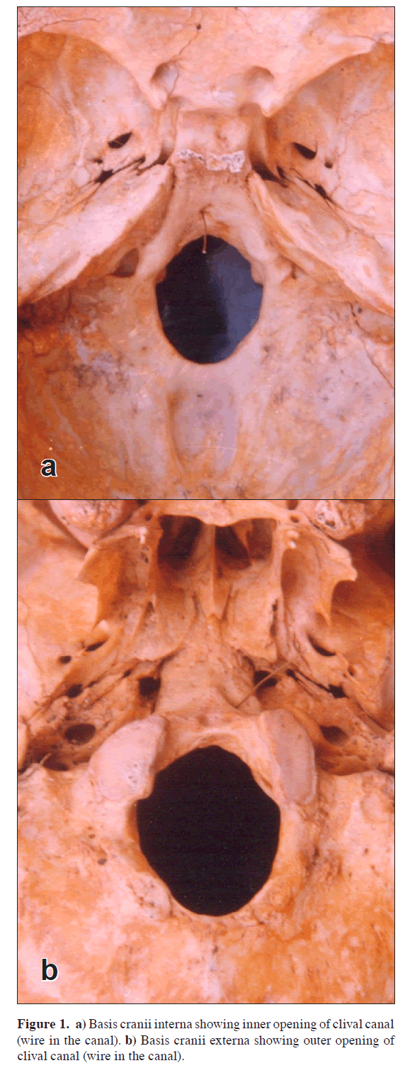
During routine examination of the skulls, of bone processing lab, in the department of Anatomy of Chhatrapati Shahuji Maharaj Medical University Lucknow, India, a bony canal was found in the clivus of dry, adult human skull of unknown sex. The inner opening of canal was located in the middle of clivus 6 mm above the anterior margin of foramen magnum. The canal was running downwards, forwards and laterally. The other end of the canal was situated anterolateral to the pharyngeal tubercle of basilar part of occipital bone, 7 mm in front of foramen magnum on the left side. A bristle was inserted to determine the length and patency of the canal. The canal was 6 mm long and the width of the clivus canal was approximately 1 mm. No other significant bony anomaly was noted (Figure 1).
Discussion
Presence of any canal in the clivus is a rare occurrence. Jalsovec and Vinter noticed a 5 mm long median bony canal in the posterior one third of clivus in a postmortem human skull with its both ends opening in the basis cranii interna. They proposed that this canal is more likely due to the persistence of remnant of notochord [1]. Nayak et al. observed a transverse bony canal in the middle third of the clivus of an adult male skull. They proposed two possible explanations for the occurrence of this canal that it may contain the vein connecting the two inferior petrosal sinuses or the canal is a remnant of first true somite [2]. Zhang and Yen found three cases of bony canal on the lower half of the clival surface out of 100 adult cranial bases studied. They assigned the term “inferior median clival canal” to this variant [3].
In the present case, the bony canal in the clivus is traversing through the basilar part of occipital bone from its superior to inferior surface. Considering the direction and location of the canal, there could be two possible explanations for its formation. It could be because of a connecting vein between basilar plexus and pharyngeal venous plexus or it might have contained the remnant of notochord.
The skeletal axis is first formed in third week of intrauterine life by non-segmental notochord which is not only limited to the region in which vertebrae will replace it but extends into the head as far as the caudal limit of hypophysis cerebri. The notochord serves as a frame work around which a blastemal or mesenchymal model is formed. The blastemal skull appears at the end of the first month and the first mass appears in the occipital region, outlining the basilar part of occipital bone. Chondrification of cranium begins in the second month. The first cartilaginous foci appear in the occipital plate, one on each side of the notochord (parachordal cartilages). These later fuse at about the end of seventh week around the oblique transit of notochord. The notochord traverses the occipital plate obliquely, being at first near its dorsal surface and then lying ventrally where it comes in close relationship of epithelium of dorsal wall of pharynx. It then re-enters the cranial base and runs ventrally to end just caudal to the hypophysis. Ossification commences before the chondrocranium has fully developed. The ossification of the basilar part starts around sixth week of intrauterine life [4].
In the present case as the canal is directed downwards and forwards from dorsal to ventral aspect of the basilar part of occipital bone it can be inferred that this canal may be due to the persistence of remnant of notochord but as the canal is not in the median plane and is directed laterally, the possibility of presence of notochord in the canal becomes lower.
The clivus is related to basilar and pharyngeal venous plexuses. The venous system generally develops in the 4th and the 5th week of fetal development, while the development of skeleton occurs later. It is possible that these two systems interfered, so that a venous channel connecting the developing basilar and pharyngeal plexuses was surrounded by ossifying elements, which then resulted in its enclosure forming a clival canal. We believe that in the present case more likely a venous communication existed between the basilar and pharyngeal venous plexuses, which led to the formation of this bony canal.
The canal of the clivus might interfere with the neurosurgical operations in the clival region and possibly provoke symptoms of the basilar artery as well as of the basilar plexus [1]. This canal may be confused for a fracture of clivus during cervical trauma in CT scan. Knowledge of its topography and potential presence in the adult may aid the clinician in interpretation of imaging of this region. Furthermore, misinterpretation of this canal might cause emotional and legal complications for the clinicians [2].
References
- Jalsovec D, Vinter I. Clinical significance of a bony canal of the clivus. Eur Arch Otorhinolaryngol. 1999; 256: 160–161.
- Nayak SR, Saralaya V, Prabhu LV, Pai MM. A mysterious clival canal and its importance. Neuroanatomy. 2007; 6: 34–35.
- Zhang WH, Yen WC. A new bony canal on the clival surface of the occipital bone. Acta Anat (Basel). 1987; 128: 63–66.
- Williams PL, Warwick R. Gray’s Anatomy. 36th Ed., Churchill Livingstone, New York. 1980; 138–143.
Navneet Kumar Chauhan*, Jyoti Chopra, Anita Rani, Archana Rani and Ajay Kumar Srivastava
Department of Anatomy, Chhatrapati Shahuji Maharaj Medical University, Lucknow, India
- *Corresponding Author:
- Dr. Navneet Kumar Chauhan
Associate Professor, Department of Anatomy, Chhatrapati Shahuji Maharaj Medical University (Upgraded King George’s Medical College) Lucknow, 226003, U.P, India
Tel: +91 941 5083580
E-mail: navneetchauhan@yahoo.com
Date of Received: December 19th, 2009
Date of Accepted: July 11th, 2010
Published Online: August 9th, 2010
© Int J Anat Var (IJAV). 2010; 3: 112–113.
Abstract
Clivus is a gradual slopping process behind the dorsum sellae that runs obliquely backwards. An unusual 6 mm long and 1 mm wide bony canal was observed on the lower one third of clivus in an adult human dry skull. The internal end of the canal was opening in the midline. The canal was directed downwards, forwards and laterally. The external opening was present antero-lateral to the pharyngeal tubercle on the left side. Presence of any canal in the clivus is a rare occurrence. There could be two possible explanations for its formation. It could be because of presence of a connecting vein or it might have contained the remnant of notochord. We believe that in the present case more likely a venous communication existed between the basilar and pharyngeal venous plexuses, which led to the formation of this bony canal. The canal of the clivus might interfere with the neurosurgical operations in the clival region or can be confused for a fracture of clivus.
-Keywords
clivus, clival canal, occipital bone, notochord remnant
Introduction
The clivus (Latin: slope) is a curved sloppy surface between the dorsum sellae of sphenoid and foramen magnum of occipital bone in human skull. In the present study, a bony canal was observed in the clivus. There are very few reports in literature about the presence of a bony passage in the clivus. These bony canals if present may contain communicating veins between extracranial and intracranial venous plexuses or remnant of notochord during life. The knowledge of this canal has a great importance for neurosurgeons, radiologists and anatomists.
Case Report
During routine examination of the skulls, of bone processing lab, in the department of Anatomy of Chhatrapati Shahuji Maharaj Medical University Lucknow, India, a bony canal was found in the clivus of dry, adult human skull of unknown sex. The inner opening of canal was located in the middle of clivus 6 mm above the anterior margin of foramen magnum. The canal was running downwards, forwards and laterally. The other end of the canal was situated anterolateral to the pharyngeal tubercle of basilar part of occipital bone, 7 mm in front of foramen magnum on the left side. A bristle was inserted to determine the length and patency of the canal. The canal was 6 mm long and the width of the clivus canal was approximately 1 mm. No other significant bony anomaly was noted (Figure 1).
Discussion
Presence of any canal in the clivus is a rare occurrence. Jalsovec and Vinter noticed a 5 mm long median bony canal in the posterior one third of clivus in a postmortem human skull with its both ends opening in the basis cranii interna. They proposed that this canal is more likely due to the persistence of remnant of notochord [1]. Nayak et al. observed a transverse bony canal in the middle third of the clivus of an adult male skull. They proposed two possible explanations for the occurrence of this canal that it may contain the vein connecting the two inferior petrosal sinuses or the canal is a remnant of first true somite [2]. Zhang and Yen found three cases of bony canal on the lower half of the clival surface out of 100 adult cranial bases studied. They assigned the term “inferior median clival canal” to this variant [3].
In the present case, the bony canal in the clivus is traversing through the basilar part of occipital bone from its superior to inferior surface. Considering the direction and location of the canal, there could be two possible explanations for its formation. It could be because of a connecting vein between basilar plexus and pharyngeal venous plexus or it might have contained the remnant of notochord.
The skeletal axis is first formed in third week of intrauterine life by non-segmental notochord which is not only limited to the region in which vertebrae will replace it but extends into the head as far as the caudal limit of hypophysis cerebri. The notochord serves as a frame work around which a blastemal or mesenchymal model is formed. The blastemal skull appears at the end of the first month and the first mass appears in the occipital region, outlining the basilar part of occipital bone. Chondrification of cranium begins in the second month. The first cartilaginous foci appear in the occipital plate, one on each side of the notochord (parachordal cartilages). These later fuse at about the end of seventh week around the oblique transit of notochord. The notochord traverses the occipital plate obliquely, being at first near its dorsal surface and then lying ventrally where it comes in close relationship of epithelium of dorsal wall of pharynx. It then re-enters the cranial base and runs ventrally to end just caudal to the hypophysis. Ossification commences before the chondrocranium has fully developed. The ossification of the basilar part starts around sixth week of intrauterine life [4].
In the present case as the canal is directed downwards and forwards from dorsal to ventral aspect of the basilar part of occipital bone it can be inferred that this canal may be due to the persistence of remnant of notochord but as the canal is not in the median plane and is directed laterally, the possibility of presence of notochord in the canal becomes lower.
The clivus is related to basilar and pharyngeal venous plexuses. The venous system generally develops in the 4th and the 5th week of fetal development, while the development of skeleton occurs later. It is possible that these two systems interfered, so that a venous channel connecting the developing basilar and pharyngeal plexuses was surrounded by ossifying elements, which then resulted in its enclosure forming a clival canal. We believe that in the present case more likely a venous communication existed between the basilar and pharyngeal venous plexuses, which led to the formation of this bony canal.
The canal of the clivus might interfere with the neurosurgical operations in the clival region and possibly provoke symptoms of the basilar artery as well as of the basilar plexus [1]. This canal may be confused for a fracture of clivus during cervical trauma in CT scan. Knowledge of its topography and potential presence in the adult may aid the clinician in interpretation of imaging of this region. Furthermore, misinterpretation of this canal might cause emotional and legal complications for the clinicians [2].
References
- Jalsovec D, Vinter I. Clinical significance of a bony canal of the clivus. Eur Arch Otorhinolaryngol. 1999; 256: 160–161.
- Nayak SR, Saralaya V, Prabhu LV, Pai MM. A mysterious clival canal and its importance. Neuroanatomy. 2007; 6: 34–35.
- Zhang WH, Yen WC. A new bony canal on the clival surface of the occipital bone. Acta Anat (Basel). 1987; 128: 63–66.
- Williams PL, Warwick R. Gray’s Anatomy. 36th Ed., Churchill Livingstone, New York. 1980; 138–143.


