A case of anomalous flexor carpi radialis brevis muscle and its clinical significance
2 Department of Anatomy, University of Otago, Christchurch, New Zealand
3 Department of Orthopaedic Surgery and Musculoskeletal Medicine, University of Otago, Christchurch, New Zealand, Email: kieserdavid@gmail.com
Received: 17-Oct-2017 Accepted Date: Oct 26, 2017; Published: 31-Oct-2017
Citation: Trowbridge S, Vidakovic H, Hammer N, et al. A case of anomalous flexor carpi radialis brevis muscle and its clinical significance. Int J Anat Var. 2017;10(4): 91-93.
This open-access article is distributed under the terms of the Creative Commons Attribution Non-Commercial License (CC BY-NC) (http://creativecommons.org/licenses/by-nc/4.0/), which permits reuse, distribution and reproduction of the article, provided that the original work is properly cited and the reuse is restricted to noncommercial purposes. For commercial reuse, contact reprints@pulsus.com
Abstract
Aim: To present an atypical case of flexor carpi radialis brevis (FCRB) and its impact on minimally invasive procedures to the volar distal radius.
Methods: Minimally invasive volar plate fixation was performed on 16 standard embalmed cadaveric forearms in the University of Otago anatomy department. The forearms were then dissected to analyse the effect on the local anatomy and the position of the metalware.
Results: One case of a FCRB was identified, with an atypical split tendon and insertion into the transverse carpal ligament and base of the index and middle metacarpal. The surgical procedure caused radial muscle fibre tearing of the FCRB.
Conclusions: The FCRB is a rare anomalous muscle of the wrist. Its presence has a detrimental effect on minimally invasive procedures to the volar distal radius. Surgeons operating on the distal forearm should be aware of this anatomical variation to prevent inadvertent injury.
Keywords
Volar plate; Distal radius; Flexor carpi radialis brevis; FCRB; Tendon
Introduction
Flexor carpi radialis brevis (FCRB) is a rare anomalous muscle of the wrist. It was first described by this name in the mid-19th Century by John Wood, an anatomy demonstrator at King’s College, London in a report on ‘variations in human Myology’ for the Royal Society [1]. In the 21st Century its presence has been described mostly as a coincidental finding during cadaveric and operative dissection, and rarely as a symptomatic painful mass in the forearm [2-4].
FCRB has an estimated incidence of 2.6 - 7.5% [5]. It originates from the volar-radial surface of the distal radius, distal to flexor pollicis longs (FPL) and proximal to the insertion of pronator quadratus (PQ). The insertion is typically the index or middle metacarpal base. However, significant variability has been described, including the ring metacarpal base, radial carpal bones, and palmar fascia [6]. It is innervated by the anterior interosseous nerve (AIN) and its usual passage past the wrist is neighbouring the tendon of flexor carpi radialis (FCR), through its osteo-fibrous tunnel in the groove of the trapezium [5].
Previous case studies have encountered FCRB during a volar approach for fixation of the distal radius [5]. Indeed, Yoon-Min and colleagues indicated the clinical importance of being aware of FCRB due to the trend towards volar plate fixation for distal radius fractures [5]. However, there is a further trend towards minimally invasive, tissue preserving techniques through smaller incisions. These approaches are performed based on predicted anatomy and therefore anomalous structures are at risk, and assumed anatomical landmarks can be rendered unreliable.
Therefore, this report aims at describing a case of FCRB and its unique anatomical variation, and discussing its impact on minimally invasive volar plating of the distal radius.
Methods
Ethics approval was attained from the University of Otago, Human Ethics Committee (H17/065). While alive, all donors gave their informed consent to the donation of their bodies for teaching and research purposes, and experiments were conducted in line with the declaration of Helsinki.
A cadaveric study on 16 New Zealand European upper limbs investigating a minimally invasive technique for volar plate fixation of the distal radius was performed. The procedure involved a 2 cm transverse volar-radial incision centred over the flexor carpi radialis (FCR) tendon and based 4-5 cm proximal to the wrist crease. After the skin incision, the subcutaneous fat is bluntly dissected to expose the fascia encasing the FCR tendon. This fascia is longitudinally incised on the radial side of the FCR tendon. The FCR is then retracted toward the ulnar, while the bed of the FCR sheath is longitudinally incised. Flexor pollicis longus (FPL) is then identified and bluntly dissected along its radial border before being retracted toward the ulnar to expose the pronator quadratus (PQ). PQ is then incised in line with its fibres and submuscularly released from the underlying radius to allow passage of a volar plate, which is subsequently secured with screws.
After plate osteo-synthesis a volar dissection was performed in all samples to assess the quality of fixation and integrity of deep structures.
Results
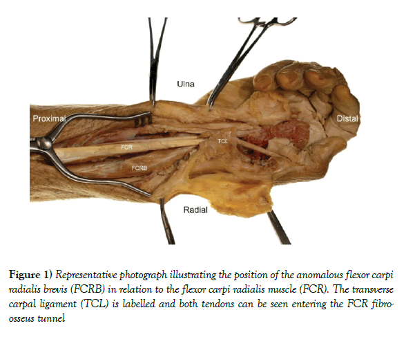
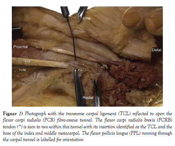
In one sample, the right upper limb of a 91 year-old male, an anomalous muscle was encountered dorso-radial to the FCR tendon at the level of the surgical approach (Figure 1). With complete dissection, a bipennate muscle with a short tendon originating from the volar surface of the radius, proximal to PQ, was identified. This muscle had a split tendon with a complex insertion into the base of the index and middle metacarpal, as well as a slip of tendon into the transverse carpal ligament (Figure 2).
Figure 2) Photograph with the transverse carpal ligament (TCL) reflected to open the flexor carpi radialis (FCR) fibro-osseus tunnel. The flexor carpi radialis brevis (FCRB) tendon (*) is seen to run within this tunnel with its insertion identified as the TCL and the base of the index and middle metacarpal. The flexor pollicis longus (FPL) running through the carpal tunnel is labelled for orientation
Innervation was likely attained from a branch of the AIN that arose proximal and superficial to PQ and penetrated the deep surface of the muscle. Its vascular supply was not clear, but primarily arose from the radial artery and anterior interosseus artery. There were no other local anatomical abnormalities identified and the PQ was not underdeveloped.
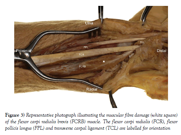
In this case, the approach utilised for minimally invasive fixation led to damage to the radial border of this muscle with tearing of radial sided muscle fibres (Figure 3).
Discussion
This case illustrates an anomalous muscle consistent with a flexor carpi radialis brevis (FCRB). Its origin, position, innervation and blood supply are consistent with previous reports on this muscle, but with an atypical, and previously undescribed split tendon inserting into the transverse carpal ligament and base of the index and middle metacarpal. There was no evidence of under development of PQ, which has commonly been described in previous studies [2].
Distal radius fractures are one of the most common fractures and are becoming an ever-increasing burden in aging populations due to increased falls and osteoporosis [7,8]. These injuries can be treated by manipulation and casting, external fixation, k-wiring or plate fixation [8]. Volar plating offers the greatest biomechanical advantage to most distal radius fractures and is therefore commonly performed [8-10]. However, this involves a large incision, with significant soft tissue mobilisation and PQ elevation [11]. Thus minimally invasive techniques to perform this procedure have been advocated [11-13].
This case has highlighted the translational importance of this anatomical variation and illustrated that the presence of a FCRB can have a detrimental effect on minimally invasive surgery of the volar distal radius. Surgeons should therefore be aware of this anatomical variation to prevent injury during the surgical procedure. If visualisation is limited, we would advocate extension of the incision to aid exposure. However, surgeons should recognise this anomalous muscle because the deep landmarks, particularly the identification of flexor pollicis longus (FPL), will be obscured.
Some FCRB muscles have been reported to present as painful forearm masses, however, most remain asymptomatic and incidentally identified [14-15]. Although the clinical signs of a FCRB are not described, previous authors have serendipitously identified this anatomical variation intra-operatively and successfully utilized it during base of thumb reconstruction for first carpo-metacarpal osteoarthritis rather than using the FCR tendon [16]. It has also been recognised on ultra-sound and MRI imaging as a normal appearing small muscle arising from the radius and inserting into the trapezium or base of index and middle metacarpals [14-18]. However, its relative rarity1, [16] and the lack of higher-level evidence to support its use in hand and wrist reconstruction surgery, we feel negates routine cross-sectional imaging for pre-operative planning.
This study is limited to a single case in a cadaveric study. We would support the intra-operative difficulties in living tissue being described as standard embalmed cadaveric tissue offers limited soft tissue pliability. Unfortunately the contra-lateral limb was not available for this study and therefore we were unable to determine if this was a symmetrical finding. Further research is also required to render the true incidence of FCRB and its bilaterality in patients. In addition, the ethnic variances are unknown and therefore warrant further investigation.
Conclusion
The FCRB is a rare anomalous muscle of the wrist. Its presence has a detrimental effect on minimally invasive procedures to the volar distal radius. Surgeons operating on the distal forearm should be aware of this anatomical variation to prevent inadvertent injury.
REFERENCES
- Wood J. Variations in human myology observed during the winter session of 1866-1867 at King’s College, London. Proc R Soc Lond. 1867;15:518-46.
- Dodds SD. A flexor carpi radialis brevis muscle with an anomalous origin on the distal radius. J Hand Surg Am. 2006;31:1507-10.
- Kang L, Carter T, Wolfe SW. The flexor carpi radialis brevis muscle: an anomalous flexor of the wrist and hand. A case report. J Hand Surg Am. 2006;31:1511-3.
- Peers SC, Kaplan FT. Flexor carpi radialis brevis muscle presenting as a painful forearm mass: case report. J Hand Surg Am. 2008;33:1878-81.
- Lee YM, Song SW, Sur YJ, et al. Flexor carpi radialis brevis: An unusual anomalous muscle of the wrist. Clin Orthop Surg. 2014;6:361-4.
- Carleton A. Flexor carpi radialis brevis vel profundus. J Anat. 1935;69:292-3.
- Nellans KW, Kowalski E, Chung KC. The epidemiology of distal radius fractures. Hand Clin. 2012;28:113-25.
- Hove LM, Lindau T, Holmer P. Distal radius fractures: Current concepts. Springer, Berlin. 2014.
- Liporace FA, Gupta S, Jeong GK, et al. A biomechanical comparison of a dorsal 3.5 mm T-plate and a volar fixed-angle plate in a model of dorsally unstable distal radius fractures. JOT. 2005;19:187-91.
- Levin SM, Nelson CO, Botts JD, et al. Biomechanical evaluation of volar locking plates for distal radius fractures. Hand. 2008;3:55-60.
- Sen MK, Strauss N, Harvey EJ. Minimally invasive plate osteosynthesis of distal radius fractures using a pronator sparing approach. Tech Hand up Extrem Surg. 2008;12:2-6.
- Imatani J, Noda T, Morito Y, et al. Minimally invasive plate osteosynthesis for comminuted fractures of the metaphysis of the radius. J Hand Surg Eu. 2005;30:220-5.
- Zenke Y, Sakai A, Oshige T, et al. Clinical results of volar locking plate for distal radius fractures: Conventional versus minimally invasive plate osteosynthesis. JOT. 2011;25:425-31.
- Peers SC, Kaplan FTD. Flexor carpi radialis brevis muscle presenting as a painful forearm mass: Case report. J Hand Surg Am. 2008;33:1878-81.
- Urigo C, Schenkel MC, Beaulieu JY, et al. Painful flexor carpi radialis brevis muscle: An ultrasound and magnetic resonance imaging assessment. J Ultrasound Med. 2017.
- Duncan SFM, McCormack RR, Roberts CC. Characteristics of the flexor carpi radialis brevis tendon on magnetic resonance imaging and its use in basal joint arthroplasty. Radiol Case Rep. 2006;1:24-6.
- Chong SJ, Al-Ani S, Pinto C, et al. Bilateral flexor carpi radialis brevis and unilateral flexor carpi ulnaris brevis muscle: case report. J Hand Surg Am. 2009;34:1868-71.
- Smith J, Kaker S. Combined flexor carpi radialis tear and flexor carpi radialis brevis tendinopathy identified by ultrasound: a case report. PM&R. 2014;6:956-9.


