A Case Report on Anatomical Variation During Neck Dissection
Received: 03-Oct-2022, Manuscript No. ijav-22-5519; Editor assigned: 05-Oct-2022, Pre QC No. ijav-22-5519 (PQ); Accepted Date: Oct 24, 2022; Reviewed: 19-Oct-2022 QC No. ijav-22-5519; Revised: 24-Oct-2022, Manuscript No. ijav-22-5519 (R); Published: 31-Oct-2022, DOI: 10.37532/1308-4038.15(10).224
Citation: Hvizdosova N. A Case Report on Anatomical Variation during Neck Dissection. Int J Anat Var. 2022;15(10):231-231.
This open-access article is distributed under the terms of the Creative Commons Attribution Non-Commercial License (CC BY-NC) (http://creativecommons.org/licenses/by-nc/4.0/), which permits reuse, distribution and reproduction of the article, provided that the original work is properly cited and the reuse is restricted to noncommercial purposes. For commercial reuse, contact reprints@pulsus.com
Abstract
We describe an uncommon termination of the right external jugular vein, which drains straight into the cephalic vein and runs in front of the medial third of the clavicle, on the right side of the neck during routine dissection in a 72-year-old female cadaver. Intervention radiography, intravenous infusion, and surgical operations affecting the head and neck region all require a thorough understanding of the anatomical differences in this area of the human body.
Keywords
External jugular vein; Cephalic vein; Cadaver
INTRODUCTION
It is important to understand the various types of variations of the venous tree in the lower neck given the advanced technology and instrumentation used in medical practice, precise interventional radiology, and the implantation of an artificial cardiac pacemaker [1], in addition to orthopedic surgeons dealing with fracture and dislocation of the clavicle.
The external jugular vein (EJV), which is formed anatomically when face veins join retro mandibular and posterior auricular veins, travels deep to superficial fascia and platysma muscle along its path. Both (EJVs) pierce the investing layer of deep fascia lateral to the sternoclavicular joint before terminating in subclavian veins (SV) in the lower posterior triangle of the neck [2].
CASE REPORT
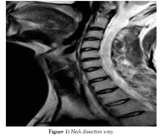
At Chatham University (Pittsburgh, Pennsylvania), the Department of Biology regularly conducts graduate-level human corpse dissection labs where we found an uncommon unilateral variation in the course of the right EJV in a 72-year-old female as shown in Figure 1.
The following conclusions were made:
• The EJV crosses the clavicle and joins the terminal portion of the cephalic vein, making these vessels on the right appear larger than those on the left (CV). Near the omohyoid muscle, the transverse cervical vein (TCV) is situated superficially (OH). Deep dissection of the axilla reveals axillary vein angiogenesis as brachial veins continue to join the cephalic vein proximally, and a massive subscapular vein replaces the axillary vein.
DISCUSSION
Numerous EJV variants have been documented in earlier research. The position and EJV confluence were both aberrant in earlier versions that were detected. Regarding confluences, it was observed that the EJV split into two loops, which then reconnected before draining into the SV [3-4].
In a different instance, the EJV structure was different because the vessel split into two vessels that did not reunite before draining into the SV, and the two vessels were larger than the typical EJV [5].
Variations in the veins draining into the EJV have also been noted, resulting in the retromandibular vein remaining as a single trunk with a link to the facial vein rather than dividing into anterior and posterior divisions [5].
CONCLUSION
Previous occurrences involving the EJV have revealed numerous anatomical differences. In this instance, we saw that the EJV drained directly into the CV and that the TCV was situated close to the OH. Additionally, the axillary vein is replaced by a large subscapular vein, and the brachial veins drain into the CV. Medical professionals should take into account variations in the head and neck region’s vasculature when performing procedures and interventions. The EJV and CV are both positioned superficially to the clavicle in the variations described in this report, which puts them at risk of injury during both a clavicular fracture and the surgical repair.
ACKNOWLEDGEMENT
None
REFERENCES
- Sarikov R, Juodzbalys G. Inferior alveolar nerve injury after mandibular third molar extraction: a literature review. J Oral Maxillofac Res. 29; 5(4):e1.
- Jones AV. Craig GT. Franklin CD. Range and demographics of odontogenic cysts diagnosed in the UK over a 30 year period. J Oral Pathol Med. 35(8):500-700.
- Rood JP, Shehab N. The radiological prediction of inferior alveolar nerve injury during third molar surgery. Br J Oral Maxillofac Surg. 28(1):20-25.
- Maegawa H, Sano K, Kitagawa Y. Preoperative assessment of the relationship between the mandibular third molar and mandibular canal by axial computed tomography. Oral Surg Oral Med Oral Pathol Oral Radiol Endod. 96(5):639-46.
- Tantanapornkul W, Kiyoshi O, Yoshikuni F, Masashi Y, et al. A comparative study of cone-beam computed tomography and conventional panoramic radiography in assessing the topographic relationship between the mandibular canal and impacted third molars. Oral Surg Oral Med Oral Pathol Oral Radiol Endod. 103(2):253-259.
Indexed at, Google Scholar, Crossref
Indexed at, Google Scholar, Crossref
Indexed at, Google Scholar, Crossref
Indexed at, Google Scholar, Crossref


