A large sternal foramen
Suba Ananthi Kumarasamy1* and Reena Agrawal2
1Department of Anatomy, Chettinad Hospital and Research Institute, Kelambakkam, Tamil Nadu, India
2Department of Anatomy, Meenakshi Medical College & Research Institute, Kancheepuram, India
- *Corresponding Author:
- Dr. Suba Ananthi Kumarasamy, MS
Department of Anatomy, Chettinad Hospital and Research Institute Kelambakkam, Kanchipuram District Tamil Nadu, 603103, India
Tel: +91 9840717911
E-mail: subavejasara@gmail.com
Date of Received: March 28th, 2011
Date of Accepted: November 20th, 2011
Published Online: December 15th 2011
© Int J Anat Var (IJAV). 2011; 4: 195–196.
[ft_below_content] =>Keywords
mesosternum, foramen, incomplete fusion
Introduction
Sternum is formed from bilateral mesenchymatous condensations, sternal plates, which begin in the dorsolateral region of body wall. These plates undergo chondrification, move ventrally towards each other from both sides, and they eventually fuse together across the midline in a craniocaudal direction. Mesosternum (body) ossifies from 4 sternebrae. Sternal foramen, of varying size and form, may occur between third and fourth sternebrae due to incomplete fusion. [1]. Foramina in sternum are reported in manubrium, body (more common) and in xiphisternum [2,3].
Case Report
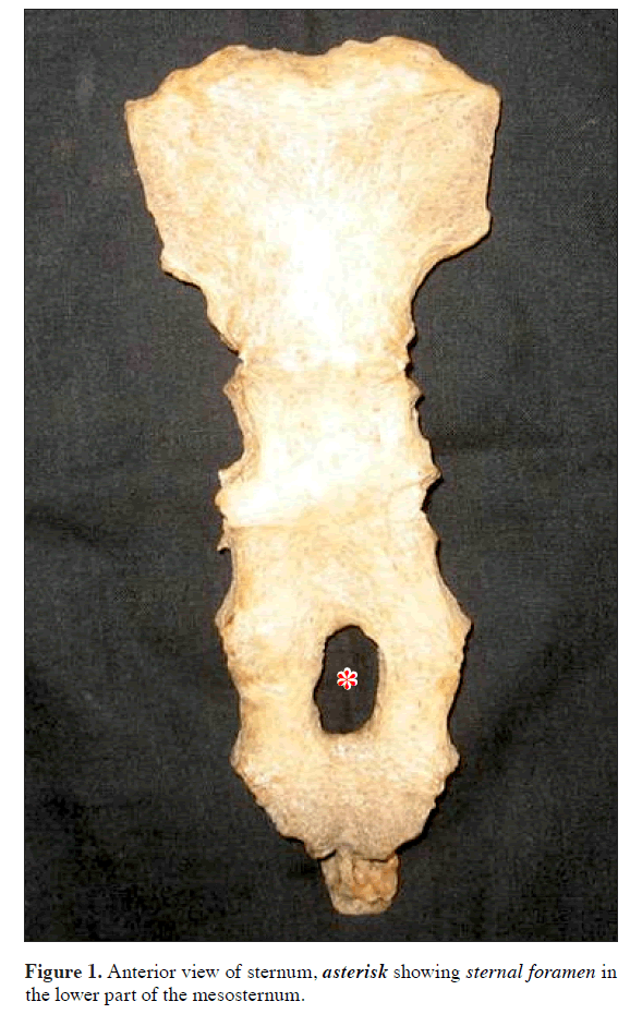
During routine osteology classes for undergraduate students in the Department of Anatomy, Meenakshi Medical College, Tamilnadu, India, we observed a sternum with a large oval foramen in lower one third of the body (Figure 1). The length and the width of the sternal foramen were 20.81 mm and 11.42 mm respectively, measured by using digital caliper.
Discussion
Sternal foramen is a congenital defect at the lower third of the sternum, usually asymptomatic. Sternal foramen may be associated with sternal sclerotic bands [4], sternal clefts with displacement of the heart or other midline abnormalities. Sternal foramen associated with accessory fissures on left lung were reported by using high-resolution computed tomography [5].
The incidence of sternal foramen was evaluated as 4.3% on the chest CT by Stark [6], 6.7% in autopsy cases by Cooper [3], 6.6% by Moore et al. [2] and 4.5% by Yekeler E et al. [4]. Aktan and Savas observed it in 5.1% of Turkish population [5]
The size of sternal foramina ranged between 2 and 16 mm, with mean of 6.5 mm [4]. But the sternal foramen in our study measured to be 20.8 mm and 11.4 mm, a larger size reported so far. Foramina in sternum were misinterpreted as acquired lesions, like gunshot wounds [7].
Serious complications following sternal puncture for bone marrow biopsy [8] or acupuncture [9] have been reported in the literature. Fatal cardiac tamponade following sternal puncture in the inferior part of the sternum with a congenital sternal foramen was reported. Therefore, awareness of the presence of sternal variations and anomalies is important to prevent these fatal complications by avoiding the inferior part of the sternal body during bone marrow aspiration. When sternal puncture is planned in corpus sterni region, radiographs should be taken to rule out this variation to avoid fatal complications.
References
- Breathnach AS. Frazer’s Anatomy of the Human Skeleton. 5th Ed., London, J & A Churchill Ltd. 1958; 53–54.
- Moore MK, Stewart JH, McCormick WF. Anomalies of the human chest plate area: radiographic findings in a large autopsy population. Am J Forensic Med Pathol. 1988; 9: 348–354.
- Cooper PD, Stewart JH, McCormick WF. Development and morphology of the sternal foramen. Am J Forensic Med Pathol. 1988; 9: 342–347.
- Yekeler E, Tunaci M, Tunaci A, Dursun M, Acunas G. Frequency of sternal variations and anomalies evaluated by MDCT. AJR Am J Roentgenol. 2006; 186: 956–960.
- Aktan ZA, Savas R. Anatomic and HRCT demonstration of midline sternal foramina. Turk J Med Sci. 1998; 28: 511–514.
- Stark P. Midline sternal foramen; CT demonstration. J Comput Assist Tomogr. 1985; 9: 489–490.
- Taylor HL. The sternal foramen: the possible forensic misinterpretation of an anatomic abnormality. J Forensic Sci. 1974; 19: 730–734.
- Bhootra BL. Fatality following a sternal bone marrow aspiration procedure: a case report. Med Sci Law. 2004; 44: 170–172.
- Halvorsen TB, Anda SS, Naess AB, Lewang OW. Fatal cardiac tamponade after acupuncture through congenital sternal foramen. Lancet. 1995; 345: 1175.
Suba Ananthi Kumarasamy1* and Reena Agrawal2
1Department of Anatomy, Chettinad Hospital and Research Institute, Kelambakkam, Tamil Nadu, India
2Department of Anatomy, Meenakshi Medical College & Research Institute, Kancheepuram, India
- *Corresponding Author:
- Dr. Suba Ananthi Kumarasamy, MS
Department of Anatomy, Chettinad Hospital and Research Institute Kelambakkam, Kanchipuram District Tamil Nadu, 603103, India
Tel: +91 9840717911
E-mail: subavejasara@gmail.com
Date of Received: March 28th, 2011
Date of Accepted: November 20th, 2011
Published Online: December 15th 2011
© Int J Anat Var (IJAV). 2011; 4: 195–196.
Abstract
Sternal foramen is an oval defect at the lower third of the sternum, the result of incomplete fusion of multiple ossification centers. It is usually asymptomatic. We report a sternal foramen of size 20.8 x 11.4 mm in lower third of body. Knowledge of sternal foramen is important for radiologists to avoid confusion with pathological conditions. Awareness of presence of sternal variations is important to prevent fatal complications by avoiding the inferior part of the sternal body during bone marrow aspiration.
-Keywords
mesosternum, foramen, incomplete fusion
Introduction
Sternum is formed from bilateral mesenchymatous condensations, sternal plates, which begin in the dorsolateral region of body wall. These plates undergo chondrification, move ventrally towards each other from both sides, and they eventually fuse together across the midline in a craniocaudal direction. Mesosternum (body) ossifies from 4 sternebrae. Sternal foramen, of varying size and form, may occur between third and fourth sternebrae due to incomplete fusion. [1]. Foramina in sternum are reported in manubrium, body (more common) and in xiphisternum [2,3].
Case Report
During routine osteology classes for undergraduate students in the Department of Anatomy, Meenakshi Medical College, Tamilnadu, India, we observed a sternum with a large oval foramen in lower one third of the body (Figure 1). The length and the width of the sternal foramen were 20.81 mm and 11.42 mm respectively, measured by using digital caliper.
Discussion
Sternal foramen is a congenital defect at the lower third of the sternum, usually asymptomatic. Sternal foramen may be associated with sternal sclerotic bands [4], sternal clefts with displacement of the heart or other midline abnormalities. Sternal foramen associated with accessory fissures on left lung were reported by using high-resolution computed tomography [5].
The incidence of sternal foramen was evaluated as 4.3% on the chest CT by Stark [6], 6.7% in autopsy cases by Cooper [3], 6.6% by Moore et al. [2] and 4.5% by Yekeler E et al. [4]. Aktan and Savas observed it in 5.1% of Turkish population [5]
The size of sternal foramina ranged between 2 and 16 mm, with mean of 6.5 mm [4]. But the sternal foramen in our study measured to be 20.8 mm and 11.4 mm, a larger size reported so far. Foramina in sternum were misinterpreted as acquired lesions, like gunshot wounds [7].
Serious complications following sternal puncture for bone marrow biopsy [8] or acupuncture [9] have been reported in the literature. Fatal cardiac tamponade following sternal puncture in the inferior part of the sternum with a congenital sternal foramen was reported. Therefore, awareness of the presence of sternal variations and anomalies is important to prevent these fatal complications by avoiding the inferior part of the sternal body during bone marrow aspiration. When sternal puncture is planned in corpus sterni region, radiographs should be taken to rule out this variation to avoid fatal complications.
References
- Breathnach AS. Frazer’s Anatomy of the Human Skeleton. 5th Ed., London, J & A Churchill Ltd. 1958; 53–54.
- Moore MK, Stewart JH, McCormick WF. Anomalies of the human chest plate area: radiographic findings in a large autopsy population. Am J Forensic Med Pathol. 1988; 9: 348–354.
- Cooper PD, Stewart JH, McCormick WF. Development and morphology of the sternal foramen. Am J Forensic Med Pathol. 1988; 9: 342–347.
- Yekeler E, Tunaci M, Tunaci A, Dursun M, Acunas G. Frequency of sternal variations and anomalies evaluated by MDCT. AJR Am J Roentgenol. 2006; 186: 956–960.
- Aktan ZA, Savas R. Anatomic and HRCT demonstration of midline sternal foramina. Turk J Med Sci. 1998; 28: 511–514.
- Stark P. Midline sternal foramen; CT demonstration. J Comput Assist Tomogr. 1985; 9: 489–490.
- Taylor HL. The sternal foramen: the possible forensic misinterpretation of an anatomic abnormality. J Forensic Sci. 1974; 19: 730–734.
- Bhootra BL. Fatality following a sternal bone marrow aspiration procedure: a case report. Med Sci Law. 2004; 44: 170–172.
- Halvorsen TB, Anda SS, Naess AB, Lewang OW. Fatal cardiac tamponade after acupuncture through congenital sternal foramen. Lancet. 1995; 345: 1175.


