A rare bilateral neurovascular variation of the upper limb: A case report of a 97-year-old Caucasian male
Shannon Goodwin* and Alireza Jalali
Division of Clinical and Functional Anatomy, Faculty of Medicine, University of Ottawa, Ottawa, ON, Canada
- *Corresponding Author:
- Shannon Goodwin
Prosector Division of Clinical and Functional Anatomy Faculty of Medicine University of Ottawa 451 Smyth Rd, Ottawa, ON Canada
Tel: +1 (613) 562-5800 ext
E-mail: sgoodwi@uottawa.ca
Date of Received: August 16th, 2016
Date of Accepted: January 1st, 2017
Published Online: January 18th, 2017
© Int J Anat Var (IJAV). 2016; 9: 46–49.
[ft_below_content] =>Keywords
variant, left radial artery, left radial recurrent artery, right superior ulnar collateral artery, musculocutaneous nerve
Introduction
Numerous review articles have described variations to the branching pattern of the vasculature [3,8,10] and innervation [1,4] in the upper limbs of cadavers. The classification of the arterial patterns that we used for this report corresponded to McCormick et al.’s [8] description of radial artery origin proximal to the intercondylar line. Multiple names have been given to this variant artery: Drizenko et al. [3] named it a high-origin radial artery, whereas Rodriquez et al. [10] called it the brachioradial artery. Such variations are additionally known to be relatively common, with McCormick et al. [8] reporting that departures from the typical arterial branching pattern in the upper limb occurred in 30.77% of cadavers, with bilateral variations in 6.32%. McCormick et al. [8] also reported that a radial artery of high origin accounted for 77% of all upper-limb variations. Rodriquez-Niedenfuhr [10] reported that the instances of high-origin radial artery occurred in 14.27% of all specimens, and of those, 65.5% originated from the brachial artery. Our arterial variation, consisting of the radial recurrent artery originating from the ulnar artery on the left and the superior ulnar collateral artery originating from the variant high-origin radial artery on the right, was not reported in these arterial variation review articles [3,8,10]. Additionally, to our knowledge, this variation has not been reported in the numerous original case studies published in the scientific literature.
There are a multitude of articles reporting on the variations of the brachial plexus. Variations to the musculocutaneous and median nerve were reported in 43% [4] to 46.4% [1] of cadavers, with a communicating branch between the two nerves in 43% [4]. Additionally, the absence of normal musculocutaneous innervation to the coracobrachialis was found in 11.1% [4] to 13.7% [1] of cadavers.
Case Report
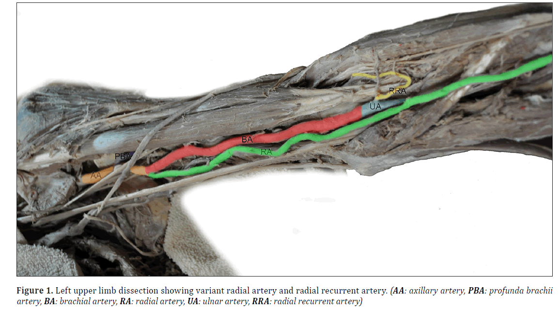
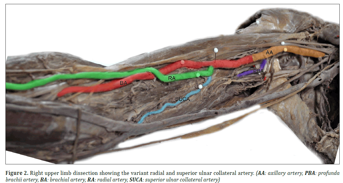
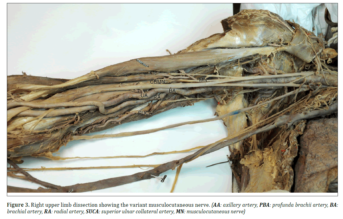
In this case report, we found bilateral variations of the typical branching pattern of the upper limb vasculature as well as a unilateral variation in the branching pattern of the brachial plexus. The left radial artery originated from the brachial artery 4.5 cm distal to the axillary-brachial junction, and the left radial recurrent artery originated from the ulnar artery in the cubital fossa (Figure 1). The right radial artery originated from the brachial artery 8 cm distal to the axillary-brachial junction, and the right superior ulnar collateral artery originated from the variant radial artery 1 cm below its origin from the brachial artery (Figure 2). Unlike usual anatomy, the musculocutaneous nerve of the brachial plexus did not pierce the coracobrachialis muscle in either limb. Furthermore, the right musculocutaneous nerve provided a communicating branch to the median nerve and provided two branches to the biceps brachii muscle before continuing along its usual course as the lateral cutaneous nerve of the forearm (Figure 3).
The practical importance of radial artery variations is evident when considering the plethora of clinical applications of this knowledge. Radial artery harvesting has become a popular surgical technique and is frequently used in procedures such as coronary bypass surgery. In addition to the radial nerve, oral and maxillofacial surgeons may also harvest the radial recurrent artery during forearm free flap procedures to repair soft tissue defects of the oral cavity during the surgical resection of tongue and mouth cancer, among other procedures [7]. Consideration of a variant radial artery may be important to the cardiac surgeon with regard to its influence on the transradial coronary procedural outcome [6]. Variations of the radial artery may be of consideration to thoracic surgeons due to the increasing use of radial artery grafts in coronary artery bypass surgery [2]. The superficial position of the variant radial artery at the area of the elbow may expose it to vulnerability or injury.
Variations to the musculocutaneous and median nerve may be important not only for the interpretation of clinical neurophysiology but also during examinations and procedures. For example, knowledge of such a variation is important to clinicians performing peripheral nerve blocks and axillary nerve blocks [9].
Materials and Methods
Dissection of a 97-year-old male cadaver was performed during the 2009/2010 academic year to teach the 1st- and 2nd-year medical students at the University of Ottawa. Variations to the origin of the radial artery, variant radial recurrent artery, superior collateral ulnar artery and musculocutaneous nerve were found. The complete arterial branching pattern in the neck and upper limbs was documented.
The right and left subclavian arteries become the axillary artery after crossing the first rib. The axillary artery follows its typical distribution pattern by branching off the superior thoracic, thoracoacromial, lateral thoracic, subscapular, anterior and posterior circumflex humeral arteries. At the inferior border of the teres major, the axillary artery changes names to become the brachial artery. On the medial surface of the left brachial artery at the axillary-brachial junction, a variant the left radial artery was found to originate from the brachial artery. The left radial artery descended along the medial side of the brachial artery, providing a small branch to the brachialis muscle 3 cm distal to its origin. The variant artery descended medially to the brachial artery, crossing the brachial artery 6 cm distal to its origin and following a superficial course along the lateral surface of the forearm. The brachial artery followed its typical course, descending along the medial border of the biceps brachii muscle. The left radial recurrent artery normally originates from the radial artery, but in this case report, it was derived from the brachial artery at the point of its typical division into the radial and ulnar arteries near the neck of the radius. The left radial recurrent artery followed its typical course to ascend behind the brachioradialis muscle and anterior to the supinator and brachialis muscles to anastomose with the radial collateral branch of the profundi brachii artery. The left brachial artery divided into terminal branches (the ulnar and common interosseous artery), which both followed their typical course in the forearm.
The right radial artery originated from the brachial artery 8 cm distal to the axillary-brachial junction, and the superior ulnar collateral artery originated from the radial artery 1 cm below its variant origin from the brachial artery. The variant radial artery descended along the medial border of the brachial artery, crossing the brachial artery proximal to the bicipital aponeurosis and following a superficial course along the lateral surface of the forearm. The superior ulnar collateral artery originated from the variant radial artery to follow its typical course along the border of the medial head of the triceps muscle to anastomose with the posterior ulnar recurrent and inferior collateral arteries.
In this case report, the brachial plexus followed its typical pattern of arrangement in the roots, trunks and cords dividing to form the terminal branches. The musculocutaneous nerve typically pierces the coracobrachialis, innervating it as well as the biceps and brachialis, and then continues as the lateral cutaneous nerve of the forearm. In this case report, the right musculocutaneous nerve did not pierce the coracobrachialis muscle. The coracobrachialis muscle was supplied by a small branch off of the posterior cord. The musculocutaneous nerve provided a communicating branch to the median nerve prior to providing two branches to the biceps brachii muscle. We were unable to find a branch to the brachialis muscle, which continued its usual course as the lateral cutaneous nerve of the forearm.
Discussion
If examined separately, each variation can be classified as a common occurrence, but when examined in combination, they constitute a rare bilateral anatomical upper limb variation with significance for anatomists, surgeons, anesthesiologists, and radiologists.
References
- Choi D, Rodríguez-Niedenführ M, Vázquez T, Parkin I, Sañudo JR. Patterns of connections between the musculocutaneous and median nerves in axillar and arm. Clinical Anatomy. 2002;15:11-17.
- Chong CF, De Souza A. Significance of radial artery anomalies in coronary artery bypass graft surgery. The Journal of Thoracic and Cardiovascular Surgery. 2008:135(6);1389-1390.
- Drizenko A, Maynou C, Mestdagh H, Mauroy B, Bailleul JP. Variations of the radial artery in man. Surg Radiol Anat. 2000;22:299-303.
- Guerri-Guttenberg RA, Ingolotti M. Classifying musculocutaneous nerve variations. Clinical Anatomy. 2009;22:671-683.
- Kerr, AT. The brachial plexus of nerves in man, the variations in its formation and branches. Am J Anat. 1918;23:285-395.
- Lo TS, Nolan J, Fountzopoulos E, Behan M, Butler R, Hetherington SL, Vijayalakshmi K, Rajagopal R, Fraser D, Zaman A, Hildick-Smith D. Radial artery anomaly and its influence on transradial coronary procedural outcome. Heart. 2009:95(5);410-415.
- Martin-Granizo R, Gomez F, Sanchez-Cuellar A. An unusual anomaly of the radial artery with potential significance to the forearm free flap. Journal of Cranio-Maxillofacial Surgery. 2002;30(3):189-191.
- McCormick L, Cauldwell E, Anson B. Brachial and antebrachial arterial patterns. Surgery, Gynechology and Obstetrics. 1953;96:43-54.
- Orebaugh SL, Williams BA. Brachial plexus: normal and variant. The Scientific World Journal. 2009;9:300-312.
- Rodríguez-Niedenführ M, Vázquez T, Nearn L, Ferreira B, Parkin I, Sañudo JR. Variations of the upper limb revisited: a morphological and statistical study, with a review of the literature. J Anat. 2001;199:547-566.
Shannon Goodwin* and Alireza Jalali
Division of Clinical and Functional Anatomy, Faculty of Medicine, University of Ottawa, Ottawa, ON, Canada
- *Corresponding Author:
- Shannon Goodwin
Prosector Division of Clinical and Functional Anatomy Faculty of Medicine University of Ottawa 451 Smyth Rd, Ottawa, ON Canada
Tel: +1 (613) 562-5800 ext
E-mail: sgoodwi@uottawa.ca
Date of Received: August 16th, 2016
Date of Accepted: January 1st, 2017
Published Online: January 18th, 2017
© Int J Anat Var (IJAV). 2016; 9: 46–49.
Abstract
During a routine dissection of a 97-year-old male cadaver, rare upper-limb neurovascular variations were found. We found bilateral variations to the radial arteries, variants to the superior collateral ulnar and radial recurrent arteries, and a variation of the musculocutaneous nerve. The combination of these variants constitutes a unique variation, which, to our knowledge, has not been reported in the available literature to date. Variations in the upper-limb neurovasculature are significant for anesthesiologists, surgeons, radiologists, anatomists, cell biologists and medical students.
-Keywords
variant, left radial artery, left radial recurrent artery, right superior ulnar collateral artery, musculocutaneous nerve
Introduction
Numerous review articles have described variations to the branching pattern of the vasculature [3,8,10] and innervation [1,4] in the upper limbs of cadavers. The classification of the arterial patterns that we used for this report corresponded to McCormick et al.’s [8] description of radial artery origin proximal to the intercondylar line. Multiple names have been given to this variant artery: Drizenko et al. [3] named it a high-origin radial artery, whereas Rodriquez et al. [10] called it the brachioradial artery. Such variations are additionally known to be relatively common, with McCormick et al. [8] reporting that departures from the typical arterial branching pattern in the upper limb occurred in 30.77% of cadavers, with bilateral variations in 6.32%. McCormick et al. [8] also reported that a radial artery of high origin accounted for 77% of all upper-limb variations. Rodriquez-Niedenfuhr [10] reported that the instances of high-origin radial artery occurred in 14.27% of all specimens, and of those, 65.5% originated from the brachial artery. Our arterial variation, consisting of the radial recurrent artery originating from the ulnar artery on the left and the superior ulnar collateral artery originating from the variant high-origin radial artery on the right, was not reported in these arterial variation review articles [3,8,10]. Additionally, to our knowledge, this variation has not been reported in the numerous original case studies published in the scientific literature.
There are a multitude of articles reporting on the variations of the brachial plexus. Variations to the musculocutaneous and median nerve were reported in 43% [4] to 46.4% [1] of cadavers, with a communicating branch between the two nerves in 43% [4]. Additionally, the absence of normal musculocutaneous innervation to the coracobrachialis was found in 11.1% [4] to 13.7% [1] of cadavers.
Case Report
In this case report, we found bilateral variations of the typical branching pattern of the upper limb vasculature as well as a unilateral variation in the branching pattern of the brachial plexus. The left radial artery originated from the brachial artery 4.5 cm distal to the axillary-brachial junction, and the left radial recurrent artery originated from the ulnar artery in the cubital fossa (Figure 1). The right radial artery originated from the brachial artery 8 cm distal to the axillary-brachial junction, and the right superior ulnar collateral artery originated from the variant radial artery 1 cm below its origin from the brachial artery (Figure 2). Unlike usual anatomy, the musculocutaneous nerve of the brachial plexus did not pierce the coracobrachialis muscle in either limb. Furthermore, the right musculocutaneous nerve provided a communicating branch to the median nerve and provided two branches to the biceps brachii muscle before continuing along its usual course as the lateral cutaneous nerve of the forearm (Figure 3).
The practical importance of radial artery variations is evident when considering the plethora of clinical applications of this knowledge. Radial artery harvesting has become a popular surgical technique and is frequently used in procedures such as coronary bypass surgery. In addition to the radial nerve, oral and maxillofacial surgeons may also harvest the radial recurrent artery during forearm free flap procedures to repair soft tissue defects of the oral cavity during the surgical resection of tongue and mouth cancer, among other procedures [7]. Consideration of a variant radial artery may be important to the cardiac surgeon with regard to its influence on the transradial coronary procedural outcome [6]. Variations of the radial artery may be of consideration to thoracic surgeons due to the increasing use of radial artery grafts in coronary artery bypass surgery [2]. The superficial position of the variant radial artery at the area of the elbow may expose it to vulnerability or injury.
Variations to the musculocutaneous and median nerve may be important not only for the interpretation of clinical neurophysiology but also during examinations and procedures. For example, knowledge of such a variation is important to clinicians performing peripheral nerve blocks and axillary nerve blocks [9].
Materials and Methods
Dissection of a 97-year-old male cadaver was performed during the 2009/2010 academic year to teach the 1st- and 2nd-year medical students at the University of Ottawa. Variations to the origin of the radial artery, variant radial recurrent artery, superior collateral ulnar artery and musculocutaneous nerve were found. The complete arterial branching pattern in the neck and upper limbs was documented.
The right and left subclavian arteries become the axillary artery after crossing the first rib. The axillary artery follows its typical distribution pattern by branching off the superior thoracic, thoracoacromial, lateral thoracic, subscapular, anterior and posterior circumflex humeral arteries. At the inferior border of the teres major, the axillary artery changes names to become the brachial artery. On the medial surface of the left brachial artery at the axillary-brachial junction, a variant the left radial artery was found to originate from the brachial artery. The left radial artery descended along the medial side of the brachial artery, providing a small branch to the brachialis muscle 3 cm distal to its origin. The variant artery descended medially to the brachial artery, crossing the brachial artery 6 cm distal to its origin and following a superficial course along the lateral surface of the forearm. The brachial artery followed its typical course, descending along the medial border of the biceps brachii muscle. The left radial recurrent artery normally originates from the radial artery, but in this case report, it was derived from the brachial artery at the point of its typical division into the radial and ulnar arteries near the neck of the radius. The left radial recurrent artery followed its typical course to ascend behind the brachioradialis muscle and anterior to the supinator and brachialis muscles to anastomose with the radial collateral branch of the profundi brachii artery. The left brachial artery divided into terminal branches (the ulnar and common interosseous artery), which both followed their typical course in the forearm.
The right radial artery originated from the brachial artery 8 cm distal to the axillary-brachial junction, and the superior ulnar collateral artery originated from the radial artery 1 cm below its variant origin from the brachial artery. The variant radial artery descended along the medial border of the brachial artery, crossing the brachial artery proximal to the bicipital aponeurosis and following a superficial course along the lateral surface of the forearm. The superior ulnar collateral artery originated from the variant radial artery to follow its typical course along the border of the medial head of the triceps muscle to anastomose with the posterior ulnar recurrent and inferior collateral arteries.
In this case report, the brachial plexus followed its typical pattern of arrangement in the roots, trunks and cords dividing to form the terminal branches. The musculocutaneous nerve typically pierces the coracobrachialis, innervating it as well as the biceps and brachialis, and then continues as the lateral cutaneous nerve of the forearm. In this case report, the right musculocutaneous nerve did not pierce the coracobrachialis muscle. The coracobrachialis muscle was supplied by a small branch off of the posterior cord. The musculocutaneous nerve provided a communicating branch to the median nerve prior to providing two branches to the biceps brachii muscle. We were unable to find a branch to the brachialis muscle, which continued its usual course as the lateral cutaneous nerve of the forearm.
Discussion
If examined separately, each variation can be classified as a common occurrence, but when examined in combination, they constitute a rare bilateral anatomical upper limb variation with significance for anatomists, surgeons, anesthesiologists, and radiologists.
References
- Choi D, Rodríguez-Niedenführ M, Vázquez T, Parkin I, Sañudo JR. Patterns of connections between the musculocutaneous and median nerves in axillar and arm. Clinical Anatomy. 2002;15:11-17.
- Chong CF, De Souza A. Significance of radial artery anomalies in coronary artery bypass graft surgery. The Journal of Thoracic and Cardiovascular Surgery. 2008:135(6);1389-1390.
- Drizenko A, Maynou C, Mestdagh H, Mauroy B, Bailleul JP. Variations of the radial artery in man. Surg Radiol Anat. 2000;22:299-303.
- Guerri-Guttenberg RA, Ingolotti M. Classifying musculocutaneous nerve variations. Clinical Anatomy. 2009;22:671-683.
- Kerr, AT. The brachial plexus of nerves in man, the variations in its formation and branches. Am J Anat. 1918;23:285-395.
- Lo TS, Nolan J, Fountzopoulos E, Behan M, Butler R, Hetherington SL, Vijayalakshmi K, Rajagopal R, Fraser D, Zaman A, Hildick-Smith D. Radial artery anomaly and its influence on transradial coronary procedural outcome. Heart. 2009:95(5);410-415.
- Martin-Granizo R, Gomez F, Sanchez-Cuellar A. An unusual anomaly of the radial artery with potential significance to the forearm free flap. Journal of Cranio-Maxillofacial Surgery. 2002;30(3):189-191.
- McCormick L, Cauldwell E, Anson B. Brachial and antebrachial arterial patterns. Surgery, Gynechology and Obstetrics. 1953;96:43-54.
- Orebaugh SL, Williams BA. Brachial plexus: normal and variant. The Scientific World Journal. 2009;9:300-312.
- Rodríguez-Niedenführ M, Vázquez T, Nearn L, Ferreira B, Parkin I, Sañudo JR. Variations of the upper limb revisited: a morphological and statistical study, with a review of the literature. J Anat. 2001;199:547-566.


