A rare instance of a tendinous interconnection between flexor hallucis longus and flexor digitorum accessorius
Upendra Kumar Gupta1 and Nazim Nasir2*
1Department of Anatomy, NIMS Medical College, NIMS University Jaipur, India
2Department of Anatomy, King Khalid University, College of Applied Medical Sciences, Abha, KSA
- *Corresponding Author:
- Dr. Nazim Nasir
Assistant Professor, Department of Anatomy, King Khalid University, College of Applied Medical Sciences, Abha, KSA
Tel: +966 (501) 617207
E-mail: sahil9792003@gmail.com
Date of Received: July 27th, 2011
Date of Accepted: August 1st, 2012
Published Online: February 4th, 2013
© Int J Anat Var (IJAV). 2013; 6: 18–19.
[ft_below_content] =>Keywords
flexor digitorum accessorius, flexor digitorum longus, flexor hallucis longus, sole of the foot, clinical significance
Introduction
A number of variants or accessory muscles are found in the human body. Foot and ankle are the sites where variations are seen occasionally [1]. Most of them are reported in tarsal tunnel –the peroneocalcaneus internus, the tibiocalcaneus internus, the accessory soleus and the flexor digitorum accessorius (FDA) [1]. FDA is also named “accessorius ad accessorium”, “accessorius secundus” and “pronator pedis”, and has been described as a very variable slip, both in its origin and insertion [2]. Usually FDA is poorly described in books and it is not so popular and continues to be unduly ignored, therefore poorly characterized [3]. But, after the report of Sammarco and Stephens, in which they first described tarsal tunnel syndrome due to the presence of FDA, this interesting variation has attracted renewed attention not only to anatomists, but also to clinicians [1]. Usually FDA arises by two heads, with the long plantar ligament situated deeply in the interval between the two heads. The medial head is larger and more fleshy and is attached to the medial concave surface of the calcaneus, below the groove for the tendon of flexor hallucis longus (FHL). The lateral head is flat and tendinous and is attached to the calcaneus distal to the lateral process of the tuberosity, and to the long plantar ligament. The muscle belly inserts into the tendon of flexor digitorum longus (FDL) at the point where it is bound by a fibrous slip to the tendon of FHL and where it divides into its four tendons. By pulling on the tendons of FDL, FDA provides a means of flexing the lateral four toes in any position of the ankle joint [4].
In this report we describe a rare case of FDA receiving a fibrous slip from FHL exactly at the point where normally FDL receives this fibrous connection. This is the first report of such a variation in Indian population.
Case Report
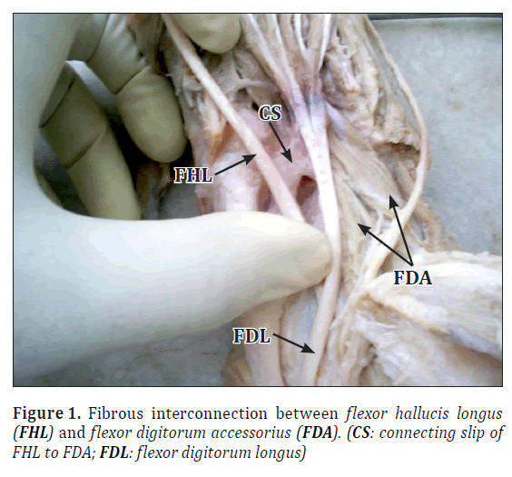
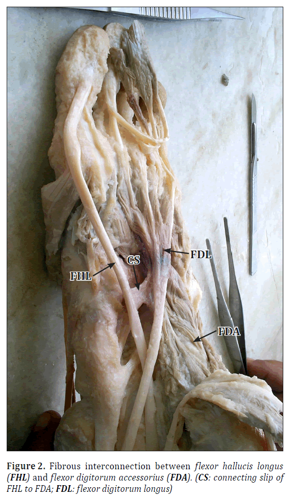
A case of a direct fibrous interconnection between FHL and FDA has been encountered during routine anatomical dissections of a left lower extremity of an adult male cadaver. This was the only instance among 12 dissected lower extremities of the 6 cadavers in the Department of Anatomy at NIMS Medical College, Jaipur (Figures 1, 2).
The aberrant fibrous connection of tendon of the FHL arose in the middle of the sole and ran medially to get attached to medial border of FDA 50 mm distal to the anterior end of the medial tubercle of calcaneus (Figure 1). The connecting slip was 13 mm in length and 10 mm in width.
Discussion
In the sole of the foot, the tendon of FHL passes forward in the second layer like a bowstring. It crosses the tendon of FDL from lateral to medial, curving obliquely superior to it. At the crossing point (knot of Henry) it gives off two strong slips to the medial two divisions of the tendons of FDL [4]. FDA could be a predisposing factor that may contribute to both tarsal tunnel syndrome and flexor hallucis syndrome [1,5–6].
Thus the line of pull of FHL is straightened by FDA indirectly through the FDL. In this case the tendon FHL is giving a slip of attachment directly to the FDA. This variation although not entrapping any nerve or vessel, thus, not responsible for any clinical symptom, but, may be the factor which facilitate the functioning of big toe during take off or tip toe position of the foot where FHL plays major role. Its line of pull is directly straightened by FDA and not through FDL.
References
- Sammarco GJ, Stephens MM. Tarsal tunnel syndrome caused by the flexor digitorum accessorius longus. A case report. J Bone Joint Surg Am. 1990; 72: 453–454.
- Jaijesh P, Shenoy M, Anuradha L, Chithralekha KK. Flexor accessorius longus: A rare variation of the deep extrinsic digital flexors of the leg and its phylogenetic significance. Indian J Plast Surg. 2006; 39: 169–171.
- Hwang SH, Hill RV. An unusual variation of the flexor digitorum accessorius longus muscle-its anatomy and clinical significance. Anat Sci Int. 2009; 84: 257–263.
- Standring S, ed. Gray’s Anatomy. The Anatomical Basis of Clinical Practice. 40th Ed., Expert Consult. 2011; 1423, 1453.
- Eberle CF, Moran B, Gleason T. The accessory flexor digitorum longus as a cause of Flexor Hallucis Syndrome. Foot Ankle Int. 2002; 23: 51–55.
- Sammarco GJ, Conti SF. Tarsal tunnel syndrome caused by an anomalous muscle. J Bone Joint Surg Am. 1994; 76: 1308–1314.
Upendra Kumar Gupta1 and Nazim Nasir2*
1Department of Anatomy, NIMS Medical College, NIMS University Jaipur, India
2Department of Anatomy, King Khalid University, College of Applied Medical Sciences, Abha, KSA
- *Corresponding Author:
- Dr. Nazim Nasir
Assistant Professor, Department of Anatomy, King Khalid University, College of Applied Medical Sciences, Abha, KSA
Tel: +966 (501) 617207
E-mail: sahil9792003@gmail.com
Date of Received: July 27th, 2011
Date of Accepted: August 1st, 2012
Published Online: February 4th, 2013
© Int J Anat Var (IJAV). 2013; 6: 18–19.
Abstract
During routine anatomical dissection a rare case of a flexor digitorum accessorius (FDA) muscle connected through fibrous slip to flexor hallucis longus (FHL) was observed. The muscle belly of FDA inserted into the tendon of flexor digitorum longus (FDL) at the point where it received a fibrous slip from the tendon of FHL and where it divided into its four tendons. In this case the tendon of FHL instead of connecting to the tendon of FDL connected directly to the medial head of FDA muscle. The possible clinical implications of this may be with the more efficient functioning of the foot in take off and tip toe positions.
-Keywords
flexor digitorum accessorius, flexor digitorum longus, flexor hallucis longus, sole of the foot, clinical significance
Introduction
A number of variants or accessory muscles are found in the human body. Foot and ankle are the sites where variations are seen occasionally [1]. Most of them are reported in tarsal tunnel –the peroneocalcaneus internus, the tibiocalcaneus internus, the accessory soleus and the flexor digitorum accessorius (FDA) [1]. FDA is also named “accessorius ad accessorium”, “accessorius secundus” and “pronator pedis”, and has been described as a very variable slip, both in its origin and insertion [2]. Usually FDA is poorly described in books and it is not so popular and continues to be unduly ignored, therefore poorly characterized [3]. But, after the report of Sammarco and Stephens, in which they first described tarsal tunnel syndrome due to the presence of FDA, this interesting variation has attracted renewed attention not only to anatomists, but also to clinicians [1]. Usually FDA arises by two heads, with the long plantar ligament situated deeply in the interval between the two heads. The medial head is larger and more fleshy and is attached to the medial concave surface of the calcaneus, below the groove for the tendon of flexor hallucis longus (FHL). The lateral head is flat and tendinous and is attached to the calcaneus distal to the lateral process of the tuberosity, and to the long plantar ligament. The muscle belly inserts into the tendon of flexor digitorum longus (FDL) at the point where it is bound by a fibrous slip to the tendon of FHL and where it divides into its four tendons. By pulling on the tendons of FDL, FDA provides a means of flexing the lateral four toes in any position of the ankle joint [4].
In this report we describe a rare case of FDA receiving a fibrous slip from FHL exactly at the point where normally FDL receives this fibrous connection. This is the first report of such a variation in Indian population.
Case Report
A case of a direct fibrous interconnection between FHL and FDA has been encountered during routine anatomical dissections of a left lower extremity of an adult male cadaver. This was the only instance among 12 dissected lower extremities of the 6 cadavers in the Department of Anatomy at NIMS Medical College, Jaipur (Figures 1, 2).
The aberrant fibrous connection of tendon of the FHL arose in the middle of the sole and ran medially to get attached to medial border of FDA 50 mm distal to the anterior end of the medial tubercle of calcaneus (Figure 1). The connecting slip was 13 mm in length and 10 mm in width.
Discussion
In the sole of the foot, the tendon of FHL passes forward in the second layer like a bowstring. It crosses the tendon of FDL from lateral to medial, curving obliquely superior to it. At the crossing point (knot of Henry) it gives off two strong slips to the medial two divisions of the tendons of FDL [4]. FDA could be a predisposing factor that may contribute to both tarsal tunnel syndrome and flexor hallucis syndrome [1,5–6].
Thus the line of pull of FHL is straightened by FDA indirectly through the FDL. In this case the tendon FHL is giving a slip of attachment directly to the FDA. This variation although not entrapping any nerve or vessel, thus, not responsible for any clinical symptom, but, may be the factor which facilitate the functioning of big toe during take off or tip toe position of the foot where FHL plays major role. Its line of pull is directly straightened by FDA and not through FDL.
References
- Sammarco GJ, Stephens MM. Tarsal tunnel syndrome caused by the flexor digitorum accessorius longus. A case report. J Bone Joint Surg Am. 1990; 72: 453–454.
- Jaijesh P, Shenoy M, Anuradha L, Chithralekha KK. Flexor accessorius longus: A rare variation of the deep extrinsic digital flexors of the leg and its phylogenetic significance. Indian J Plast Surg. 2006; 39: 169–171.
- Hwang SH, Hill RV. An unusual variation of the flexor digitorum accessorius longus muscle-its anatomy and clinical significance. Anat Sci Int. 2009; 84: 257–263.
- Standring S, ed. Gray’s Anatomy. The Anatomical Basis of Clinical Practice. 40th Ed., Expert Consult. 2011; 1423, 1453.
- Eberle CF, Moran B, Gleason T. The accessory flexor digitorum longus as a cause of Flexor Hallucis Syndrome. Foot Ankle Int. 2002; 23: 51–55.
- Sammarco GJ, Conti SF. Tarsal tunnel syndrome caused by an anomalous muscle. J Bone Joint Surg Am. 1994; 76: 1308–1314.


