A rare variation of trifurcation of right femoral artery
Parasa Savithri*
Department of Anatomy, Guntur Medical College, Guntur, Andhra Pradesh, India
- *Corresponding Author:
- Dr. Parasa Savithri, MD
Associate Professor, Department of Anatomy, Guntur Medical College, Guntur – 522 004, Andhra Pradesh, India
Tel: +91 (934) 6642682
E-mail: savithrichukkaparasa@gmail.com
Date of Received: October 22nd, 2011
Date of Accepted: September 2nd, 2012
Published Online: January 13th, 2013
© Int J Anat Var (IJAV). 2013; 6: 4–6.
[ft_below_content] =>Keywords
femoral artery, profunda femoris artery, lateral circumflex femoral artery
Introduction
The anatomy of the vessels of lower limb has long received attention from various scientists and author. The femoral artery enters the thigh at a point midway between the anterior superior iliac spine and the pubic symphysis. Here it lies on the psoas major tendon, which separates the artery from capsule of the hip joint. This is the position where its pulsation can be felt and where it can be entered for arterial catheterization. Cannulation, the femoral artery is second only to the radial as the site of choice for the placement of an arterial line. Its superficial position below the inguinal ligament makes it easily accessible. The most common complications include retroperitoneal hemorrhage and perforation of the gut and arterio-venous fistula. The lateral circumflex femoral artery occasionally arises from femoral artery [1]. An expansile swelling lying along the course of the femoral artery that fluctuates in time with the pulse rate should make the diagnosis of aneurysm of the femoral artery certain [2].The lateral and medial femoral circumflex arteries are typically the largest branches of the profunda femoris although they do not always arise from this vessel; either or both may arise as branches of the femoral artery itself above the origin of the profunda femoris. In any case, they originate in the femoral triangle whether they arise from the profunda femoris or from femoral artery, they may share a common stem or have separate origins [3].
Case Report
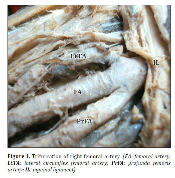
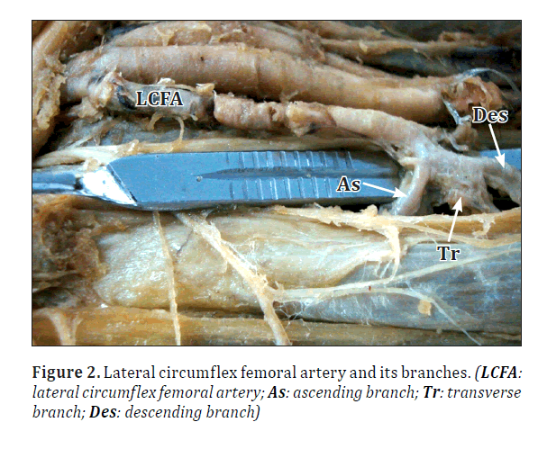
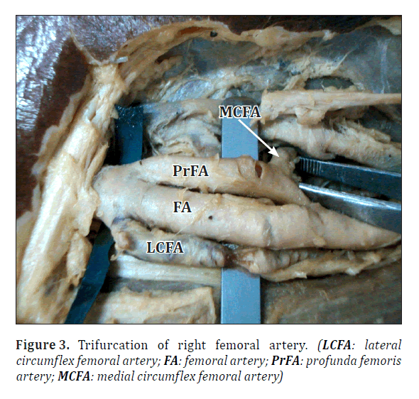
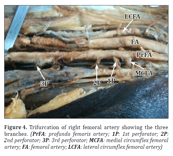
During the routine dissections for the undergraduate students batch 2011–2012 in Guntur Medical College, Guntur, Andra Pradesh, on a middle-aged male embalmed cadaver, we noticed a rare variation of trifurcation of right femoral artery. Variation was noticed as the trifurcation of right femoral artery approximately 7 mm below the inguinal ligament. The trifurcated arteries from lateral to medial side were lateral circumflex femoral artery, femoral artery and profunda femoris artery (Figure 1). On tracing lateral circumflex femoral artery with an external diameter of 5mm, giving the branches of ascending, transverse, descending and muscular arteries (Figure 2). The femoral artery had its usual course and continued as popliteal artery. The profunda femoris artery originating from medial side of femoral artery or medial separation of femoral artery as profunda femoris artery initially was superficial to the femoral vein coursed downwards posteriorly relating posteromedial to the femoral artery. The medial circumflex femoral artery originated as a thin branch and it was dividing into transverse, ascending branches (Figure 3. The profunda femoris artery had given rise to 3 perforating arteries, 2 nutrient arteries. The 1st nutrient artery was accompanying the 1st perforating artery, 2nd nutrient artery accompanying 3rd perforating artery and continued as 4th perforating artery (Figure 4).
Discussion
Statistics on the origins of the circumflex arteries differ somewhat, reported and reviewed by several workers, the incidence of different types apparently varies with race. Lipshutz found that the vessels on the two sides of the same body differed in origin in 60%of cases. He also reported a difference in incidence of the various types of origin according to side; but Williams and his co-workers reporting a much larger series, failed to substantiate this. Williams and his colleagues described varying origins of two circumflexes from the profunda femoris and the femoral artery. They summarized the findings on 979 white persons as presenting the several different types of origins in the percentages. A common stem from the femoral for both circumflexes has been reported in Negros and Japanese but not white persons [3–7].The circumflex femoral arteries are variable, both arising from the deep femoral artery in only 56 % of cases [7], and the lateral circumflex femoral artery has an origin from the femoral artery in 14% of cases [8]. Prevalence of origin of lateral circumflex femoral artery from femoral including common stem is 13.2% of cases [9]. Angiographs reported prevalence of origin of lateral circumflex femoral artery from femoral artery including common stem in 21.4% [10]. Prevalence of origin of lateral circumflex femoral artery from femoral artery including common stem is 22.7% of cases [11]. The femoral artery develops from a capillary plexus in the ventral aspect of the thigh, establishes communication proximally with the external iliac artery, and distally joins with the axis artery [12].
Conclusion
The profunda femoris artery acts as a collateral vessel in the occlusion of the femoral artery and for this important function, it has a large caliber. Accurate knowledge of anatomical variations regarding origins of profunda femoris, medial circumflex, lateral circumflex femoral arteries are important for clinicians in the present modern era of interventional radiology. These variations also provide facilities in vascular surgery and angiographic applications which may reduce the complications. The anatomical knowledge of level of origin is important in avoiding iatrogenic femoral arterio-venous fistula formed during puncture of femoral artery.
References
- Sinnatamby CS. Last’s Anatomy Regional and Applied. 10th Ed., Churchill Livingstone. 1999; 114–115.
- Snell RS. Clinical Anatomy. 7th Ed., Lippincott Williams & Wilkins. 2004; 626–629.
- Hollinshed HW. Anatomy for Surgeons. Vol. 3, New York, Hoeber & Harper. 1958; 737–739.
- Lipshutz BB. Studies on the blood vascular tree I. A composite study of the femoral artery. Anat Rec. 1916; 10: 361.
- Ming-Tzu P. Origin of deep and circumflex femoral group of arteries in the Chinese. Am J Phys Anthropol. 1937; 22: 417–424.
- Auburtin G. Die bieden arteriae circummflexae femoris des menschen. Anat Anz. 1905; 27: 247–269.
- Williams GD, Martin CH, McIntire LR. Origin of the deep and circumflex femoral group of arteries. Anat Rec. 1934; 60: 189–196.
- Woodburne RT. Essentials of Human Anatomy. 2nd Ed., Oxford University Press. 1961; 544–545.
- Choi SW, Park JY, Hur MS, Park HD, Kang HJ, Hu KS, Kim HJ. An anatomic assessment on perforators of the lateral circumflex femoral artery for anterolateral thigh flap. J Craniofac Surg. 2007; 18: 866–871.
- Fukuda H, Ashida M, Ishii R, Abe S, Ibukuro K. Anatomical variants of the lateral femoral circumflex artery: an angiographic study. Surg Radiol Anat. 2005; 27: 260–264.
- Uzel M, Tanyeli E, Yildirim M. An anatomical study of the origins of the lateral circumflex femoral artery in the Turkish population. Folia Morphol (Warsz). 2008; 67: 226–230.
- Datta AK. Essentials of Human Embryology. 4th Ed., Calcutta, Current Books International. 2000; 196.
Parasa Savithri*
Department of Anatomy, Guntur Medical College, Guntur, Andhra Pradesh, India
- *Corresponding Author:
- Dr. Parasa Savithri, MD
Associate Professor, Department of Anatomy, Guntur Medical College, Guntur – 522 004, Andhra Pradesh, India
Tel: +91 (934) 6642682
E-mail: savithrichukkaparasa@gmail.com
Date of Received: October 22nd, 2011
Date of Accepted: September 2nd, 2012
Published Online: January 13th, 2013
© Int J Anat Var (IJAV). 2013; 6: 4–6.
Abstract
The femoral artery, the primary artery of thigh is continuation of external iliac artery. The anatomical knowledge of variations of femoral artery and its branches is important as it is frequently accessed by surgeons and radiologists. The femoral artery is generally preferred and easily accessible for catheterization in number of investigations and angiographies. The awareness of variation in height of origin of profunda femoris artery and the distribution of its branches is of great importance. Knowledge of variation of lateral circumflex femoral artery is important when undertaking clinical procedures in the femoral region and hip joint replacement. It can be used as an anterolateral thigh flap in coronary artery bypass grafting. The lateral circumflex femoral artery flap is used for the reconstruction of large defects in the face secondary to gunshot wounds. During the routine dissections for the undergraduate students batch 2011–2012 in Guntur Medical College, on a middle-aged male cadaver we noticed a rare variation of trifurcation of femoral artery. The trifurcation was approximately 7 mm below the inguinal ligament. The trifurcating arteries were lateral circumflex femoral artery, femoral artery, profunda femoris artery. Lateral circumflex femoral artery with an external diameter of 5 mm, further tracing down branching in to ascending, transverse, descending and muscular arteries. The femoral artery had the usual course and continued as popliteal artery and external diameter was 9 mm. The profunda femoris artery was medially separated from femoral artery first laid lateral to femoral vein and passed downwards giving origin to medial circumflex femoral artery, 3 perforating arteries, 2 nutrient arteries and continued as 4th perforating artery.
-Keywords
femoral artery, profunda femoris artery, lateral circumflex femoral artery
Introduction
The anatomy of the vessels of lower limb has long received attention from various scientists and author. The femoral artery enters the thigh at a point midway between the anterior superior iliac spine and the pubic symphysis. Here it lies on the psoas major tendon, which separates the artery from capsule of the hip joint. This is the position where its pulsation can be felt and where it can be entered for arterial catheterization. Cannulation, the femoral artery is second only to the radial as the site of choice for the placement of an arterial line. Its superficial position below the inguinal ligament makes it easily accessible. The most common complications include retroperitoneal hemorrhage and perforation of the gut and arterio-venous fistula. The lateral circumflex femoral artery occasionally arises from femoral artery [1]. An expansile swelling lying along the course of the femoral artery that fluctuates in time with the pulse rate should make the diagnosis of aneurysm of the femoral artery certain [2].The lateral and medial femoral circumflex arteries are typically the largest branches of the profunda femoris although they do not always arise from this vessel; either or both may arise as branches of the femoral artery itself above the origin of the profunda femoris. In any case, they originate in the femoral triangle whether they arise from the profunda femoris or from femoral artery, they may share a common stem or have separate origins [3].
Case Report
During the routine dissections for the undergraduate students batch 2011–2012 in Guntur Medical College, Guntur, Andra Pradesh, on a middle-aged male embalmed cadaver, we noticed a rare variation of trifurcation of right femoral artery. Variation was noticed as the trifurcation of right femoral artery approximately 7 mm below the inguinal ligament. The trifurcated arteries from lateral to medial side were lateral circumflex femoral artery, femoral artery and profunda femoris artery (Figure 1). On tracing lateral circumflex femoral artery with an external diameter of 5mm, giving the branches of ascending, transverse, descending and muscular arteries (Figure 2). The femoral artery had its usual course and continued as popliteal artery. The profunda femoris artery originating from medial side of femoral artery or medial separation of femoral artery as profunda femoris artery initially was superficial to the femoral vein coursed downwards posteriorly relating posteromedial to the femoral artery. The medial circumflex femoral artery originated as a thin branch and it was dividing into transverse, ascending branches (Figure 3. The profunda femoris artery had given rise to 3 perforating arteries, 2 nutrient arteries. The 1st nutrient artery was accompanying the 1st perforating artery, 2nd nutrient artery accompanying 3rd perforating artery and continued as 4th perforating artery (Figure 4).
Discussion
Statistics on the origins of the circumflex arteries differ somewhat, reported and reviewed by several workers, the incidence of different types apparently varies with race. Lipshutz found that the vessels on the two sides of the same body differed in origin in 60%of cases. He also reported a difference in incidence of the various types of origin according to side; but Williams and his co-workers reporting a much larger series, failed to substantiate this. Williams and his colleagues described varying origins of two circumflexes from the profunda femoris and the femoral artery. They summarized the findings on 979 white persons as presenting the several different types of origins in the percentages. A common stem from the femoral for both circumflexes has been reported in Negros and Japanese but not white persons [3–7].The circumflex femoral arteries are variable, both arising from the deep femoral artery in only 56 % of cases [7], and the lateral circumflex femoral artery has an origin from the femoral artery in 14% of cases [8]. Prevalence of origin of lateral circumflex femoral artery from femoral including common stem is 13.2% of cases [9]. Angiographs reported prevalence of origin of lateral circumflex femoral artery from femoral artery including common stem in 21.4% [10]. Prevalence of origin of lateral circumflex femoral artery from femoral artery including common stem is 22.7% of cases [11]. The femoral artery develops from a capillary plexus in the ventral aspect of the thigh, establishes communication proximally with the external iliac artery, and distally joins with the axis artery [12].
Conclusion
The profunda femoris artery acts as a collateral vessel in the occlusion of the femoral artery and for this important function, it has a large caliber. Accurate knowledge of anatomical variations regarding origins of profunda femoris, medial circumflex, lateral circumflex femoral arteries are important for clinicians in the present modern era of interventional radiology. These variations also provide facilities in vascular surgery and angiographic applications which may reduce the complications. The anatomical knowledge of level of origin is important in avoiding iatrogenic femoral arterio-venous fistula formed during puncture of femoral artery.
References
- Sinnatamby CS. Last’s Anatomy Regional and Applied. 10th Ed., Churchill Livingstone. 1999; 114–115.
- Snell RS. Clinical Anatomy. 7th Ed., Lippincott Williams & Wilkins. 2004; 626–629.
- Hollinshed HW. Anatomy for Surgeons. Vol. 3, New York, Hoeber & Harper. 1958; 737–739.
- Lipshutz BB. Studies on the blood vascular tree I. A composite study of the femoral artery. Anat Rec. 1916; 10: 361.
- Ming-Tzu P. Origin of deep and circumflex femoral group of arteries in the Chinese. Am J Phys Anthropol. 1937; 22: 417–424.
- Auburtin G. Die bieden arteriae circummflexae femoris des menschen. Anat Anz. 1905; 27: 247–269.
- Williams GD, Martin CH, McIntire LR. Origin of the deep and circumflex femoral group of arteries. Anat Rec. 1934; 60: 189–196.
- Woodburne RT. Essentials of Human Anatomy. 2nd Ed., Oxford University Press. 1961; 544–545.
- Choi SW, Park JY, Hur MS, Park HD, Kang HJ, Hu KS, Kim HJ. An anatomic assessment on perforators of the lateral circumflex femoral artery for anterolateral thigh flap. J Craniofac Surg. 2007; 18: 866–871.
- Fukuda H, Ashida M, Ishii R, Abe S, Ibukuro K. Anatomical variants of the lateral femoral circumflex artery: an angiographic study. Surg Radiol Anat. 2005; 27: 260–264.
- Uzel M, Tanyeli E, Yildirim M. An anatomical study of the origins of the lateral circumflex femoral artery in the Turkish population. Folia Morphol (Warsz). 2008; 67: 226–230.
- Datta AK. Essentials of Human Embryology. 4th Ed., Calcutta, Current Books International. 2000; 196.


