A variant of extensor medii proprius: A case report
Vic Holmes1*, Hao (Howe) Liu2,3, Armando Rosales3 and Claire Kirchhoff3
1Department of Physician Assistant Studies,University of North Texas Health Science Center, Fort Worth, TX, USA.
2 Department of Physical Therapy,University of North Texas Health Science Center, Fort Worth, TX, USA.
3Department of Cell Biology & Anatomy,University of North Texas Health Science Center, Fort Worth, TX, USA.
- *Corresponding Author:
- Vic Holmes, PA
Dept. of Physician Assistant Studies University of North Texas Health Science Center ,3500 Camp Bowie Blvd. Fort Worth, TX 76107, USA.
Tel: +1 (817) 735-2188
E-mail: vic.holmes@unthsc.edu
Date of Received: April 24th, 2014
Date of Accepted: August 9th, 2014
Published Online: December 20th, 2015
© Int J Anat Var (IJAV). 2015; 8: 32–33.
[ft_below_content] =>Keywords
variation,extensor digitorum muscle,extensor expansion
Introduction
Variations of human hand extensor muscles have been previously described [1]. The extensor medii proprius (EMP) is one such variation reported with an incidence ranging from 0.8% (2 out of 263 specimens) [2], to 10.3% (6 out of 58 dissected hands) [3]. The EMP often co-exists with the extensor indicis muscle or its variant, the extensor indicis proprius on one hand or both hands [1,4,5]. Based on these reports, the EMP originates from the distal 1/3 of the ulna and inserts on the dorsal aponeurosis (extensor expansion) of the 3rd manual digit [1,3–5]. We present a case where the EMP originates from the lunate, an origination not previously described. We propose that this variant be termed extensor medii proprius brevis (EMPB). We also present the innervation of this variant of EMP as observed in this case.
Case Report
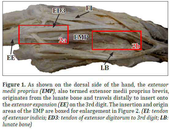
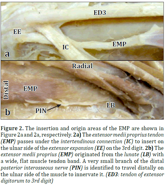
During routine dissection in a gross anatomy laboratory, an unusual EMP muscle was found in the left hand of a 67-year-old female cadaver with “stroke” listed as the cause of death. As shown in Figures 1 and 2, the tendon and muscle belly of the EMP originated at the rostrodorsal surface of the lunate bone. The muscle belly was thin and about 1.5 centimeters wide. It traveled distally superficial to the dorsal surface of the 3rd dorsal interosseous muscle. At the distal one-fourth of the muscle, the EMP became a slender tendon passing deep to the intertendinous connection between the 2nd and 3rd tendon slips of the extensor digitorum muscle (EDM). The tendon of the EMP continued traveling distally ulnar to the 2nd EDM tendon slip, deep to the transverse fibers of the extensor expansion, superficially crossed the 2nd metacarpophalangeal joint, and finally inserted into the rostrodorsal surface of the proximal phalanx of the third digit (Figure 2a). A branch from the posterior interosseous nerve was traced distally and medially for approximately one-fourth of the EMP muscle’s length before turning laterally deep to the muscle to innervate it (Figure 2b).
Figure 1: As shown on the dorsal side of the hand, the extensor medii proprius (EMP), also termed extensor medii proprius brevis, originates from the lunate bone and travels distally to insert onto the extensor expansion (EE) on the 3rd digit. The insertion and origin areas of the EMP are boxed for enlargement in Figure 2. (EI: tendon of extensor indicis; ED3: tendon of extensor digitorum to 3rd digit; LB: lunate bone)
Figure 2: The insertion and origin areas of the EMP are shown in Figure 2a and 2a, respectively. 2a) The extensor medii proprius tendon (EMP) passes under the intertendinous connection (IC) to insert on the ulnar side of the extensor expansion (EE) on the 3rd digit. 2b) The extensor medii proprius (EMP) originated from the lunate (LB) with a wide, flat muscle tendon band. A very small branch of the distal posterior interosseous nerve (PIN) is identified to travel distally on the ulnar side of the muscle to innervate it. (ED3: tendon of extensor digitorum to 3rd digit)
Discussion
In this case report, some aspects of the identified EMP are similar to previous reports [1-5]. Four important differences are presented in this case. First, the EMP originates at the lunate, which means the muscle does not cross the radiocarpal joint as described in previous reports. The muscle belly is found only in the dorsum of the hand rather than the posterior forearm. We therefore propose that this variant could be designated as the extensor medii proprius brevis (EMPB). Second, the co-occurrence of the EMP with the extensor indicis proprius or related muscles was not observed in this case. Third, the EMP tendon travels deep to the intertendinous connection between the 2nd and 3rd tendon slips of the extensor digitorum muscle. As such, the intertendinous connection might play a role in preventing the EMP tendon from bowstringing during contraction. Finally, among relevant reports [1–5], this is the first time that the EMP is found to be innervated by a branch from the posterior interosseous nerve.
As described by Straus [6], the dorsal (extensor) muscle mass on the forearm differentiates into three different muscle portions (radial, superficial, and deep). These three portions then further differentiate into the extensor muscles of the forearm. The superficial and deep portions will develop into the muscles that cross the metacarpophalangeal (MCP) joints –this may include the EMP (or the EMPB in this case) as a muscle which crosses the MCP. Based on the location of EMP, phylogenetic comparisons suggest that the EMP may be an evolutionary remnant of a normal developmental arrangement [3]; EMPB may develop from a similar trajectory due to its similarities with EMP.
In functional terms, the EMP/EMPB may act as an accessory MCP extensor. Due to its small size, however, the impact of EMP/EMPB on MCP extension may be negligible. Clinicians should still be aware of this variation, since swelling or tenderness of the muscle may lead to misdiagnoses of ganglion cysts or adipose tumors around this area of the dorsal hand.
Acknowledgement
The authors wish to thank those who have made an anatomical gift to the University of North Texas Health Science Center Willed Body Program.
References
- Tan ST, Smith PJ. Anomalous extensor muscles of the hand: a review. J Hand Surg Am. 1999;24: 449–455.
- Cauldwell EW, Anson BJ, Wright RR. The extensor indicis proprius muscle: a study of 263 consecutive specimens. Q Bull Northwest Univ Med School. 1943; 17: 267–269.
- Von Schroeder HP, Botte MJ. The extensor medii proprius and anomalous extensor tendons to the long finger. J Hand Surg Am. 1991; 16: 1141–1145.
- Cigali BS, Kutoglu T, Cikmaz S. Musculus extensor digiti medii proprius and musculus extensor digitorum brevis manus – a case report of a rare variation. Anat Histol Embryol.2002; 31: 126–127.
- Komiyama M, Nwe TM, Toyota N, Shimada Y. Variations of the extrensor indicis muscle and tendon. J Hand Surg Br. 1999; 24: 5: 575–578.
- Straus WL The phylogeny of the human forearm extensors. Hum Biol. 1941; 13: 23–50,204–238.
Vic Holmes1*, Hao (Howe) Liu2,3, Armando Rosales3 and Claire Kirchhoff3
1Department of Physician Assistant Studies,University of North Texas Health Science Center, Fort Worth, TX, USA.
2 Department of Physical Therapy,University of North Texas Health Science Center, Fort Worth, TX, USA.
3Department of Cell Biology & Anatomy,University of North Texas Health Science Center, Fort Worth, TX, USA.
- *Corresponding Author:
- Vic Holmes, PA
Dept. of Physician Assistant Studies University of North Texas Health Science Center ,3500 Camp Bowie Blvd. Fort Worth, TX 76107, USA.
Tel: +1 (817) 735-2188
E-mail: vic.holmes@unthsc.edu
Date of Received: April 24th, 2014
Date of Accepted: August 9th, 2014
Published Online: December 20th, 2015
© Int J Anat Var (IJAV). 2015; 8: 32–33.
Abstract
A variant of the extensor medii proprius (EMP) was identified during routine dissection of the left hand of a 67-year-old female cadaver. The flat, fleshy muscle originated from the lunate bone, narrowed into a flat tendon near the 3rd metacarpophalangeal joint, and continued distally to insert on the extensor expansion of the 3rd digit. A branch from the posterior interosseous nerve was traced to the EMP. We propose that this previously unreported variation be termed extensor medii proprius brevis (EMPB).
-Keywords
variation,extensor digitorum muscle,extensor expansion
Introduction
Variations of human hand extensor muscles have been previously described [1]. The extensor medii proprius (EMP) is one such variation reported with an incidence ranging from 0.8% (2 out of 263 specimens) [2], to 10.3% (6 out of 58 dissected hands) [3]. The EMP often co-exists with the extensor indicis muscle or its variant, the extensor indicis proprius on one hand or both hands [1,4,5]. Based on these reports, the EMP originates from the distal 1/3 of the ulna and inserts on the dorsal aponeurosis (extensor expansion) of the 3rd manual digit [1,3–5]. We present a case where the EMP originates from the lunate, an origination not previously described. We propose that this variant be termed extensor medii proprius brevis (EMPB). We also present the innervation of this variant of EMP as observed in this case.
Case Report
During routine dissection in a gross anatomy laboratory, an unusual EMP muscle was found in the left hand of a 67-year-old female cadaver with “stroke” listed as the cause of death. As shown in Figures 1 and 2, the tendon and muscle belly of the EMP originated at the rostrodorsal surface of the lunate bone. The muscle belly was thin and about 1.5 centimeters wide. It traveled distally superficial to the dorsal surface of the 3rd dorsal interosseous muscle. At the distal one-fourth of the muscle, the EMP became a slender tendon passing deep to the intertendinous connection between the 2nd and 3rd tendon slips of the extensor digitorum muscle (EDM). The tendon of the EMP continued traveling distally ulnar to the 2nd EDM tendon slip, deep to the transverse fibers of the extensor expansion, superficially crossed the 2nd metacarpophalangeal joint, and finally inserted into the rostrodorsal surface of the proximal phalanx of the third digit (Figure 2a). A branch from the posterior interosseous nerve was traced distally and medially for approximately one-fourth of the EMP muscle’s length before turning laterally deep to the muscle to innervate it (Figure 2b).
Figure 1: As shown on the dorsal side of the hand, the extensor medii proprius (EMP), also termed extensor medii proprius brevis, originates from the lunate bone and travels distally to insert onto the extensor expansion (EE) on the 3rd digit. The insertion and origin areas of the EMP are boxed for enlargement in Figure 2. (EI: tendon of extensor indicis; ED3: tendon of extensor digitorum to 3rd digit; LB: lunate bone)
Figure 2: The insertion and origin areas of the EMP are shown in Figure 2a and 2a, respectively. 2a) The extensor medii proprius tendon (EMP) passes under the intertendinous connection (IC) to insert on the ulnar side of the extensor expansion (EE) on the 3rd digit. 2b) The extensor medii proprius (EMP) originated from the lunate (LB) with a wide, flat muscle tendon band. A very small branch of the distal posterior interosseous nerve (PIN) is identified to travel distally on the ulnar side of the muscle to innervate it. (ED3: tendon of extensor digitorum to 3rd digit)
Discussion
In this case report, some aspects of the identified EMP are similar to previous reports [1-5]. Four important differences are presented in this case. First, the EMP originates at the lunate, which means the muscle does not cross the radiocarpal joint as described in previous reports. The muscle belly is found only in the dorsum of the hand rather than the posterior forearm. We therefore propose that this variant could be designated as the extensor medii proprius brevis (EMPB). Second, the co-occurrence of the EMP with the extensor indicis proprius or related muscles was not observed in this case. Third, the EMP tendon travels deep to the intertendinous connection between the 2nd and 3rd tendon slips of the extensor digitorum muscle. As such, the intertendinous connection might play a role in preventing the EMP tendon from bowstringing during contraction. Finally, among relevant reports [1–5], this is the first time that the EMP is found to be innervated by a branch from the posterior interosseous nerve.
As described by Straus [6], the dorsal (extensor) muscle mass on the forearm differentiates into three different muscle portions (radial, superficial, and deep). These three portions then further differentiate into the extensor muscles of the forearm. The superficial and deep portions will develop into the muscles that cross the metacarpophalangeal (MCP) joints –this may include the EMP (or the EMPB in this case) as a muscle which crosses the MCP. Based on the location of EMP, phylogenetic comparisons suggest that the EMP may be an evolutionary remnant of a normal developmental arrangement [3]; EMPB may develop from a similar trajectory due to its similarities with EMP.
In functional terms, the EMP/EMPB may act as an accessory MCP extensor. Due to its small size, however, the impact of EMP/EMPB on MCP extension may be negligible. Clinicians should still be aware of this variation, since swelling or tenderness of the muscle may lead to misdiagnoses of ganglion cysts or adipose tumors around this area of the dorsal hand.
Acknowledgement
The authors wish to thank those who have made an anatomical gift to the University of North Texas Health Science Center Willed Body Program.
References
- Tan ST, Smith PJ. Anomalous extensor muscles of the hand: a review. J Hand Surg Am. 1999;24: 449–455.
- Cauldwell EW, Anson BJ, Wright RR. The extensor indicis proprius muscle: a study of 263 consecutive specimens. Q Bull Northwest Univ Med School. 1943; 17: 267–269.
- Von Schroeder HP, Botte MJ. The extensor medii proprius and anomalous extensor tendons to the long finger. J Hand Surg Am. 1991; 16: 1141–1145.
- Cigali BS, Kutoglu T, Cikmaz S. Musculus extensor digiti medii proprius and musculus extensor digitorum brevis manus – a case report of a rare variation. Anat Histol Embryol.2002; 31: 126–127.
- Komiyama M, Nwe TM, Toyota N, Shimada Y. Variations of the extrensor indicis muscle and tendon. J Hand Surg Br. 1999; 24: 5: 575–578.
- Straus WL The phylogeny of the human forearm extensors. Hum Biol. 1941; 13: 23–50,204–238.


