A variant origin of cystic artery from superior mesenteric artery
George Joseph Lufukuja*
Department of Anatomy and Histology, St. Francis University College of Health and Allied Sciences, Ifakara, Morogoro, Tanzania
- *Corresponding Author:
- George Joseph Lufukuja
Assistant Lecturer, Dept. of Anatomy and Histology, St. Francis University College of Health and Allied Sciences, P.O. Box: 175 Ifakara, Morogoro, Tanzania
Tel: +255 717 090 572
E-mail: lufukuja70@yahoo.co.uk
Date of Received: February 21st, 2013
Date of Accepted: October 21st, 2013
Published Online: June 1st, 2014
© Int J Anat Var (IJAV). 2014; 7: 30–31.
[ft_below_content] =>Keywords
cystic artery, gallbladder, common hepatic duct, common bile duct
Introduction
The cystic artery (CA) arises from the right hepatic artery in the Calot’s triangle in the cysto-hepatic angle immediately to the right of common hepatic duct. After it has passed behind the common hepatic duct and anterior to the cystic duct, it reaches the superior aspect of the neck of gallbladder to divide in to superficial and deep branches. The superficial branch ramifies on the gallbladder to divide in to superficial and deep branches. The superficial branch ramifies on the inferior aspect of gallbladder and the deep branch on the superior aspect; these branches anastomose over the surface of body and fundus [1]. Sometimes there can be more than one cystic artery supplying the gallbladder or the artery may arise from other arteries like the left hepatic artery, the common hepatic artery, the gastroduodenal artery, the superior pancreaticoduodenal artery and the superior mesenteric artery [2].
Case Report
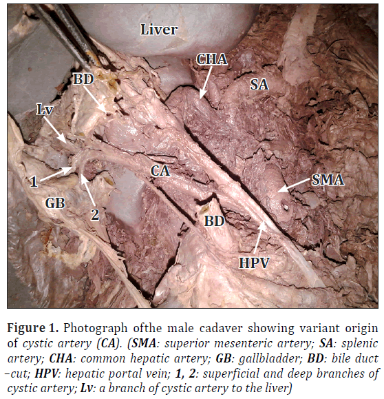
In the routine dissection of 43-year-old male cadaver, we found a very rare origin of the CA, which was taking origin from superior mesenteric artery (Figure 1). It passed posterior to the common hepatic duct to reach the superior aspect of the neck of the gallbladder and divided into superficial and deep branches that supplied the gallbladder and continued to the right lobe of liver. This kind of variant origin of CA, according to our knowledge is rarely reported in anatomical literature.
Figure 1: Photograph ofthe male cadaver showing variant origin of cystic artery (CA). (SMA: superior mesenteric artery; SA: splenic artery; CHA: common hepatic artery; GB: gallbladder; BD: bile duct –cut; HPV: hepatic portal vein; 1, 2: superficial and deep branches of cystic artery; Lv: a branch of cystic artery to the liver)
Discussion
The CA is known to exhibit variations in its origin and branching pattern. This is attributed to the developmental changes occurring in the primitive ventral splanchnic arteries [3]. In most of the cases, the CA arises from the right hepatic artery. The other origins include superior mesenteric artery through its inferior pancreatico-duodenal branch [2]. During development, the extrahepatic biliary system arises from an intestinal diverticulum, which carries a rich supply of vessels from the aorta, coeliac trunk and superior mesenteric artery. Later most of these vessels are absorbed, leaving in place the mature vascular system. As the pattern of absorption is highly variable, it is not unusual for the cystic artery and its branches to derive from any other artery in the vicinity [2].
In a case reported by Veena et al., two cystic arteries were reported to arise from the right hepatic artery to supply the two surfaces of gallbladder separately [1]. Considering the fact that the diseases of the extra hepatic biliary apparatus often need surgical intervention, it is very important to know anatomy of the same, which is frequently unusual [1]. The CA originating from the middle hepatic artery at a distance of about 1 cm from its origin was also reported [3]. In the 160 Sudanese cases investigated, the origin of the cystic artery was from the right hepatic artery in 78%, from the common hepatic artery in 17%, from the left hepatic artery in 2% and from the gastroduodenal artery in 3%. No cases arising from other arteries were noted [4].
Knowledge of the different anatomical variations of the arterial supply of the gallbladder, liver and stomach is of great importance in hepatobiliary and gastric surgical procedures [5]. The arterial variations should not be ignored and with an accurate knowledge on the anatomical variations, many operative and postoperative complications can be avoided. The knowledge on the CT variations would enable the radiologists in protecting the important vessels prior to transcatheter therapies, and also in preventing inadvertent injuries. The anatomy of the hepatic artery is of great importance in hepatic surgeries, especially in liver transplantation and in many radiological procedures, to ensure complete arterializations of the grafts, which can prevent necrosis of the liver parenchyma postoperatively [6].
Acknowledgement
My gratitude and thank you goes to the following people and organizations who made this study possible:
• To my Almighty God, for guiding me and giving me the strength and belief that to him nothing is impossible,
• To my parents, Joseph M. Lufukuja and Maria P.Ng’wakami, for their encouragement and for believing in me,
•To my wife Rachel, and Son Joseph junior for their immeasurable support throughout this case report writing.
References
- Pai V, Subhash LP, Anupama D, Nagaraj DN. Dual cystic artery – a case report. Anatomica Karnataka. 2011; 5: 52–53.
- Vishnumaya G, Bhagath KP, Vasavi RG, Thejodhar P. Anomalous origin of cystic artery from gastroduodenal artery – A case report. Int J Morphol. 2008; 26: 75–76.
- Hlaing KP, Thwin SS, Shwe N. Unique origin of the cystic artery. Singapore Med J. 2011; 52: e262–264.
- Bakheit MA. Prevalence of variations of the cystic artery in the Sudanese. East Mediterr Health J. 2009; 15: 1308–1312.
- Loukas M, Fergurson A, Louis RG Jr, Colborn GL. Multiple variations of the hepatobiliary vasculature including double cystic arteries, accessory left hepatic artery and hepatosplenic trunk: a case report. Surg Radiol Anat. 2006; 28: 525–528.
- Chandrachar JK, Shetty S, Ivan AS. Variations in the branching pattern of the coeliac trunk. J Clin Diagn Res. 2012; 6: 1289–1291.
George Joseph Lufukuja*
Department of Anatomy and Histology, St. Francis University College of Health and Allied Sciences, Ifakara, Morogoro, Tanzania
- *Corresponding Author:
- George Joseph Lufukuja
Assistant Lecturer, Dept. of Anatomy and Histology, St. Francis University College of Health and Allied Sciences, P.O. Box: 175 Ifakara, Morogoro, Tanzania
Tel: +255 717 090 572
E-mail: lufukuja70@yahoo.co.uk
Date of Received: February 21st, 2013
Date of Accepted: October 21st, 2013
Published Online: June 1st, 2014
© Int J Anat Var (IJAV). 2014; 7: 30–31.
Abstract
The cystic artery usually arises from the right hepatic artery. It usually passes posterior to the common hepatic duct and anterior to the cystic duct to reach the superior aspect of the neck of the gallbladder and divides into superficial and deep branches. The superficial branch ramifies on the inferior aspect of the body of the gallbladder, the deep branch on the superior aspect. In our case we observed a variant origin of cystic artery from the superior mesenteric artery. Knowledge of the variant vascular anatomy of the sub-hepatic region is important for hepatobiliary surgeons in limiting operative complications due to unexpected bleeding.
-Keywords
cystic artery, gallbladder, common hepatic duct, common bile duct
Introduction
The cystic artery (CA) arises from the right hepatic artery in the Calot’s triangle in the cysto-hepatic angle immediately to the right of common hepatic duct. After it has passed behind the common hepatic duct and anterior to the cystic duct, it reaches the superior aspect of the neck of gallbladder to divide in to superficial and deep branches. The superficial branch ramifies on the gallbladder to divide in to superficial and deep branches. The superficial branch ramifies on the inferior aspect of gallbladder and the deep branch on the superior aspect; these branches anastomose over the surface of body and fundus [1]. Sometimes there can be more than one cystic artery supplying the gallbladder or the artery may arise from other arteries like the left hepatic artery, the common hepatic artery, the gastroduodenal artery, the superior pancreaticoduodenal artery and the superior mesenteric artery [2].
Case Report
In the routine dissection of 43-year-old male cadaver, we found a very rare origin of the CA, which was taking origin from superior mesenteric artery (Figure 1). It passed posterior to the common hepatic duct to reach the superior aspect of the neck of the gallbladder and divided into superficial and deep branches that supplied the gallbladder and continued to the right lobe of liver. This kind of variant origin of CA, according to our knowledge is rarely reported in anatomical literature.
Figure 1: Photograph ofthe male cadaver showing variant origin of cystic artery (CA). (SMA: superior mesenteric artery; SA: splenic artery; CHA: common hepatic artery; GB: gallbladder; BD: bile duct –cut; HPV: hepatic portal vein; 1, 2: superficial and deep branches of cystic artery; Lv: a branch of cystic artery to the liver)
Discussion
The CA is known to exhibit variations in its origin and branching pattern. This is attributed to the developmental changes occurring in the primitive ventral splanchnic arteries [3]. In most of the cases, the CA arises from the right hepatic artery. The other origins include superior mesenteric artery through its inferior pancreatico-duodenal branch [2]. During development, the extrahepatic biliary system arises from an intestinal diverticulum, which carries a rich supply of vessels from the aorta, coeliac trunk and superior mesenteric artery. Later most of these vessels are absorbed, leaving in place the mature vascular system. As the pattern of absorption is highly variable, it is not unusual for the cystic artery and its branches to derive from any other artery in the vicinity [2].
In a case reported by Veena et al., two cystic arteries were reported to arise from the right hepatic artery to supply the two surfaces of gallbladder separately [1]. Considering the fact that the diseases of the extra hepatic biliary apparatus often need surgical intervention, it is very important to know anatomy of the same, which is frequently unusual [1]. The CA originating from the middle hepatic artery at a distance of about 1 cm from its origin was also reported [3]. In the 160 Sudanese cases investigated, the origin of the cystic artery was from the right hepatic artery in 78%, from the common hepatic artery in 17%, from the left hepatic artery in 2% and from the gastroduodenal artery in 3%. No cases arising from other arteries were noted [4].
Knowledge of the different anatomical variations of the arterial supply of the gallbladder, liver and stomach is of great importance in hepatobiliary and gastric surgical procedures [5]. The arterial variations should not be ignored and with an accurate knowledge on the anatomical variations, many operative and postoperative complications can be avoided. The knowledge on the CT variations would enable the radiologists in protecting the important vessels prior to transcatheter therapies, and also in preventing inadvertent injuries. The anatomy of the hepatic artery is of great importance in hepatic surgeries, especially in liver transplantation and in many radiological procedures, to ensure complete arterializations of the grafts, which can prevent necrosis of the liver parenchyma postoperatively [6].
Acknowledgement
My gratitude and thank you goes to the following people and organizations who made this study possible:
• To my Almighty God, for guiding me and giving me the strength and belief that to him nothing is impossible,
• To my parents, Joseph M. Lufukuja and Maria P.Ng’wakami, for their encouragement and for believing in me,
•To my wife Rachel, and Son Joseph junior for their immeasurable support throughout this case report writing.
References
- Pai V, Subhash LP, Anupama D, Nagaraj DN. Dual cystic artery – a case report. Anatomica Karnataka. 2011; 5: 52–53.
- Vishnumaya G, Bhagath KP, Vasavi RG, Thejodhar P. Anomalous origin of cystic artery from gastroduodenal artery – A case report. Int J Morphol. 2008; 26: 75–76.
- Hlaing KP, Thwin SS, Shwe N. Unique origin of the cystic artery. Singapore Med J. 2011; 52: e262–264.
- Bakheit MA. Prevalence of variations of the cystic artery in the Sudanese. East Mediterr Health J. 2009; 15: 1308–1312.
- Loukas M, Fergurson A, Louis RG Jr, Colborn GL. Multiple variations of the hepatobiliary vasculature including double cystic arteries, accessory left hepatic artery and hepatosplenic trunk: a case report. Surg Radiol Anat. 2006; 28: 525–528.
- Chandrachar JK, Shetty S, Ivan AS. Variations in the branching pattern of the coeliac trunk. J Clin Diagn Res. 2012; 6: 1289–1291.


