Abnormal formation and communication of external jugular vein
Satheesha Nayak B1*, Soumya KV2
1Department of Anatomy, Melaka Manipal Medical College (Manipal Campus) Manipal, India.
2Department of Mathematics, Manipal Institute of Technology, Manipal, India.
- *Corresponding Author:
- Satheesha Nayak B., MSc. PhD.
Associate Professor of Anatomy, Melaka Manipal Medical College (Manipal Campus), International Centre for Health Sciences, Madhav Nagar, Manipal, Udupi District, Karnataka State, 576 104, India.
Tel: +91 820 2571905
E-mail: nayaksathish@yahoo.com
Date of Received: July 5th, 2008
Date of Accepted: August 27th, 2008
Published Online: August 29th, 2008
© IJAV. 2008; 1: 15–16.
[ft_below_content] =>Keywords
variation, retromandibular vein, cephalic vein, facial vein, neck
Introduction
External jugular vein collects most of the blood from the exterior of the cranium and the deep part of the face. It is normally formed by the union of posterior division of retromandibular vein and the posterior auricular vein. It starts at the level of the angle of the mandible, just below the apex of parotid gland and runs vertically down in the superficial fascia till a point just above midpoint of clavicle. On its course, it crosses superficial to the sternocleidomastoid muscle obliquely. Just above the midpoint of clavicle, it pierces the deep cervical fascia and opens into the subclavian vein. It usually receives the occipital, posterior external jugular, anterior jugular and transverse cervical veins.
The cephalic vein is the longest vein of the upper limb. It starts at the anatomical snuff box as the continuation of the lateral end of the dorsal venous arch. It winds around the lateral border of the forearm and ascends along the lateral border in the superficial fascia until the lower border of pectoralis major muscle. Here it pierces the deep fascia and runs through the deltopectoral groove. At the deltopectoral triangle, it pierces the clavipectoral fascia and terminates in the axillary vein. It communicates with the basilic vein through the median cubital vein at the cubital fossa.
Case Report
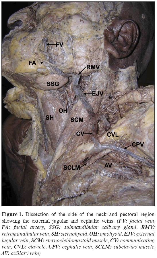
During regular dissections for undergraduate medical students, we observed the following venous variations in the head and neck region in an adult male cadaver. The retromandibular vein did not divide into two divisions. It joined with the facial vein to form the external jugular vein (Figure 1). It then descended down vertically superficial to the sternocleidomastoid muscle till the midpoint of the clavicle and terminated into the subclavian vein after piercing the deep fascia. The posterior auricular vein was absent. The terminal part of the cephalic vein passed between the clavicle and the subclavius muscle before opening into the axillary vein (Figure 1). There was a large communicating vein that connected cephalic vein with the external jugular vein. The communicating vein crossed superficial to the clavicle (Figure 1). There were no other notable variations in the cadaver.
Figure 1: Dissection of the side of the neck and pectoral region showing the external jugular and cephalic veins. (FV: facial vein, FA: facial artery, SSG: submandibular salivary gland, RMV: retromandibular vein, SH: sternohyoid, OH: omohyoid, EJV: external jugular vein, SCM: sternocleidomastoid muscle, CV: communicating vein, CVL: clavicle, CPV: cephalic vein, SCLM: subclavius muscle, AV: axillary vein)
Discussion
The external jugular vein is one of the variable veins. It shows more variations at its origin than termination. Bergman et al., [1] have listed several variations of the external jugular vein. The variations that they have noted include:
a. Its formation merely by the posterior auricular vein
b. Receiving the facial, lingual, or the cephalic veins as tributaries
c. Passing over the clavicle and open into the cephalic, subclavian, or internal jugular veins
d. Doubling of the vein
e. Formation of an annulus around the clavicle
f. Receiving lingual vein as its tributary.
In a study conducted on 89 dissected adult cadavers, the external jugular vein received facial vein in 9% of cases [2]. The formation of external jugular vein by union of the entire retromandibular vein and facial vein [3], and a case of facial vein continuing as external jugular vein have also been reported [4]. The variations of cephalic veins are also well known and their clinical implication has been documented. The cephalic vein may be small or absent. It may be accompanied by an accessory cephalic vein [1]. A supraclavicular course of the cephalic vein has also been noted [5].
The superficial veins, especially the external jugular vein and cephalic vein are increasingly being utilized for cannulation to conduct diagnostic procedures or intravenous therapies. While the subclavian or axillary vein can be safely and successfully punctured in the majority of cases, some device implanters still prefer cut down to the cephalic vein as the initial approach to venous access for transvenous placement of pacemaker or defibrillator leads out of concern for the risk of pneumothorax, subclavian crush, and other possible complications. The complications related to the use of external jugular or cephalic veins for cannulation are relatively less than that of internal jugular vein. It is important to know about the communication between the cephalic vein and external jugular veins across the clavicle. The catheter can easily pass into this communicating vein and miss the intended direction. The cephalic vein running between subclavius muscle and clavicle, and the communicating vein running superficial to clavicle may bleed profusely in case of fracture of clavicle. Ultrasound-guided venipuncture is a viable possibility in cases of variations in the patterns of superficial veins, and their knowledge is also important for surgeons doing reconstructive surgery.
References
- Bergman RA, Afifi AK, Miyauchi R. Illustrated encyclopedia of human anatomic variation: Opus II: Cardiovascular system: Veins: Head, neck, and thorax. http://www.anatomyatlases.org/AnatomicVariants/Cardiovascular/Text/Veins/JugularExternal.shtml. (accessed on July 4, 2008).
- Gupta V, Tuli A, Choudhry R, Agarwal S, Mangal A. Facial vein draining into external jugular vein in humans: its variations, phylogenetic retention and clinical relevance. Surg. Radiol. Anat. 2003; 25: 36–41.
- Yadav S, Ghosh SK, Anand C. Variations of superficial veins of head and neck. J. Anat. Soc. India. 2000: 49; 61–62.
- Vollala VR, Bolla SR, Pamidi N. Important vascular anomalies of face and neck – a cadaveric study with clinical implications. Firat Tip Dergisi. 2008: 13;123–126.
- Lau EW, Liew R, Harris S. An unusual case of the cephalic vein with a supraclavicular course. Pacing Clin. Electrophysiol. 2007: 30; 719–720.
Satheesha Nayak B1*, Soumya KV2
1Department of Anatomy, Melaka Manipal Medical College (Manipal Campus) Manipal, India.
2Department of Mathematics, Manipal Institute of Technology, Manipal, India.
- *Corresponding Author:
- Satheesha Nayak B., MSc. PhD.
Associate Professor of Anatomy, Melaka Manipal Medical College (Manipal Campus), International Centre for Health Sciences, Madhav Nagar, Manipal, Udupi District, Karnataka State, 576 104, India.
Tel: +91 820 2571905
E-mail: nayaksathish@yahoo.com
Date of Received: July 5th, 2008
Date of Accepted: August 27th, 2008
Published Online: August 29th, 2008
© IJAV. 2008; 1: 15–16.
Abstract
Knowledge of variations in the origin, course and termination of external jugular vein may be important for surgeons, radiologists, and plastic surgeons. In this report, we present a variation in the origin of the external jugular vein and its abnormal communication with the cephalic vein. The external jugular vein was formed by the union of facial and retromandibular veins. Its course and termination were normal but it communicated with the cephalic vein through a large communicating vein, which crossed superficial to clavicle. The retromandibular vein did not divide into two divisions and the posterior auricular vein was absent. The terminal part of cephalic vein was sandwiched between the clavicle and subclavius muscle.
-Keywords
variation, retromandibular vein, cephalic vein, facial vein, neck
Introduction
External jugular vein collects most of the blood from the exterior of the cranium and the deep part of the face. It is normally formed by the union of posterior division of retromandibular vein and the posterior auricular vein. It starts at the level of the angle of the mandible, just below the apex of parotid gland and runs vertically down in the superficial fascia till a point just above midpoint of clavicle. On its course, it crosses superficial to the sternocleidomastoid muscle obliquely. Just above the midpoint of clavicle, it pierces the deep cervical fascia and opens into the subclavian vein. It usually receives the occipital, posterior external jugular, anterior jugular and transverse cervical veins.
The cephalic vein is the longest vein of the upper limb. It starts at the anatomical snuff box as the continuation of the lateral end of the dorsal venous arch. It winds around the lateral border of the forearm and ascends along the lateral border in the superficial fascia until the lower border of pectoralis major muscle. Here it pierces the deep fascia and runs through the deltopectoral groove. At the deltopectoral triangle, it pierces the clavipectoral fascia and terminates in the axillary vein. It communicates with the basilic vein through the median cubital vein at the cubital fossa.
Case Report
During regular dissections for undergraduate medical students, we observed the following venous variations in the head and neck region in an adult male cadaver. The retromandibular vein did not divide into two divisions. It joined with the facial vein to form the external jugular vein (Figure 1). It then descended down vertically superficial to the sternocleidomastoid muscle till the midpoint of the clavicle and terminated into the subclavian vein after piercing the deep fascia. The posterior auricular vein was absent. The terminal part of the cephalic vein passed between the clavicle and the subclavius muscle before opening into the axillary vein (Figure 1). There was a large communicating vein that connected cephalic vein with the external jugular vein. The communicating vein crossed superficial to the clavicle (Figure 1). There were no other notable variations in the cadaver.
Figure 1: Dissection of the side of the neck and pectoral region showing the external jugular and cephalic veins. (FV: facial vein, FA: facial artery, SSG: submandibular salivary gland, RMV: retromandibular vein, SH: sternohyoid, OH: omohyoid, EJV: external jugular vein, SCM: sternocleidomastoid muscle, CV: communicating vein, CVL: clavicle, CPV: cephalic vein, SCLM: subclavius muscle, AV: axillary vein)
Discussion
The external jugular vein is one of the variable veins. It shows more variations at its origin than termination. Bergman et al., [1] have listed several variations of the external jugular vein. The variations that they have noted include:
a. Its formation merely by the posterior auricular vein
b. Receiving the facial, lingual, or the cephalic veins as tributaries
c. Passing over the clavicle and open into the cephalic, subclavian, or internal jugular veins
d. Doubling of the vein
e. Formation of an annulus around the clavicle
f. Receiving lingual vein as its tributary.
In a study conducted on 89 dissected adult cadavers, the external jugular vein received facial vein in 9% of cases [2]. The formation of external jugular vein by union of the entire retromandibular vein and facial vein [3], and a case of facial vein continuing as external jugular vein have also been reported [4]. The variations of cephalic veins are also well known and their clinical implication has been documented. The cephalic vein may be small or absent. It may be accompanied by an accessory cephalic vein [1]. A supraclavicular course of the cephalic vein has also been noted [5].
The superficial veins, especially the external jugular vein and cephalic vein are increasingly being utilized for cannulation to conduct diagnostic procedures or intravenous therapies. While the subclavian or axillary vein can be safely and successfully punctured in the majority of cases, some device implanters still prefer cut down to the cephalic vein as the initial approach to venous access for transvenous placement of pacemaker or defibrillator leads out of concern for the risk of pneumothorax, subclavian crush, and other possible complications. The complications related to the use of external jugular or cephalic veins for cannulation are relatively less than that of internal jugular vein. It is important to know about the communication between the cephalic vein and external jugular veins across the clavicle. The catheter can easily pass into this communicating vein and miss the intended direction. The cephalic vein running between subclavius muscle and clavicle, and the communicating vein running superficial to clavicle may bleed profusely in case of fracture of clavicle. Ultrasound-guided venipuncture is a viable possibility in cases of variations in the patterns of superficial veins, and their knowledge is also important for surgeons doing reconstructive surgery.
References
- Bergman RA, Afifi AK, Miyauchi R. Illustrated encyclopedia of human anatomic variation: Opus II: Cardiovascular system: Veins: Head, neck, and thorax. http://www.anatomyatlases.org/AnatomicVariants/Cardiovascular/Text/Veins/JugularExternal.shtml. (accessed on July 4, 2008).
- Gupta V, Tuli A, Choudhry R, Agarwal S, Mangal A. Facial vein draining into external jugular vein in humans: its variations, phylogenetic retention and clinical relevance. Surg. Radiol. Anat. 2003; 25: 36–41.
- Yadav S, Ghosh SK, Anand C. Variations of superficial veins of head and neck. J. Anat. Soc. India. 2000: 49; 61–62.
- Vollala VR, Bolla SR, Pamidi N. Important vascular anomalies of face and neck – a cadaveric study with clinical implications. Firat Tip Dergisi. 2008: 13;123–126.
- Lau EW, Liew R, Harris S. An unusual case of the cephalic vein with a supraclavicular course. Pacing Clin. Electrophysiol. 2007: 30; 719–720.


