Abnormal superior trunk formation of brachial plexus: a case report and review of literature
Singh R*
Department of Anatomy, AIIMS, Rishikesh, Uttrakhand, India.
- *Corresponding Author:
- Dr. Rajani Singh
Additional Professor and HOD, Department of Anatomy, AIIMS, Rishikesh 249201, Uttrakhand, India.
Tel: 0135-2452932
E-mail: nani_sahayal@rediffmail.com
Citation: Singh R. Abnormal superior trunk formation of brachial plexus: a case report and review of literature. Int J Anat Var. 2017;10(3):45-6.
Copyright: This open-access article is distributed under the terms of the Creative Commons Attribution Non-Commercial License (CC BY-NC) (http://creativecommons.org/licenses/by-nc/4.0/), which permits reuse, distribution and reproduction of the article, provided that the original work is properly cited and the reuse is restricted to noncommercial purposes. For commercial reuse, contact reprints@pulsus.com.
[ft_below_content] =>Keywords
Brachial plexus; Variation; Trunk; Anatomist
Introduction
Brachial plexus is complicated nerve network formed by roots C5, C6, C7, C8 and T1. Upper trunk is formed by union of C5 and C6. C7 continues as middle trunk. C8 and T1 fuse together to form lower trunk. Each of these trunks divides into anterior and posterior divisions. Anterior divisions of upper and middle trunk joined to form lateral cord, anterior division of lower trunk continue as medial cord. Posterior divisions of these entire three trunks fuse together to form posterior cord. In the present study rare variant formation of trunks was observed which has several clinical repercussions.
Case Report
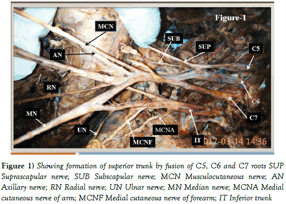
During dissection of right upper limb of female cadaver of 70 years old fixed in 10% formaline solution for teaching of undergraduate medical students variant formation of trunk of brachial plexus was observed. C5, C6 fused to form a trunk and then C7 fused with this trunk to form superior trunk (Figure 1).
Figure 1: Showing formation of superior trunk by fusion of C5, C6 and C7 roots SUP Suprascapular nerve; SUB Subscapular nerve; MCN Musculocutaneous nerve; AN Axillary nerve; RN Radial nerve; UN Ulnar nerve; MN Median nerve; MCNA Medial cutaneous nerve of arm; MCNF Medial cutaneous nerve of forearm; IT Inferior trunk.
C8 and T1 united together to form inferior trunk. Length of superior trunk was 4 cm. Branch from inferior trunk joined the radial nerve. Median nerve formed by union of medial root from inferior trunk and lateral root from superior trunk. Superior trunk did not divide into cords but gave all the branches which normally arose from lateral and posterior cords. These branches are musculocutaneous nerve, lateral root, radial nerve, axillary nerve, subscapular, thoracodorsal nerves. Inferior trunk gave all branches normally arise from medial cord namely ulnar nerve, medial root, medial cutaneous nerve of arm and forearm and medial pectoral nerve. There was no other abnormality in this specimen.
Discussion
Substantial literature describes variations in the formation of the trunks of the brachial plexuses [1-4]. Shetty et al, [1] dissected 44 specimens and found unilateral variations in the formation of the trunks in five cadavers (11.3%). In one cadaver (2.27%) the middle trunk was formed by union of the C7 and C8 roots and the lower trunk by T1 root. The upper and middle trunks were fused together in one specimen (2.27%). C5 root pierced the scalenus anterior before joining C6 to form the upper trunk in three specimens (6.81%).
In a study by Uysal et al [2] the superior trunk was not formed in 1% of cases, the inferior trunk in 9%. In one plexus, the superior trunk was formed by the ventral rami of the C4 and C5 nerves. In another case, the inferior trunk was formed by the ventral rami of the T1 and T2 nerves.
Formation of the upper trunk of the brachial plexus by the C5, C6 and C7 roots is very rare and is associated with absence of the middle trunk, which can also be interpreted as anatomical fusion of the upper and middle trunks. One such abnormal upper trunk was reported by Nayak et al [3]. Our case is little different from this in the fact the in our case a trunk was formed by fusion of C5 and C6 roots and then C7 fused with this trunk to form superior trunk. Matejcik [4] reported a bilateral case of fusion of the upper and middle trunks. Unilateral upper trunk variations have also been reported [2,4]. James et al, [5] reported a case of a 55-yearold female cadaver in which the superior trunk on the left side was absent. In this case the ventral rami of the C5 and C6 nerve roots did not join to form the superior trunk but divided independently into anterior and posterior divisions which, joined to form the lateral and posterior cords, respectively [5].
Also the C7, C8 and T1 nerve roots have been shown to form the inferior trunk with absence of the middle trunk [4]. Knowledge of variations in the formation of brachial plexus is very useful for neurosurgeons. It has great value in the surgical treatment of tumors of nerve sheaths such as schwannomas and neurofibromas. Orthopedic procedures of the cervical spine also need a thorough knowledge of the normal and variant formations of the brachial plexus. As variations in the roots and trunks of the brachial plexus can be identified by ultrasound examinations, hence these should be screened before planning surgery in this area [6]. Although variations of the brachial plexus might not affect the normal functioning of the patient’s limb, they are very important in clinical neurosurgery and orthopedic procedures. Certain variations of the brachial plexus like formation of superior trunk as in present case may cause an unpredictable spread of local anesthetic [7] or at times undesired blockade distribution. Possible implications of abnormal formation of superior trunk as in present study also include the inability to perform selective blockade, misidentification of structures, inability to obtain a motor response, or paresthesia due to the aberrant course of the plexus [8]. Thus knowledge of variations in the formation of trunks of the brachial plexus is of paramount importance for anesthetists, surgeons, radiologists and anatomists.
References
- Shetty SD, Nayak BS, Madahv V, et al. A Study on the Variations in the Formation of the Trunks of brachial Plexus. Int J Morphol. 2011;29:555-8.
- Uysal II, Seker M, Karabulut AK, et al. Brachial plexus variations in human fetuses. Neurosurgery. 2003;53:676-84.
- Nayak S, Somayaji N, Vollala VR, et al. A rare variation in the formation of the upper trunk of the brachial plexus - a case report. Neuroanatomy. 2005;4:37-8.
- Matejcik V. Anatomic variations in the brachial plexus trunks and nerve roots. Rozhl Chir. 2003;82:456-9.
- James V, Shannon G, Maxwell H, et al. Brachial plexus variation characterized by the absence of the superior trunk. Neuroanatomy. 2009;8:4-6.
- Royse CE, Sha S, Soeding PF, et al. Anatomical study of the brachial plexus using surface ultrasound. Anaesth. Intensive Care. 2006;34:203-10.
- Abrahams MS, Panzer O, Atchabahian A, et al. Case report: limitation of local anesthetic spread during ultrasound-guided interscalene block-description of an anatomic variant with clinical correlation. Reg Anesth Pain Med. 2008;33:357-9.
- Aggarwal A, Sahni D, Kaur H, et al. A Rare Anatomical Variation of the Brachial Plexus: Single Cord Anomaly. Anesthesia & Analgesia. 2012; 114:466-70.
Singh R*
Department of Anatomy, AIIMS, Rishikesh, Uttrakhand, India.
- *Corresponding Author:
- Dr. Rajani Singh
Additional Professor and HOD, Department of Anatomy, AIIMS, Rishikesh 249201, Uttrakhand, India.
Tel: 0135-2452932
E-mail: nani_sahayal@rediffmail.com
Citation: Singh R. Abnormal superior trunk formation of brachial plexus: a case report and review of literature. Int J Anat Var. 2017;10(3):45-6.
Copyright: This open-access article is distributed under the terms of the Creative Commons Attribution Non-Commercial License (CC BY-NC) (http://creativecommons.org/licenses/by-nc/4.0/), which permits reuse, distribution and reproduction of the article, provided that the original work is properly cited and the reuse is restricted to noncommercial purposes. For commercial reuse, contact reprints@pulsus.com.
Abstract
Brachial plexus is a complicated network of nerves formed by fusion and splitting. Variations in the brachial plexus occur at the level of roots, trunks, cords and branches emanating from these structures. We observed a rare variation of formation of upper trunk of brachial plexus in upper right limb of female cadaver aged 70 years. This variation was observed in one limb out of total 28 limbs constituting 3.6%. The rare finding has great diagnostic and therapeutic implications and is of utmost use to anestheologists, radiologists, orthopaedic surgeons and anatomists.
-Keywords
Brachial plexus; Variation; Trunk; Anatomist
Introduction
Brachial plexus is complicated nerve network formed by roots C5, C6, C7, C8 and T1. Upper trunk is formed by union of C5 and C6. C7 continues as middle trunk. C8 and T1 fuse together to form lower trunk. Each of these trunks divides into anterior and posterior divisions. Anterior divisions of upper and middle trunk joined to form lateral cord, anterior division of lower trunk continue as medial cord. Posterior divisions of these entire three trunks fuse together to form posterior cord. In the present study rare variant formation of trunks was observed which has several clinical repercussions.
Case Report
During dissection of right upper limb of female cadaver of 70 years old fixed in 10% formaline solution for teaching of undergraduate medical students variant formation of trunk of brachial plexus was observed. C5, C6 fused to form a trunk and then C7 fused with this trunk to form superior trunk (Figure 1).
Figure 1: Showing formation of superior trunk by fusion of C5, C6 and C7 roots SUP Suprascapular nerve; SUB Subscapular nerve; MCN Musculocutaneous nerve; AN Axillary nerve; RN Radial nerve; UN Ulnar nerve; MN Median nerve; MCNA Medial cutaneous nerve of arm; MCNF Medial cutaneous nerve of forearm; IT Inferior trunk.
C8 and T1 united together to form inferior trunk. Length of superior trunk was 4 cm. Branch from inferior trunk joined the radial nerve. Median nerve formed by union of medial root from inferior trunk and lateral root from superior trunk. Superior trunk did not divide into cords but gave all the branches which normally arose from lateral and posterior cords. These branches are musculocutaneous nerve, lateral root, radial nerve, axillary nerve, subscapular, thoracodorsal nerves. Inferior trunk gave all branches normally arise from medial cord namely ulnar nerve, medial root, medial cutaneous nerve of arm and forearm and medial pectoral nerve. There was no other abnormality in this specimen.
Discussion
Substantial literature describes variations in the formation of the trunks of the brachial plexuses [1-4]. Shetty et al, [1] dissected 44 specimens and found unilateral variations in the formation of the trunks in five cadavers (11.3%). In one cadaver (2.27%) the middle trunk was formed by union of the C7 and C8 roots and the lower trunk by T1 root. The upper and middle trunks were fused together in one specimen (2.27%). C5 root pierced the scalenus anterior before joining C6 to form the upper trunk in three specimens (6.81%).
In a study by Uysal et al [2] the superior trunk was not formed in 1% of cases, the inferior trunk in 9%. In one plexus, the superior trunk was formed by the ventral rami of the C4 and C5 nerves. In another case, the inferior trunk was formed by the ventral rami of the T1 and T2 nerves.
Formation of the upper trunk of the brachial plexus by the C5, C6 and C7 roots is very rare and is associated with absence of the middle trunk, which can also be interpreted as anatomical fusion of the upper and middle trunks. One such abnormal upper trunk was reported by Nayak et al [3]. Our case is little different from this in the fact the in our case a trunk was formed by fusion of C5 and C6 roots and then C7 fused with this trunk to form superior trunk. Matejcik [4] reported a bilateral case of fusion of the upper and middle trunks. Unilateral upper trunk variations have also been reported [2,4]. James et al, [5] reported a case of a 55-yearold female cadaver in which the superior trunk on the left side was absent. In this case the ventral rami of the C5 and C6 nerve roots did not join to form the superior trunk but divided independently into anterior and posterior divisions which, joined to form the lateral and posterior cords, respectively [5].
Also the C7, C8 and T1 nerve roots have been shown to form the inferior trunk with absence of the middle trunk [4]. Knowledge of variations in the formation of brachial plexus is very useful for neurosurgeons. It has great value in the surgical treatment of tumors of nerve sheaths such as schwannomas and neurofibromas. Orthopedic procedures of the cervical spine also need a thorough knowledge of the normal and variant formations of the brachial plexus. As variations in the roots and trunks of the brachial plexus can be identified by ultrasound examinations, hence these should be screened before planning surgery in this area [6]. Although variations of the brachial plexus might not affect the normal functioning of the patient’s limb, they are very important in clinical neurosurgery and orthopedic procedures. Certain variations of the brachial plexus like formation of superior trunk as in present case may cause an unpredictable spread of local anesthetic [7] or at times undesired blockade distribution. Possible implications of abnormal formation of superior trunk as in present study also include the inability to perform selective blockade, misidentification of structures, inability to obtain a motor response, or paresthesia due to the aberrant course of the plexus [8]. Thus knowledge of variations in the formation of trunks of the brachial plexus is of paramount importance for anesthetists, surgeons, radiologists and anatomists.
References
- Shetty SD, Nayak BS, Madahv V, et al. A Study on the Variations in the Formation of the Trunks of brachial Plexus. Int J Morphol. 2011;29:555-8.
- Uysal II, Seker M, Karabulut AK, et al. Brachial plexus variations in human fetuses. Neurosurgery. 2003;53:676-84.
- Nayak S, Somayaji N, Vollala VR, et al. A rare variation in the formation of the upper trunk of the brachial plexus - a case report. Neuroanatomy. 2005;4:37-8.
- Matejcik V. Anatomic variations in the brachial plexus trunks and nerve roots. Rozhl Chir. 2003;82:456-9.
- James V, Shannon G, Maxwell H, et al. Brachial plexus variation characterized by the absence of the superior trunk. Neuroanatomy. 2009;8:4-6.
- Royse CE, Sha S, Soeding PF, et al. Anatomical study of the brachial plexus using surface ultrasound. Anaesth. Intensive Care. 2006;34:203-10.
- Abrahams MS, Panzer O, Atchabahian A, et al. Case report: limitation of local anesthetic spread during ultrasound-guided interscalene block-description of an anatomic variant with clinical correlation. Reg Anesth Pain Med. 2008;33:357-9.
- Aggarwal A, Sahni D, Kaur H, et al. A Rare Anatomical Variation of the Brachial Plexus: Single Cord Anomaly. Anesthesia & Analgesia. 2012; 114:466-70.


