Abrant origion and distribution of pectoral nerves with prominent ansa pectoralis and absent lateral pectoral nerve
Received: 27-Feb-2020 Accepted Date: Jun 08, 2020; Published: 15-Jun-2020
This open-access article is distributed under the terms of the Creative Commons Attribution Non-Commercial License (CC BY-NC) (http://creativecommons.org/licenses/by-nc/4.0/), which permits reuse, distribution and reproduction of the article, provided that the original work is properly cited and the reuse is restricted to noncommercial purposes. For commercial reuse, contact reprints@pulsus.com
Abstract
During the dissection of the left axilla of an 80-year-old male cadaver three anterior and one medial pectoral nerves were incidentally detected. All the three anterior pectoral nerve arose from the anterior division of the middle trunk of the brachial plexus. The upper two anterior pectoral nerves entered pectoralis major while the most inferior joined the medial pectoral nerve to form a prominent ansa pectoralis. The ansa pectorals then gave four branches that distributed to the two pectoralis muscles. Two of the ansa pectoralis branchesentered pectoralis major, one ended on pectoralis minor and one entered both muscles. No lateral pectoral nerve from the lateral cord was observed. Though the presence of multiple branches like in this case would make approach to the pectoral region difficult without damage to these nerves, it could safeguard the normal muscle function in situation of isolated nerve injuries
Keywords
Brachial plexus; Anterior pectoral nerves; Medial pectoral nerve; Ansa pectoralis.
Introduction
The pectoralis muscles are innervated by the medial and lateral pectoral nerves, which are branches of the respective cords of the brachial plexus. The medial pectoral nerve innervates both Pectoral muscles, while the lateral pectoral nerve innervates only the pectoralis major muscle [1]. According to standard textbooks, these two nerves are classically named based on their origin from the medial and lateral cords of the infraclavicular part of the brachial plexus. However, numerous studies and reports on pectoral nerves indicate that there is an extensive degree of variation in their origin, branching and distribution patterns. Though they are variable, the pectoral nerves are clinically important in that they can be involved in various diseases, traumatic and entrapment injury processes of the thorax, shoulder and axilla. During surgery, the branches of medial pectoral nerve to pectoralis major muscle can be preserved or severed during subpectoral dissection [2]. The severance of these branches to pectoralis major muscle do not cause clinically significant weakness of the muscle, therefore, the media pectoral nerve can be used as a donor nerve in nerve transfer procedures for neurosurgical treatment of nerve injuries [3]. The lateral pectoral nerve is frequently affected by mononeuropathies caused by repetitive forceful macroand micro-traumas that result in focal atrophy and weakness of the clavicular part of the pectoralis major muscle with asymmetry of the pectoral region [4]. Regional pectoral nerve block is also becoming a common practice for effective analgesia in various clinical procedures like in orthopedic treatment of shoulder problems, nerve transfer surgeries (neurotization), pectoral flap transfer, mastectomy breast implant and augmentation procedures as well as in the control of preoperative and postoperative regional pain syndromes [4,5].
Case Report
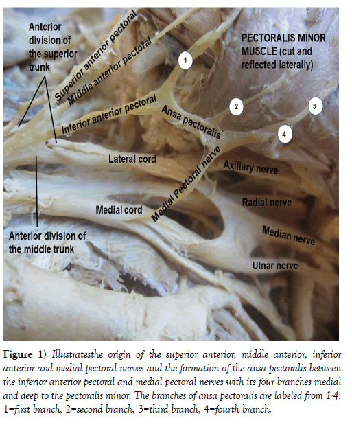
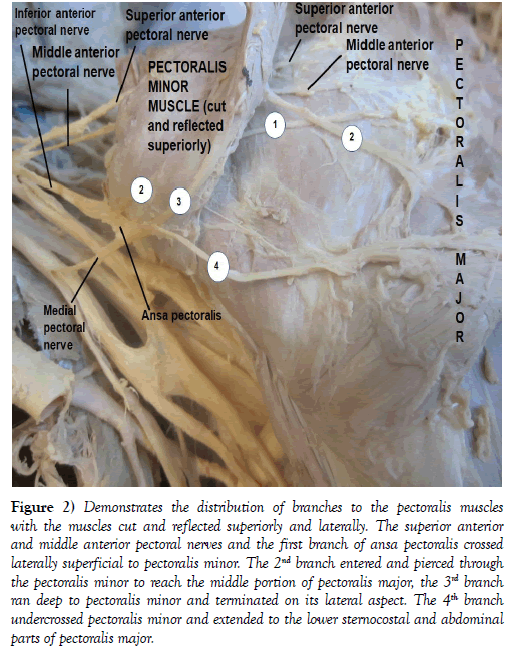
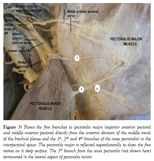
During dissection of the left axilla of an 80-year-old male cadaver, three anterior pectoral nerves (superior, middle and inferior) and a prominent ansa pectoralis formed by the union of the anterior inferior and medial pectoral nerves were incidentally noticed. These nerves were cleaned and followed farther from their origins to their termination on the pectoralis minor and pectoralis major muscles to demonstration their distribution. The nerves were designated based on their origin from the different parts of the brachial plexus for a descriptive purpose. The three anterior pectoral nerves were found to arise from the anterior division of the middle trunk of the brachial plexus and the medial pectoral nerve originated from the medial cord of the brachial plexus (Figures 1-3). According to their relationship to each other the three anterior pectoral nerves were named as superior, middle and inferior anterior pectoral nerves. The superior anterior pectoral nerve entered the clavicular part of pectoralis major and the middle terminated in the most superior fibers of the sternocostal part of pectoralis major closer to its clavicular part (Figures 1-3). The inferior anterior pectoral nerve joined the medial pectoral nerve to form a loop, the ansa pectoralis (Figures 1 and 2). Farther dissection revealed four branches of ansa pectoralis that variably distributed to the two pectoral muscles (Figures 1 and 3). The 1st branch from the ansa pectoralis crossed the medial border of pectoralis minor, ran superficial to it and entered the deep surface of superior part of the sternocostal portion of pectoralis major (Figures 1 and 2). The 2nd branch entered pectoralis minor and pierced through it to extend to the middle portion of the sternocostal part of pectoralis major (Figures 1 and 3). The 3rd branch also entered pectoralis minor along its lateral border but it terminated within the substance of the muscles (Figures 1 and 2). The 4th branch crossed laterally deep to pectoralis minor and entered the inferior portion of the sternocostal and abdominal parts of pectoralis major (Figures 1 and 2).
Figure 1: Illustratesthe origin of the superior anterior, middle anterior, inferior anterior and medial pectoral nerves and the formation of the ansa pectoralis between the inferior anterior pectoral and medial pectoral nerves with its four branches medial and deep to the pectoralis minor. The branches of ansa pectoralis are labeled from 1-4; 1=first branch, 2=second branch, 3=third branch, 4=fourth branch.
Figure 2: Demonstrates the distribution of branches to the pectoralis muscles with the muscles cut and reflected superiorly and laterally. The superior anterior and middle anterior pectoral nerves and the first branch of ansa pectoralis crossed laterally superficial to pectoralis minor. The 2nd branch entered and pierced through the pectoralis minor to reach the middle portion of pectoralis major, the 3rd branch ran deep to pectoralis minor and terminated on its lateral aspect. The 4th branch undercrossed pectoralis minor and extended to the lower sternocostal and abdominal parts of pectoralis major.
Figure 3: Shows the five branches to pectoralis major (superior anterior pectoral and middle anterior pectoral directly from the anterior division of the middle trunk of the brachial plexus and the 1st, 2nd and 4th branches of the ansa pectoralis) in the interpectoral space. The pectoralis major is reflected superolaterally to show the five nerves on it deep surface. The 3rd branch from the ansa pectoralis (not shown here) terminated in the lateral aspect of pectoralis minor.
Discussion
There are several documentations and reports that relate to the origin, number and distribution patterns of the pectoral nerves. A few of these that relate to this case report include three separate lateral pectoral nerves that arose from the lateral cord of the brachial plexus [6], three pectoral nerves that originated from the cords and division of the brachial plexus [7] and two lateral pectoral nerves that stemmed from the lateral cord of the brachial plexus [8].
The lateral pectoral nerve runs across the medial aspect of the pectoralis minor and then on the undersurface of pectoralis major along with the pectoral branch of thoracoacromial artery, innervating the proximal twothirds of pectoralis major [9]. This nerve can arise by two roots from the anterior divisions of the upper and middle trunks of the brachial plexus or by a single root from its lateral cord and has a constant course along the thoracoacromial vessels [10].
In 76% of 25 cases the medial pectoral nerve coursed through the pectoralis minor muscle as a single trunk and as divided branches in only 34% to innervate the lower portion of the pectoralis major [9]. Another study demonstrated that in 56% of the cases the medial pectoral nerve pierced the pectoralis minor as a single trunk to innervate pectoralis major, and in 44% it divided before entering pectoralis minor and its branches passed through the muscle or around its lateral border to reach pectoralis major [10]. There is also a report on a series of three pectoral nerves to the pectoralis major originated from the lateral cord of the brachial plexus. The report highlighted the usefulness of these three nerve in the pectoralis muscle flap transfer to the head and neck region [6]. However, lateral pectoral nerve arising from the lateral cord of the brachial plexus is not observe in the current case report, rather the pectoralis major is innervated by the anterior and middle anterior pectoral nerves from the anterior division of the middle trunk of the brachial plexus and three other branches from the ansa pectoralis.
An earlier study also demonstrated the presence of superior, middle and inferior pectoral nerves arising from the anterior divisions of the respective trunks of brachial plexuswith the formation of ansa pectoralis between the middle and inferior pectoral nerves [7]. Though, the designation of the nerveslooks similar to this case report, the origins, courses and distribution patterns of these nerves are completely different. The case in this report is very unique in that,all the three distinct anterior pectoral nerves (superior, middle and inferior) arose from the anterior division of the middle trunk of the brachial plexus while the medial pectoral nerve took its origin from medial cord of brachial plexus as classically described. The anterior inferior pectoral nerve joined the medial pectoral nerve to form the ansa pectoralis that farther divided into four branches. Hence, according to the finding in this case, the pectoralis major is innervated by five of these nerves (two from the anterior division of the middle trunk of the brachial plexus and three from the ansa pectoralis); while pectoralis minor is innervated by two nerves both of which are from the ansa pectoralis.
No case similar to this could be found by the extensive search of literature made during compiling this case report. Therefore, the finding in this case does not verify the classically described medial and lateral pectoral nerves to pectoralis muscles as branches of the medial and lateral cords of the brachial plexus and has no similarities to any of the documented and reported cases so far.
Conclusion
The observation in this case does not fit the classic two nerve description of nerves to the pectoralis muscles and any of the variations reported in literature. What is unique in this case is the absence of lateral pectoral nerve from the lateral cord of brachial plexus with three anterior pectoral nerves arising from the anterior division of the middle trunk of the brachial plexus and the formation of ansa pectoralis, which provided four branches that entered different portions of the two pectoralis muscles. The unusual origins, ramifications and distributions observed in this case are like a double-edged sword, which on one hand could make clinical approach to pectoral nerves difficult without a damage to them and on the other hand the innervation of pectoralis major by five distinct branches and pectoralis minor by two different branches could safeguard normal function of the muscles in isolated nerve injuries. Therefore, these nerves could possibly be used as potential donors for harvesting branches for neurotization procedures with a minimum or no effect on the muscles.
Acknowledgement
I would like to thank the donor and his families for their invaluable donation and consent for research and the department of biomedical sciences for its support. I am also grateful to Denelle Kees, Chelsey Swanson and John Opland for their uninterrupted assistance in the gross anatomy laboratory during the dissection of this cadaver.
REFERENCES
- Standring S. Gray’s Anatomy: The anatomical basis of clinical practice, 40th Ed, London, Elsevier Churchill Livingstone. 2008;821.
- Goncalves AV, Teixeira LC, Torresan R, et al. Randomized clinical trial on the preservation of medial pectoral nerve following mastectomy due to breast cancer: impact on upper limb rehabilitation. Sao Paulo Med J. 2009;127:117-21.
- Yu DW, Kim MS, Jung YJ, et al. Neurotization from two medial pectoral nerves to musculocutaneous nerve in a pediatric brachial plexus injury. J Korean Neurosurg Soc. 2012;52:267-269.
- Budak F, Ozkara B, Aydin E. Isolated lesion of lateral pectoral nerve due to repeated trauma. J Neurol Neurophysiol. 2017;8:1.
- Hassan AMA, Zanfaly HE, Biomy TA. Pre-emptive analgesia of ultrasound-guided pectoral nerve block II with dexmedetomidine-bupivacaine for control of chronic pain after modified mastectomy. Res Opin Anesth Intensive Care. 2016;3:6-13.
- Rai R, Ranade AV, Prabhu LV, et al. Accessory lateral pectoral nerve supplying the pectoralis major. Rom J Morphol Embryol. 2008; 49:577-79.
- David S, Balaguer T, Baque P, et al. The anatomy of pectoral nerves and its significance in breast augmentation, axillary dissection and pectoral flap. JPRAS. 2012; 65:1193-8.
- Nayak SB, Swamy RS, Kumar N, et al. Concurrent variation of lateral pectoral, median, and musculocutaneous nerves. J Health Allied Scs. 2016;15:11.
- Prakash KG, Saniya K. Anatomical study of pectoral nerves and its implication in surgery. JCDR. 2014; 8: AC01-AC05.
- Macchi V, Tiengo C, Porzionato A, et al. Medial and lateral pectoral nerves: course and branches. Clini Anat. 2007;20:157-162.


