Accessory belly of the first lumbrical – a case report
Mallikarjun Adibatti* and Pramod Rangsubhe
Department of Anatomy, J. J. M. Medical College, Davangere, Karnataka, India.
- *Corresponding Author:
- Mallikarjun Adibatti
Assistant Professor, Department of Anatomy, J. J. M. Medical College, Davangere-570004, Karnataka, India.
Tel: +91 944 9984305
E-mail: na_mallikarjun@rediffmail.com
Date of Received: June 24th, 2010
Date of Accepted: December 22nd, 2010
Published Online: January 27th, 2011
© Int J Anat Var (IJAV). 2011; 4: 20–21.
[ft_below_content] =>Keywords
lumbricals, intrinsic muscles, dissection, cadaver, additional slip
Introduction
Much of the versatility of the human hand depends upon its intrinsic musculature. The lumbrical muscles constitute an important part of the intrinsic musculature of the hands. Lumbricals are the four small intrinsic muscles of the hand. They arise from the four tendons of flexor digitorum profundus (FDP) in the hand and passing on the radial side of the corresponding metacarpophalangeal joint, are inserted into the dorsal digital expansion of the medial four fingers.
The first and second lumbrical muscles originate from the radial sides and palmar surfaces of the FDP of the index and middle finger, the third lumbrical from the adjacent sides of the FDP tendons of the middle and ring finger and the fourth lumbrical from the adjacent sides of the tendons of the ring and little finger. As a part of the intrinsic musculature, the lumbricals are important for delicate digital movements. They are said to flex the metacarpophalangeal joints and extend the interphalangeal joints.
These are quite unique in their position as they connect the flexors of the digits to the extensors and that both of its attachments are mobile. These play a vital role in the precision movements of the hands, along with thenar, hypothenar, and interossei muscles.
Case Report
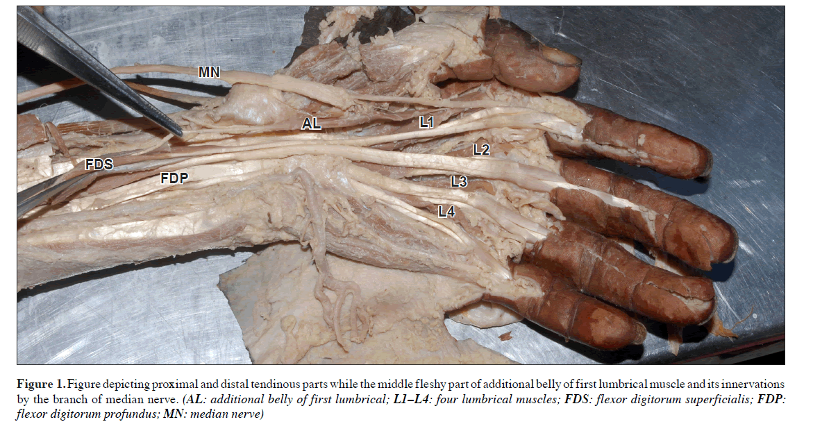
Additional slip of the origin of the first lumbrical muscle was observed in the left hand of a 60-year-old male cadaver. The first origin of the first lumbrical muscle arose from the radial side of the FDP for the index finger, while the additional slip took origin in the forearm from the radial side of the tendon of flexor digitorum superficialis (FDS) to the index finger about 32 mm proximal to the flexor retinaculum (FR). Later it joined the first lumbrical, before the latter got inserted to the dorsal digital expansion on the radial side of the index finger. This additional slip was fleshy in the middle, while it was tendinous at both the ends.
The total length of the muscle was 106 mm, out of which 70 mm was fleshy part and 36 mm was tendinous part. Proximal tendinous portion was 32 mm proximal to the FR. Distal tendinous portion was 4 mm distal to FR. Maximum width of the fleshy part was 8 mm.
The additional slip of origin was innervated by a twig from the lateral branch of the median nerve. However second, third, fourth lumbricals were as usual with respect to their origin, insertion and innervations.
Discussion
Lumbricals as a part of the intrinsic musculature are important for its delicate digital movements. Variations in the origin and insertion of the lumbricals are common [1]. In the present study we observed an additional slip of the origin of the first lumbrical originating in the forearm from the radial side of the tendon of FDS to index finger which merged with the first lumbrical for insertion into the radial side of the index finger into the dorsal digital expansion.
Figure 1: Figure depicting proximal and distal tendinous parts while the middle fleshy part of additional belly of first lumbrical muscle and its innervations by the branch of median nerve. (AL: additional belly of first lumbrical; L1–L4: four lumbrical muscles; FDS: flexor digitorum superficialis; FDP: flexor digitorum profundus; MN: median nerve)
Additional lumbricals occurring more frequently than a reduction in their number. Origin of lumbricals may be displaced proximally arising from FR, FDS, FDP or flexor pollicis longus. Accessory belly of first lumbrical may arise from flexor pollicis longus, FDS, first metacarpal, opponens pollicis or palmar carpal ligament [2].
The additional fibers from the forearm origin merged at varying point with the belly coming from the palmar origin and in no case reached the insertion of extensor expansion independently. Hence these are termed as additional forearm origin and not as double lumbricals [3]. The first lumbrical had an additional palmar origin from the tendon of FDS for the index finger [2].
FDS in mammals is homologous with the intrinsic musculature of the palm, and that it shifts its origin proximally in the forearm [4].
First lumbrical and the distal muscle belly for the index finger of the FDS have an intimate relationship with each other and have a common phylogenetic origin [5].
An additional slip of origin of the lumbrical from the FDS in the forearm, such as that described in the present study, has the potential to cause compression of the median nerve in the carpal tunnel, resulting in carpal tunnel syndrome which may be due to the incursion of the lumbrical muscles within the carpal during the finger movements or by hypertrophy of the lumbricals. Hence the clinician must be aware constantly of such possibilities, although preoperative diagnosis may be difficult.
References
- Bergman RA, Thompson SA, Afifi AK, Saadeh FA. Compendium of Human Anatomic Variation. Munich, Urban and Schwarzenberg. 1988; 13–14, 17.
- Ajmani ML. Morphological variations of lumbrical muscles in the human hand with some observations on its nerve supply. Med J Iran Hosp. 2001; 3: 20–25.
- Mehta HJ, Gardner WU. A study of lumbrical muscles in the human hand. Am J Anat. 1961; 109: 227–238.
- Haines RW. The flexor muscles of the forearm and hand in lizards and mammals. J Anat. 1950; 84: 13–29.
- Koizumi M, Kawai K, Honma S, Kodma K. Anomalous lumbrical muscles arising from the deep surface of flexor digitorum superficialis muscles in man. Ann Anat. 2002; 184: 387–392.
- Singh G, Bay BH, Yip GW, Tay S. Lumbrical muscle with an additional origin in the forearm. ANZ J Surg. 2001; 71: 301–302.
- Nayak SR, Ramanathan L, Prabhu LV, Raju S. Additional flexor muscles of the forearm: case report and clinical significance. Singapore Med J. 2007; 48: e231–233.
- Joshi SD, Joshi SS, Athavale SA. Lumbrical muscles and carpal tunnel. J Anat Soc India. 2005; 54: 12–15.
- Barbe M, Bradfield J, Donathan M, Elmaleh J. Coexistence of multiple anomalies in the carpal tunnel. Clin Anat. 2005; 180 251–259.
- Butler B, Bigley EC. Aberrant index (first) lumbrical tendinous origin associated with carpal-tunnel syndrome. A case report. J Bone Joint Surg Am. 1971; 53: 160–162.
Mallikarjun Adibatti* and Pramod Rangsubhe
Department of Anatomy, J. J. M. Medical College, Davangere, Karnataka, India.
- *Corresponding Author:
- Mallikarjun Adibatti
Assistant Professor, Department of Anatomy, J. J. M. Medical College, Davangere-570004, Karnataka, India.
Tel: +91 944 9984305
E-mail: na_mallikarjun@rediffmail.com
Date of Received: June 24th, 2010
Date of Accepted: December 22nd, 2010
Published Online: January 27th, 2011
© Int J Anat Var (IJAV). 2011; 4: 20–21.
Abstract
Out of the 20 cadavers allotted for first MBBS students dissection we focused our study on the intrinsic muscles of the hand. All the intrinsic muscles in the hands of the female cadavers were as usual. However, we observed an additional slip of origin of the first lumbrical in one hand out of the 36 hands from male cadavers examined.
-Keywords
lumbricals, intrinsic muscles, dissection, cadaver, additional slip
Introduction
Much of the versatility of the human hand depends upon its intrinsic musculature. The lumbrical muscles constitute an important part of the intrinsic musculature of the hands. Lumbricals are the four small intrinsic muscles of the hand. They arise from the four tendons of flexor digitorum profundus (FDP) in the hand and passing on the radial side of the corresponding metacarpophalangeal joint, are inserted into the dorsal digital expansion of the medial four fingers.
The first and second lumbrical muscles originate from the radial sides and palmar surfaces of the FDP of the index and middle finger, the third lumbrical from the adjacent sides of the FDP tendons of the middle and ring finger and the fourth lumbrical from the adjacent sides of the tendons of the ring and little finger. As a part of the intrinsic musculature, the lumbricals are important for delicate digital movements. They are said to flex the metacarpophalangeal joints and extend the interphalangeal joints.
These are quite unique in their position as they connect the flexors of the digits to the extensors and that both of its attachments are mobile. These play a vital role in the precision movements of the hands, along with thenar, hypothenar, and interossei muscles.
Case Report
Additional slip of the origin of the first lumbrical muscle was observed in the left hand of a 60-year-old male cadaver. The first origin of the first lumbrical muscle arose from the radial side of the FDP for the index finger, while the additional slip took origin in the forearm from the radial side of the tendon of flexor digitorum superficialis (FDS) to the index finger about 32 mm proximal to the flexor retinaculum (FR). Later it joined the first lumbrical, before the latter got inserted to the dorsal digital expansion on the radial side of the index finger. This additional slip was fleshy in the middle, while it was tendinous at both the ends.
The total length of the muscle was 106 mm, out of which 70 mm was fleshy part and 36 mm was tendinous part. Proximal tendinous portion was 32 mm proximal to the FR. Distal tendinous portion was 4 mm distal to FR. Maximum width of the fleshy part was 8 mm.
The additional slip of origin was innervated by a twig from the lateral branch of the median nerve. However second, third, fourth lumbricals were as usual with respect to their origin, insertion and innervations.
Discussion
Lumbricals as a part of the intrinsic musculature are important for its delicate digital movements. Variations in the origin and insertion of the lumbricals are common [1]. In the present study we observed an additional slip of the origin of the first lumbrical originating in the forearm from the radial side of the tendon of FDS to index finger which merged with the first lumbrical for insertion into the radial side of the index finger into the dorsal digital expansion.
Figure 1: Figure depicting proximal and distal tendinous parts while the middle fleshy part of additional belly of first lumbrical muscle and its innervations by the branch of median nerve. (AL: additional belly of first lumbrical; L1–L4: four lumbrical muscles; FDS: flexor digitorum superficialis; FDP: flexor digitorum profundus; MN: median nerve)
Additional lumbricals occurring more frequently than a reduction in their number. Origin of lumbricals may be displaced proximally arising from FR, FDS, FDP or flexor pollicis longus. Accessory belly of first lumbrical may arise from flexor pollicis longus, FDS, first metacarpal, opponens pollicis or palmar carpal ligament [2].
The additional fibers from the forearm origin merged at varying point with the belly coming from the palmar origin and in no case reached the insertion of extensor expansion independently. Hence these are termed as additional forearm origin and not as double lumbricals [3]. The first lumbrical had an additional palmar origin from the tendon of FDS for the index finger [2].
FDS in mammals is homologous with the intrinsic musculature of the palm, and that it shifts its origin proximally in the forearm [4].
First lumbrical and the distal muscle belly for the index finger of the FDS have an intimate relationship with each other and have a common phylogenetic origin [5].
An additional slip of origin of the lumbrical from the FDS in the forearm, such as that described in the present study, has the potential to cause compression of the median nerve in the carpal tunnel, resulting in carpal tunnel syndrome which may be due to the incursion of the lumbrical muscles within the carpal during the finger movements or by hypertrophy of the lumbricals. Hence the clinician must be aware constantly of such possibilities, although preoperative diagnosis may be difficult.
References
- Bergman RA, Thompson SA, Afifi AK, Saadeh FA. Compendium of Human Anatomic Variation. Munich, Urban and Schwarzenberg. 1988; 13–14, 17.
- Ajmani ML. Morphological variations of lumbrical muscles in the human hand with some observations on its nerve supply. Med J Iran Hosp. 2001; 3: 20–25.
- Mehta HJ, Gardner WU. A study of lumbrical muscles in the human hand. Am J Anat. 1961; 109: 227–238.
- Haines RW. The flexor muscles of the forearm and hand in lizards and mammals. J Anat. 1950; 84: 13–29.
- Koizumi M, Kawai K, Honma S, Kodma K. Anomalous lumbrical muscles arising from the deep surface of flexor digitorum superficialis muscles in man. Ann Anat. 2002; 184: 387–392.
- Singh G, Bay BH, Yip GW, Tay S. Lumbrical muscle with an additional origin in the forearm. ANZ J Surg. 2001; 71: 301–302.
- Nayak SR, Ramanathan L, Prabhu LV, Raju S. Additional flexor muscles of the forearm: case report and clinical significance. Singapore Med J. 2007; 48: e231–233.
- Joshi SD, Joshi SS, Athavale SA. Lumbrical muscles and carpal tunnel. J Anat Soc India. 2005; 54: 12–15.
- Barbe M, Bradfield J, Donathan M, Elmaleh J. Coexistence of multiple anomalies in the carpal tunnel. Clin Anat. 2005; 180 251–259.
- Butler B, Bigley EC. Aberrant index (first) lumbrical tendinous origin associated with carpal-tunnel syndrome. A case report. J Bone Joint Surg Am. 1971; 53: 160–162.


