Accessory muscle of the deep posterior leg: A novel case presentation
Received: 20-Sep-2017 Accepted Date: Oct 31, 2017; Published: 06-Nov-2017
Citation: Young M, Reeves R, Rosales R. Accessory muscle of the deep posterior leg: A novel case presentation. Int J Anat Var. 2017;10(4): 94-95.
This open-access article is distributed under the terms of the Creative Commons Attribution Non-Commercial License (CC BY-NC) (http://creativecommons.org/licenses/by-nc/4.0/), which permits reuse, distribution and reproduction of the article, provided that the original work is properly cited and the reuse is restricted to noncommercial purposes. For commercial reuse, contact reprints@pulsus.com
Abstract
During a routine dissection in the anatomy laboratory at The University of North Texas Health Science Center, the left leg of an 84-year-old female cadaver was found to have an additional muscle belly and tendon slip originating from the deep muscles of the posterior compartment. The small accessory muscle had two separate heads (medial and lateral) that arose in the lower third of the leg from the posterior deep fascia covering the flexor digitorum longus, tibialis posterior, and flexor hallucis longus muscles.
The accessory muscles tendon bifurcated near its terminus, and then inserted on the fascia between the deep surface of the abductor hallucis and the quadratus plantae muscles. The surrounding muscular, vascular and nervous structures followed the typical course described in standard anatomical texts.
Keywords
Variant; Additional belly; Posterior compartment; Conjoined belly
Introduction
The deep muscles of the posterior compartment have varied actions depending on their insertion points, but most share the common action of plantar flexion. The popliteus has been excluded from this discussion due to its non-involvement in this particular anatomical variation. In particular, this study is looking at the muscle group that passes through the tarsal tunnel. Those muscles include the tibialis posterior, flexor digitorum longus and flexor hallucis longus, which all share a common tendon path posterior to the medial malleolus and deep to the flexor retinaculum of the ankle before diverging to their various insertion points on the plantar surface of the foot.
Previous anatomical variations in the deep compartment include flexor digitorum accessorius longus (FDAL) [1-8] and peroneocalcaneus internus (PCI) [9]. Other variations in this area are typically outside the deep compartment and include accessorius soleus and multiple variations of the peroneal accessory muscles [10]. This report is unique since it is the first to identify an accessory muscle that inserts into the abductor hallucis muscle.
Case Report
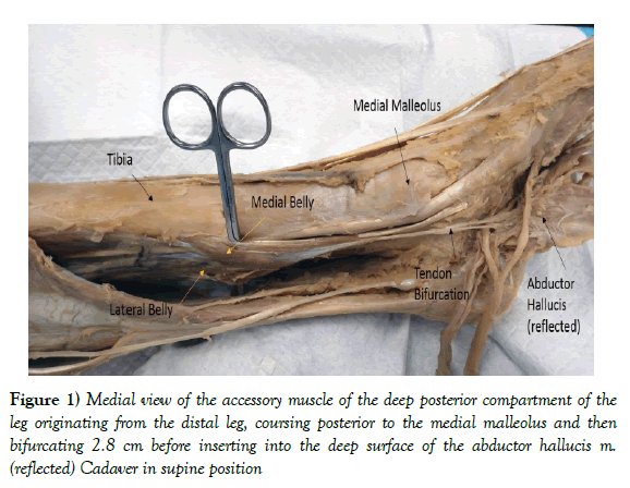
During routine dissection of an 84-year-old female cadaver in our medical human anatomy course, a small accessory muscle with two separate, conjoined heads (muscle bellies) were identified within the deep compartment of the left posterior leg. The muscle lay superficial to the deep leg muscles, originated from two separate muscle bellies (medial and lateral head), passed as a single tendon through the tarsal tunnel then emerged from the inferior aspect of the flexor retinaculum to bifurcate and insert into the abductor hallucis muscle on the medial plantar surface of the foot (Figure 1). Whereas many aberrant muscles found in routine dissections are named, this muscle poses a unique problem in naming since it originates from the deep posterior leg muscles (flexors), but inserts into an abductor of the great toe. For that reason, we will not attempt to name this unique accessory muscle at this time.
Figure 1) Medial view of the accessory muscle of the deep posterior compartment of the leg originating from the distal leg, coursing posterior to the medial malleolus and then bifurcating 2.8 cm before inserting into the deep surface of the abductor hallucis m. (reflected) Cadaver in supine position
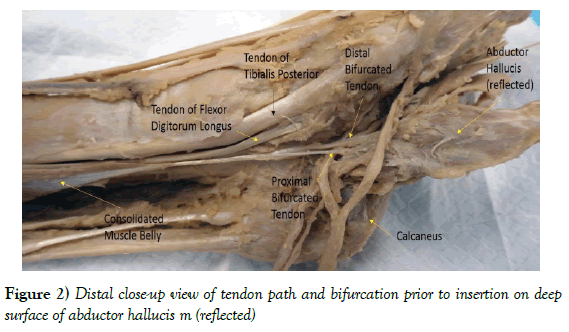
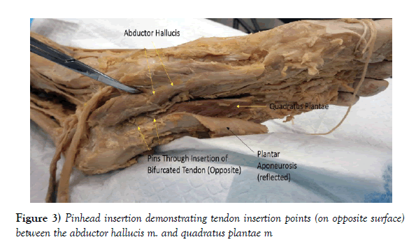
The two separate heads of the muscle were well-formed, with the origin of the lateral head attached to the deep fascia of the flexor hallucis muscle. The origin of the medial head was attached to the tibialis posterior and the flexor digitorum longus muscles, as well as a minor attachment to the medial side of the posterior tibia. The full width of muscle (both heads) at its origin was 4.0 cm. The tendon followed the course of the primary plantar flexors of the deep compartment, through the tarsal tunnel, traveling posterior to the medial malleolus before bifurcating distal to the flexor retinacula (Figure 2). Both parts of the bifurcated tendon inserted onto the deep surface of the abductor hallucis muscle near its origin at the calcaneal tubercle (Figure 3). The total length of the variant muscle was 17.2 cm.
Discussion
The compartments of the lower limb are known for having multiple interesting and pertinent anatomical variations. Most of these variations reported to date were named as some type of accessory flexor muscle based on the tendinous insertion into one of the leg or foot flexor muscles [1- 8]. From the literature, several accessory flexor muscles of the posterior leg have been observed having two heads [2,5]. From our review of the literature, the non-osseous insertion into the abductor hallucis muscle appears to be a novel variation. Novel both in the reported variant’s insertion into an abductor muscle, as well the bifurcation of the distal tendon at that insertion site. There was no apparent obstruction or particular anatomical reason that would necessitate a bifurcation of the distal tendon. The post-mortem discovery of the variant muscle makes it impossible to tell what physiological purpose it may have served. This muscle could serve to supply minor stabilization to the medial arch of the foot. However, a variant muscle that passes through the tarsal tunnel could potentially have clinical significance if it entrapped the tibial nerve distal to the ankle as recently described in another case by Georgeiv et al. [11,12]. Furthermore, the muscle still maintains educational relevance as it adds to the pool of knowledge of possible anatomical variations for clinicians and anatomists alike.
Acknowledgement
This report was made possible through the selfless gift of our donors to the Willed Body Program, Center for Anatomical Sciences at the University of North Texas Health Science Center, Fort Worth, TX.
REFERENCES
- Georgiev GP, Jelev L, Kinov P, et al. A rare instance of an accessory long flexor to the second toe. Int J Anat Var. 2009;2:108-10.
- Hwang SH, Hill RV. An unusual variation of the flexor digitorum accessories longus muscle-its anatomy and clinical significance. Anat Sci Int. 2008;84:257-63.
- Jaijesh P, Shenoy M, Anuradha L, et al. Flexor accessories longus: A rare variation of the deep extrinsic digital flexors of the leg and its phylogenic significance. Indan J Plast Surg. 2006;39:169-71.
- Deroy AR, Clause CC, Baskin ES, et al. Recognition of the flexor digitorum accessories longus. J Am Podiatr Med Assoc. 2002;92:463-6.
- Gumusalan Y, Kalaycioglu A. Bilateral accessory flexor digitorium longus muscle in man. Ann Anat. 2000;182: 573-6.
- Peterson DA, Stonson W, Larmore JR. The long accessory flexor muscle: an anatomical study. Foot Ankle Int. 1995; 16:637-640.
- Nathan H, Gloobe H, Yosipovitch Z. Flexor digitorium accessories longus. Clin Orthop Relat. 1975;113:158-61.
- Grogono BJ, Jowsey J. Flexor accessories longus: an unusual muscle anomaly. J Bone Joint Surg Br. 1965:47: 118-9.
- Seipel R, Linklater J, Pitsis G, et al. The peroneocalcaneus internus muscle: an unusual cause of posterior ankle impingement. Foot Ankle Int. 2005;26: 890-3.
- Carroll JF. Accessory Muscles of the Ankle.
- Georgiev GP. Significance of anatomical variations for clinical practice. Int J Anat Var. 2017;10:43-4.
- Georgiev GP, Landshov B, Slavchev S, et al. Tarsal tunnel syndrome caused by anomalous muscle: case report. Scripta Scienficia Medica. 2013;45:109-10.


