An axillary arch muscle – a case report
Mahendra A.Kathole*,Rajani A. Joshi and Narsimh G.Herekar
Department of Anatomy, Government Medical College, Miraj, Tal-Miraj, Dist- Sangali, Maharashtra, India.
- *Corresponding Author:
- Dr. Mahendra A. Kathole
Quarter No. 9, Building No. 1, Residential Campus Pandharpur Road, Government Medical College, Miraj Tal-Miraj, Dist-Sangali, Maharashtra, 416410, India.
Tel: +91 9960079973
E-mail: mahendra.kathole@rediffmail.com
Date of Received: September 9th, 2011
Date of Accepted: April 9th, 2012
Published Online: August 30th, 2012
© Int J Anat Var (IJAV). 2012; 5: 32–34.
[ft_below_content] =>Keywords
latissimus dorsi muscle,coracobrachialis muscle,axillary arch muscle,axillary artery
Introduction
A number of accessory muscle slip cases in the axilla arising from latissimus dorsi, pectoralis major, ribs and costal cartilages have been reported by different authors [1]. These accessory muscle slips in the axilla have been described under variety of names (e.g., chondro-epitrochlearis, dorso-epitrochlearis, etc). These variant bundles are commonly referred to as “axillary arch” muscle, regardless of their site of origin [2]. In the present case, an axillary arch muscle was found which was arising from the latissimus dorsi and was inserting into the coracobrachialis muscle.
The incidence of axillary arch muscle reported in different population groups as 7% in Japanese, 10% in Belgian, 0.25% in British Population [3] . Axillary arch muscles are common in apes.
Presence of an axillary arch muscle in humans has an immense clinical and morphological importance.
Case Report
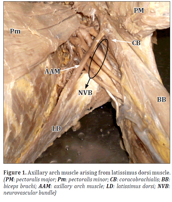
During routine dissection of the left upper limb of an 80-year-old female cadaver, we came across an additional muscular slip in the axilla. This muscular slip was arising from anterior border of latissimus dorsi, proximal to its tendon of insertion. It crossed the neurovascular bundle of axilla and was inserted into upper part of coracobrachialis, near the origin of later. It was measured 7 cm in length and 8 mm in breadth (Figure 1). It was supplied by the nerve to the latissimus dorsi.
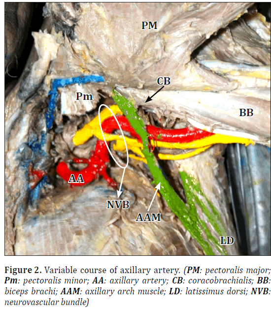
The axillary artery proximal to the arch was variably dilated. It showed an S-shaped course when the limb was placed by the side of the body, indicating variable course [4] (Figure 2).
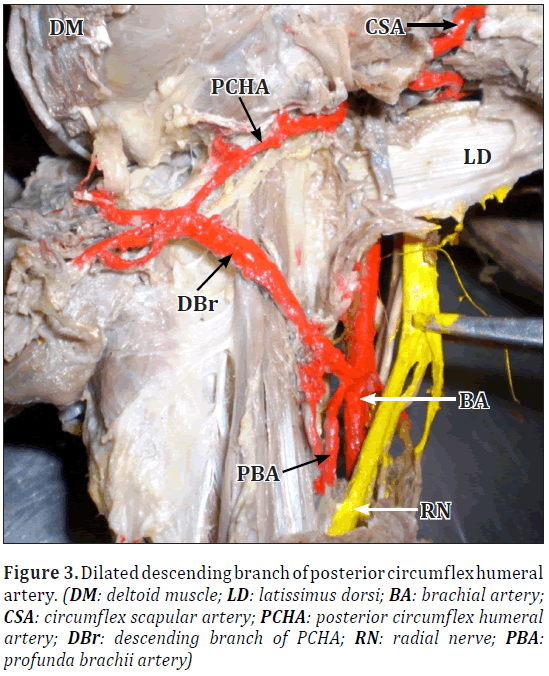
The descending branch of posterior circumflex humeral artery was variably dilated as compared to the main trunk of posterior circumflex humeral artery (Figure 3).
On the right side, latissimus dorsi was as usual and the course of axillary artery showed no variation.
Discussion
The considerable variations are found in the extent of latissimus dorsi and its attachments [5]. The presence of an axillary arch muscle, the muscular slips arising from the latissimus dorsi or pectoralis major and inserting into various sites, is reported by different authors. The different sites of insertion are flexor muscles of arm [2], coracobrachialis, biceps brachii or long head of triceps brachii, teres major, the coracoid process of scapula and the medial epicondyle of the humerus as the ‘chondroepitrochlearis’ muscle, etc. [1]. These muscular slips are commonly referred to as an “axillary arch” muscle, regardless of their site of origin [2].
The additional muscular slip found in the present case, was an axillary arch muscle (Figure 1). Its nerve supply was from nerve to latissimus dorsi (thoracodorsal nerve), thus confirming its embryological origin from latissimus dorsi muscle.
Compression of neurovascular bundle of axilla by axillary arch muscle is documented in literature. In present case, the variable dilatation of an axillary artery and its variable S-shaped course proximal to the arch, indicated severe compression by an axillary arch muscle (Figure 2).
The posterior circumflex humeral artery, a branch of axillary artery, usually gives off a descending branch which anastomoses with deltoid branch of profunda brachii artery and with the anterior circumflex humeral and acromial branches of suprascapular and thoraco-acromial arteries.
In the present case, the descending branch of posterior circumflex humeral artery was variably dilated as compared to its main trunk (Figure 3). The compression of axillary artery by arch muscle, might be affecting the blood supply to the limb, distal to the arch. This might be responsible for dilatation of descending branch of posterior circumflex humeral artery and formation of the collateral channel between axillary artery proximal to the arch muscle and branches of brachial artery distal to it.
Presence of an axillary arch muscle has immense clinical and morphological significance. The muscular arch when present causes difficulty in staging lymph nodes in malignancy cases, in axillary surgeries, shoulder instability, in cosmetic problems [6]. Accessory muscle present in axilla may be a reason of an axillary mass and can exert pressure on the neighboring neurovascular bundle or lymph nodes, thus expressing wide range of symptoms [7].
Also, the presence of an accessory muscle should be kept in mind while constructing latissimus dorsi flaps and while draining the axillary abscesses.
Embryological Origin
The limb muscles arise in situ from mesenchyme around the developing bones. This mesenchyme is derived from the somatic layer of lateral plate mesoderm. The development of skeletal muscle is divided into four separate stages [8] as follows:
First stage –the premyoblast stage, second stage –the myoblast stage, third stage –the myotube stage, fourth stage –the muscle fiber.
The myotubes are converted into muscle fibers, during the fourth stage. The muscular variation described in the present case may have developed during stages three and four of ontogenesis of muscle in the left axilla. During phase three, some muscle primordia from different layers get fused to form a single muscle. Persistence of some cells between latissimus dorsi and coracobrachialis might have formed the muscle slip, which was observed in present case.
An axillary arch muscle commonly receives its nerve supply from the medial pectoral nerve, indicating its embryological origin from the pectoral muscle mass [9]; but those closely connected to latissimus dorsi may be supplied by its nerve, the thoracodorsal nerve [2]. In the present case, muscular slip was supplied by thoracodorsal nerve, indicating its embryological origin from latissimus dorsi muscle.
References
- Dharap A. An unusually medial axillary arch muscle. J Anat. 1994; 184: 639–641.
- Hollinshead WH. Anatomy for Surgeons. Volume 3. Heber Harper, New York. 1958; 284–300.
- Pai MM, Rajanigandha, Prabhu LV, Shetty P, Narayana K. Axillary arch (of Langer): incidence, innervation, importance. Online J Health Allied Scs. 2006; 5: 1–4.
- Williams PL, ed. Gray’s Anatomy. 39th Ed., Churchill Livingstone, New York. 2005; 837–42.
- Flaherty G, O’Neill MN, Folan-Curran J. Case report: bilateral occurrence of a chondroepitrochlearis muscle. J Anat. 1999; 194: 313–315.
- Ucerler H, Ikiz ZA, Pinar Y. Clinical importance of the muscular arch of the axilla (axillopectoral muscle, Langer’s axillary arch). Acta Chir Belg. 2005; 105: 326–328.
- Turgut HB, Peker T, Gulekon N, Anil A, Karakose M. Axillopectoral muscle (Langer’s muscle). Clin Anat. 2005; 18: 220–223.
- Hamilton WJ, Mossman HW. Hamilton, Boyd and Mossman’s Human Embryology. 4th Ed., The Macmillan Press Ltd., London and Basingstoke. 1976; 557–559.
- Soubhagya RN, Latha VP, Ashwin K, Madhan KSJ, Ganesh CK. Coexistence of an axillary arch muscle (latissimocondyloideus muscle) with an unusual axillary artery branching: case report and review. Int J Morphol. 2006; 24: 147–150.
Mahendra A.Kathole*,Rajani A. Joshi and Narsimh G.Herekar
Department of Anatomy, Government Medical College, Miraj, Tal-Miraj, Dist- Sangali, Maharashtra, India.
- *Corresponding Author:
- Dr. Mahendra A. Kathole
Quarter No. 9, Building No. 1, Residential Campus Pandharpur Road, Government Medical College, Miraj Tal-Miraj, Dist-Sangali, Maharashtra, 416410, India.
Tel: +91 9960079973
E-mail: mahendra.kathole@rediffmail.com
Date of Received: September 9th, 2011
Date of Accepted: April 9th, 2012
Published Online: August 30th, 2012
© Int J Anat Var (IJAV). 2012; 5: 32–34.
Abstract
The numbers of accessory muscle slips in the axilla have been reported by different authors. During routine dissection for undergraduate medical students at Department of Anatomy, in an 80-year-old female cadaver, we came across a variant muscular slip, which arose from anterior aspect of latissimus dorsi muscle of left side and was inserted in the proximal part of coracobrachialis muscle. This variant slip of latissimus dorsi muscle is an “axillary arch muscle”. The axillary artery proximal to the arch muscle showed variable course. The compression of neurovascular bundle of axilla by an axillary arch muscle is documented in literature. Presence of an axillary arch muscle has immense clinical and morphological significance.
-Keywords
latissimus dorsi muscle,coracobrachialis muscle,axillary arch muscle,axillary artery
Introduction
A number of accessory muscle slip cases in the axilla arising from latissimus dorsi, pectoralis major, ribs and costal cartilages have been reported by different authors [1]. These accessory muscle slips in the axilla have been described under variety of names (e.g., chondro-epitrochlearis, dorso-epitrochlearis, etc). These variant bundles are commonly referred to as “axillary arch” muscle, regardless of their site of origin [2]. In the present case, an axillary arch muscle was found which was arising from the latissimus dorsi and was inserting into the coracobrachialis muscle.
The incidence of axillary arch muscle reported in different population groups as 7% in Japanese, 10% in Belgian, 0.25% in British Population [3] . Axillary arch muscles are common in apes.
Presence of an axillary arch muscle in humans has an immense clinical and morphological importance.
Case Report
During routine dissection of the left upper limb of an 80-year-old female cadaver, we came across an additional muscular slip in the axilla. This muscular slip was arising from anterior border of latissimus dorsi, proximal to its tendon of insertion. It crossed the neurovascular bundle of axilla and was inserted into upper part of coracobrachialis, near the origin of later. It was measured 7 cm in length and 8 mm in breadth (Figure 1). It was supplied by the nerve to the latissimus dorsi.
The axillary artery proximal to the arch was variably dilated. It showed an S-shaped course when the limb was placed by the side of the body, indicating variable course [4] (Figure 2).
The descending branch of posterior circumflex humeral artery was variably dilated as compared to the main trunk of posterior circumflex humeral artery (Figure 3).
On the right side, latissimus dorsi was as usual and the course of axillary artery showed no variation.
Discussion
The considerable variations are found in the extent of latissimus dorsi and its attachments [5]. The presence of an axillary arch muscle, the muscular slips arising from the latissimus dorsi or pectoralis major and inserting into various sites, is reported by different authors. The different sites of insertion are flexor muscles of arm [2], coracobrachialis, biceps brachii or long head of triceps brachii, teres major, the coracoid process of scapula and the medial epicondyle of the humerus as the ‘chondroepitrochlearis’ muscle, etc. [1]. These muscular slips are commonly referred to as an “axillary arch” muscle, regardless of their site of origin [2].
The additional muscular slip found in the present case, was an axillary arch muscle (Figure 1). Its nerve supply was from nerve to latissimus dorsi (thoracodorsal nerve), thus confirming its embryological origin from latissimus dorsi muscle.
Compression of neurovascular bundle of axilla by axillary arch muscle is documented in literature. In present case, the variable dilatation of an axillary artery and its variable S-shaped course proximal to the arch, indicated severe compression by an axillary arch muscle (Figure 2).
The posterior circumflex humeral artery, a branch of axillary artery, usually gives off a descending branch which anastomoses with deltoid branch of profunda brachii artery and with the anterior circumflex humeral and acromial branches of suprascapular and thoraco-acromial arteries.
In the present case, the descending branch of posterior circumflex humeral artery was variably dilated as compared to its main trunk (Figure 3). The compression of axillary artery by arch muscle, might be affecting the blood supply to the limb, distal to the arch. This might be responsible for dilatation of descending branch of posterior circumflex humeral artery and formation of the collateral channel between axillary artery proximal to the arch muscle and branches of brachial artery distal to it.
Presence of an axillary arch muscle has immense clinical and morphological significance. The muscular arch when present causes difficulty in staging lymph nodes in malignancy cases, in axillary surgeries, shoulder instability, in cosmetic problems [6]. Accessory muscle present in axilla may be a reason of an axillary mass and can exert pressure on the neighboring neurovascular bundle or lymph nodes, thus expressing wide range of symptoms [7].
Also, the presence of an accessory muscle should be kept in mind while constructing latissimus dorsi flaps and while draining the axillary abscesses.
Embryological Origin
The limb muscles arise in situ from mesenchyme around the developing bones. This mesenchyme is derived from the somatic layer of lateral plate mesoderm. The development of skeletal muscle is divided into four separate stages [8] as follows:
First stage –the premyoblast stage, second stage –the myoblast stage, third stage –the myotube stage, fourth stage –the muscle fiber.
The myotubes are converted into muscle fibers, during the fourth stage. The muscular variation described in the present case may have developed during stages three and four of ontogenesis of muscle in the left axilla. During phase three, some muscle primordia from different layers get fused to form a single muscle. Persistence of some cells between latissimus dorsi and coracobrachialis might have formed the muscle slip, which was observed in present case.
An axillary arch muscle commonly receives its nerve supply from the medial pectoral nerve, indicating its embryological origin from the pectoral muscle mass [9]; but those closely connected to latissimus dorsi may be supplied by its nerve, the thoracodorsal nerve [2]. In the present case, muscular slip was supplied by thoracodorsal nerve, indicating its embryological origin from latissimus dorsi muscle.
References
- Dharap A. An unusually medial axillary arch muscle. J Anat. 1994; 184: 639–641.
- Hollinshead WH. Anatomy for Surgeons. Volume 3. Heber Harper, New York. 1958; 284–300.
- Pai MM, Rajanigandha, Prabhu LV, Shetty P, Narayana K. Axillary arch (of Langer): incidence, innervation, importance. Online J Health Allied Scs. 2006; 5: 1–4.
- Williams PL, ed. Gray’s Anatomy. 39th Ed., Churchill Livingstone, New York. 2005; 837–42.
- Flaherty G, O’Neill MN, Folan-Curran J. Case report: bilateral occurrence of a chondroepitrochlearis muscle. J Anat. 1999; 194: 313–315.
- Ucerler H, Ikiz ZA, Pinar Y. Clinical importance of the muscular arch of the axilla (axillopectoral muscle, Langer’s axillary arch). Acta Chir Belg. 2005; 105: 326–328.
- Turgut HB, Peker T, Gulekon N, Anil A, Karakose M. Axillopectoral muscle (Langer’s muscle). Clin Anat. 2005; 18: 220–223.
- Hamilton WJ, Mossman HW. Hamilton, Boyd and Mossman’s Human Embryology. 4th Ed., The Macmillan Press Ltd., London and Basingstoke. 1976; 557–559.
- Soubhagya RN, Latha VP, Ashwin K, Madhan KSJ, Ganesh CK. Coexistence of an axillary arch muscle (latissimocondyloideus muscle) with an unusual axillary artery branching: case report and review. Int J Morphol. 2006; 24: 147–150.


