Analysis of the Posterior Auricular Muscle
2 Faculty for Anatomical Sciences, Lincoln Memorial University- DeBusk College of Osteopathic Medicine, Harrogate, TN, 37752, USA, Email: jmillard@vt.vcom.edu
Received: 01-Feb-2022, Manuscript No. ijav-22-4251; Editor assigned: 04-Feb-2022, Pre QC No. ijav-22-4251(PQ); Reviewed: 18-Feb-2022 QC No. ijav-22-4251; Revised: 24-Feb-2022, Manuscript No. ijav-22-4251(R); Published: 28-Feb-2022, DOI: 10.37532/ijav.2022.15(2).180
Citation: Millard JA, Beger AW, Scarborough JH, et al. Analysis of the Posterior Auricular Muscle. Int J Anat Var. 2021;15(2):158-160.
This open-access article is distributed under the terms of the Creative Commons Attribution Non-Commercial License (CC BY-NC) (http://creativecommons.org/licenses/by-nc/4.0/), which permits reuse, distribution and reproduction of the article, provided that the original work is properly cited and the reuse is restricted to noncommercial purposes. For commercial reuse, contact reprints@pulsus.com
Abstract
The posterior auricular muscle is a small skeletal muscle arising from the mastoid process and inserting onto the eminentia conchae. Many species utilize the auricular muscles to adjust the pinna to better localize sounds. In humans, the muscle is vestigial and consequently its morphological features are not well characterized in the literature. Advances in plastic surgery have identified the potential utility of this muscle in improving the outcomes of otoplasty procedures. Data from 99 left and right posterior auricular muscles from 50 formalin-fixed whole-body donors were collected. Muscle length and width were measured with digital calipers and the number of tendinous insertions was noted. Additionally, information about donor height and age at death were gathered. Descriptive statistics were calculated along with statistical tests evaluating group differences and relationships between variables. For all muscles studied, the mean muscle width and length were 8.75 mm and 27.50 mm, respectively. Most muscles (73.7%) had a single tendinous insertion, 23.2% had two distinct muscular slips inserting at unique locations, and three muscles had a triple insertion onto the concha (3.0%). The results of these findings enhance the current knowledge of the posterior auricular muscle’s characteristics, including those salient during otoplasty procedures.
Keywords
Auricular muscle; External ear; Auricular cartilage; Otoplasty; Pinnaplasty
Introduction
The compared to many muscles of the head and neck, the characteristics of the posterior auricular muscle lack a thorough description in the literature. Numerous investigations have been performed in a clinical context. Nicolétis and Guerin-Surville first described transposing the muscle for correction of valgus concha and under folding of the antihelix [1]. Many techniques involving the manipulation of the muscle have since been employed to improve aesthetic outcomes of otoplasty procedures [2-5]. The muscle has recently been explored as a useful landmark for Concho mastoid suture placement, without disturbing its attachments [6]. Agenesis of the posterior auricular muscle is associated with malposition of the auricular cartilage [7,8]. Detailed characterization of the muscle’s size and variable insertion patterns may influence clinical decision-making and improve patient outcomes.
Materials and Methods
99 left and right posterior auricular muscles from 50 formalin-fixed wholebody donors were exposed using typical dissection techniques. The donor population included 21 males and 29 females with a mean age of 78.1 years (min.= 57, max.= 97). Body donors utilized in this study were gifted to the Lincoln Memorial University Anatomical Donation Program (Harrogate, TN, USA) and the West Virginia State Anatomical Human Gift Registry (Morgantown, WV, USA). Documentation detailing donor wishes to opt into scientific research was verified by the primary author. Protocols and methods related to this study were approved by both donation programs. The muscles were exposed entirely from their origin on the mastoid process to their insertion(s) onto the medial surface of the auricular cartilage. Muscle length and width were measured with digital calipers (Neiko 01407a).
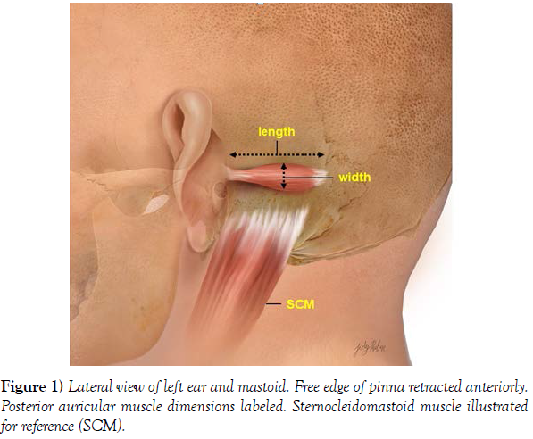
“Muscle length” was defined as the longest continuous muscular component from origin to insertion, and “muscle width” was defined as the widest point of continuous muscle fibers, not including spaces between muscular slips (Figure 1). The pinna was reflected anteriorly to reduce slack in the muscle when collecting measurements. The number of muscular slips with unique insertions was noted for each muscle. Additional data including donor age, sex, and height were collected. Statistics were calculated with IBM SPSS (v. 28.0).
Results
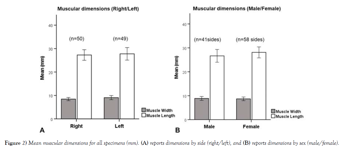
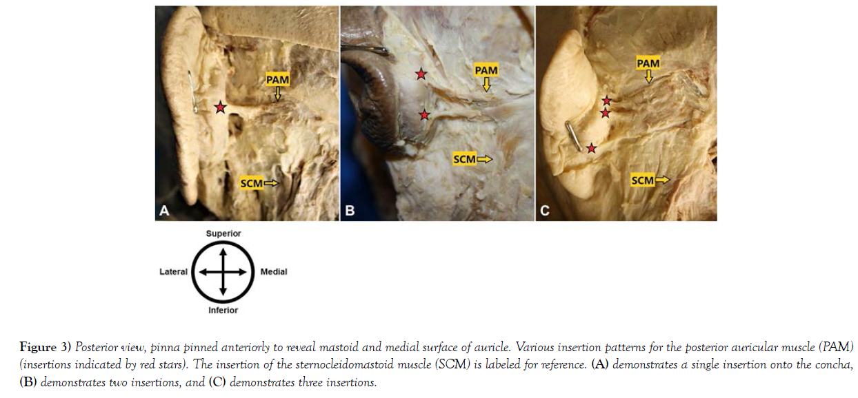
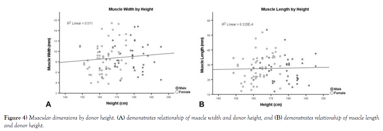
Group descriptive statistics were calculated for muscle dimensions (Figure 2). The mean muscle width and length for all specimens (n=99) were 8.76 mm (SD = 2.79) and 27.51 mm (SD = 8.60), respectively. There were no statistically significant differences in muscular dimensions between right and left sides or between male and female sexes. The origin of the posterior auricular muscle from the mastoid process was readily identifiable. Closer dissection of the muscle toward its insertion revealed one or more distinguishable tendons which inserted onto various areas of the auricular cartilage. Of all specimens examined 73.74% (73 of 99) attached to the concha via a single tendon, 23.23% (23 of 99) attached via two diverging tendons, and 3.03% (3 of 99) attached via three independent tendons (Figure 3). Chi-square tests revealed that the frequency of muscular slips did not differ by laterality (p=0.35) or sex (p=0.44) (Tables 1 and 2). Muscular width and length did not significantly correlate with donor height (r=.11, p=0.29; r=0.03, p=0.77, respectively) (Figure 4).
| Right Side | Left Side | Total | ||||
|---|---|---|---|---|---|---|
| Insertion | Frequency | % | Frequency | % | Frequency | % |
| (no.) | ||||||
| 1 slip | 40 | 80 | 33 | 67.35 | 73 | 73.74 |
| 2 slips | 9 | 18 | 14 | 28.57 | 23 | 23.23 |
| 3 slips | 1 | 2 | 2 | 4 | 3 | 3.03 |
TABEL 1 Frequency of the insertion pattern of the posterior auricular muscle on the left and right sides.
| Male | Female | Total | ||||
|---|---|---|---|---|---|---|
| Insertion (no.) | Frequency | % | Frequency | % | Frequency | % |
| 1 slip | 33 | 80.49 | 40 | 68.97 | 73 | 73.74 |
| 2 slips | 7 | 17.07 | 16 | 27.59 | 23 | 23.23 |
| 3 slips | 1 | 2.44 | 2 | 3.45 | 3 | 3.03 |
TABEL 2 Frequency of the insertion pattern of the posterior auricular muscle in male and female sexes.
Figure 3) Posterior view, pinna pinned anteriorly to reveal mastoid and medial surface of auricle. Various insertion patterns for the posterior auricular muscle (PAM) (insertions indicated by red stars). The insertion of the sternocleidomastoid muscle (SCM) is labeled for reference. (A) demonstrates a single insertion onto the concha, (B) demonstrates two insertions, and (C) demonstrates three insertions.
Discussion
Few studies have carefully examined the physical characteristics of the three muscles of the external ear. The anterior auricular muscle arises from the temporal fascia and being the smallest of the three is often difficult to distinguish. The superior auricular muscle is traditionally described as being the largest of the three and fan-shaped. Recently, Chon et al reported the average dimensions of the superior auricular muscle to be 4.5 cm by 4.9 cm [9]. Our study found the average posterior auricular muscle to be approximately three times longer than it is wide (8.76 mm by 27.51 mm), although the variability between individuals was marked (Figure 2). This result is marginally longer than reported by Oda, which found an average length of 22 mm in 25 Japanese cadavers [10]. The diminutive size of the muscle and relatively immobile nature of the human external ear renders the retraction range of the pinna insignificant, although electromyography evidence suggests a vestigial auriculomotor reflex is present in humans [11]. Interestingly, muscular dimensions did not significantly change with donor height (Figure 4). Additional anthropometric measurements specific to the cranial skeleton may serve as more appropriate predictors of the muscle’s size.
Cataloging the variable insertion patterns of the muscle is of clinical relevance. The shape of the posterior auricular muscle is distinctly different from the other two extrinsic ear muscles and is frequently characterized as “two or three fleshy fasciculi” [9, 12]. The findings of this study show the dominant muscle form is a single muscle slip (n=73, 73.74%). While not uncommon to discover multiple fleshy bellies, this was not the principal morphology in this donor population. There were no significant differences in insertion pattern between males and females, and there were no notable differences between left and right sides. Clinicians utilizing the muscle in plastic surgery and reconstructive procedures should be aware of the possibility of encountering variable insertion patterns. The location and borders of the muscle may be approximated by folding the free edge of the auricular cartilage anteriorly, which pulls the muscle taut against the skin. The single-slip tendinous insertion is the easiest to reveal with typical dissection methods; however, complete exposure of the muscle’s course is necessary to exclude the possibility of multiple insertions. A failure to identify the muscle’s complete form may result in partial manipulation or incomplete transection which could have undesirable aesthetic consequences.
Finally, the posterior auricular muscle was present in 100% of the individuals included in this study. It could not be determined if one muscle on a single individual was present or inadvertently removed during dissection. In a large study of 568 posterior auricular muscles, Sato reported the muscle was completely absent in 8.77% of males and 14.16% of females in a Japanese population [13]. Guerra et al reported that the muscle was present in 95% of specimens (38/40) in a largely white population [14]. Agenesis of the extrinsic ear muscles may contribute to hypermobility of the auricle, causing individuals to seek surgical interventions. Clinicians who regularly employ the posterior auricular muscle in otoplasty should be aware of the possibility that the muscle may be absent.
Conclusion
The posterior auricular muscle has proven useful in clinical settings. Despite its clinical utility, fundamental descriptions of the muscle have remained insufficient in the literature. There are many limitations to this study. Individuals included in this donor population were largely from the Appalachian region of the United States and were overwhelmingly white. Results from this report should be interpreted with caution when attempting to generalize them to other populations. The average donor age in this study was 78.1 years. Age-related changes to the musculoskeletal system may have influenced these results, although this is a common limitation of cadaveric studies. Limitations notwithstanding, this study provides a robust analysis of the posterior auricular muscle’s size and insertion patterns, which could prove useful in clinical decision-making and patient outcomes.
Acknowledgement
The authors would like to thank the body donors and their families for participating in the advancement of scientific knowledge. The authors would also like to extend our gratitude to Elaine Powers and Savannah Easter ling of the Edward Via College of Osteopathic Medicine – VA campus library for their expertise in locating research materials. Finally, the authors would like to thank Judy Rubin (jrubinvisuals.com) for her illustrative contributions to this work
REFERENCES
- Nicolétis C, Guerin-Surville H. Prominent ears: transposition of the posterior auricular muscle on the scapha: A new technique. Aesthetic Plast Surg. 1978;2:295-302.
- Yazici I, Findikçioğlu F, Ozmen S et al. Posterior auricular muscle flap as an adjunct to otoplasty. Aesthetic Plast Surg. 2009;33(4):527-532.
- Azad S, Edwin A, Kumar PV. Posterior auricular muscle – a useful adjunct in otoplasty. Br J Plast Surg. 2003;56(7): 722-723.
- Scuderi N, Tenna S, Bitonti A et al. Repositioning of posterior auricular muscle combined with conventional otoplasty: a personal technique. J Plast Reconstr Aesthet Surg. 2007; 60(2): 201-204.
- Ersen B, Sarialtin Y, Cihantimur B et al. A new otoplasty procedure: combination of perichondrio-apido-dermal flap, posterior auricular muscle transpositioning and cartilage suturing to decrease the post-operative complication rates. Eur J Plast Surg. 2018; 41: 557-562.
- Stephen C, Lowrie AG. The posterior auricular muscle: a useful anatomical landmark for otoplasty. J Laryngol Otol. 2017; 131: 465-467.
- Zerin M, Van Allen MI, Smith DW. Intrinsic auricular muscles and auricular form. Pediatrics. 1982; 69(1): 91-93.
- Hoogbergen MM, Schuurman AH, Rijnders W et al. Auricular hypermobility due to agenesis of the extrinsic muscles. Plast Reconstr Surg. 1996; 98(5): 869-871.
- Chon BH, Blandford AD, Hwang CJ et al. Dimensions, Function and Applications of the Auricular Muscle in Facial Plastic Surgery. Aesthetic Plast Surg. 2021; 45(1): 309-314.
- Oda K. Pri la muskoloj orelauriko ce japanoj. Acta Anatomica Nipponica. 1935; 8: 329.
- Strauss DJ, Corona-Strauss FI, Schroeer A et al. Vestigial auriculomotor activity indicates the direction of auditory attention in humans. Elife. 2020; 9: 545-536.
- Goss CM. Gray’s anatomy – Anatomy of the human body, 28th Edition Philadelphia, PA, USA, Lea & Febiger. 1966; 384.
- Sato S. Statistical studies on the exceptional muscles of the Kyushu-Japanese. Kurume Med J. 1968; 15(2): 69-82.
- Guerra AB, Metzinger SE, Metzinger RC. Variability of the postauricular muscle complex: analysis of 40 hemicadaver dissections. Arch Facial Plast Surg. 2004; 6(5): 342-347.
Google Scholar, Crossref, Indexed at
Google Scholar, Crossref, Indexed at
Google Scholar, Crossref, Indexed at
Google Scholar, Crossref, Indexed at
Google Scholar, Crossref, Indexed at
Google Scholar, Crossref, Indexed at
Google Scholar, Crossref, Indexed at
Google Scholar, Crossref, Indexed at
Google Scholar, Crossref, Indexed at
Google Scholar, Crossref, Indexed at


