Anatomical variations in the arterial supply of liver
Narayanaperumal Mugunthan1*, Danabalan Devi Jansirani1, Christilda Felicia2 and Jayaram Anbalagan1
1Department of Anatomy, Mahatma Gandhi Medical College & Research Institute, Pillaiyarkuppam, Puducherry, India.
2SRM Medical College Hospital and Research Centre, SRM Nagar, Potheri, Kattankolathur, Chennai, India.
- *Corresponding Author:
- Dr. Narayanaperumal Mugunthan
Assistant Professor of Anatomy, Mahatma Gandhi Medical College & Research Institute, Pillaiyarkuppam, Cuddlaore-Pondy Main Road, Puducherry – 607402, India
Tel: +91 944 3118932
E-mail: dr.mugunth111 @gmail.com
Date of Received: September 20th, 2011
Date of Accepted: August 18th, 2012
Published Online: December 15th, 2012
© Int J Anat Var (IJAV). 2012; 5: 107–109.
[ft_below_content] =>Keywords
liver, hepatic artery, replaced right hepatic artery, superior mesenteric artery, accessory left hepatic artery
Introduction
The right hepatic artery (RHA) and left hepatic artery (LHA) normally arises from proper hepatic artery. The middle hepatic artery (MHA) arises either from right or left hepatic artery. After its origin RHA crosses anterior to the portal vein from left to right and passes behind the common hepatic duct to enter the Calot’s triangle where it lies to the left of the cystic duct. As it approaches the cystic duct, it gives off the cystic artery and then turns upwards behind the right hepatic duct to enter the right lobe of liver. The LHA takes origin from the proper hepatic artery. It passes anterior and to the left of the portal vein. Then it ascends upwards to reach the left lobe of the liver.
This “text book” arrangement of common hepatic artery arising from celiac trunk leading to proper, left and right hepatic arteries was found only in 55% of cases [1,2]. Variations in the hepatic artery and its branches are exceedingly common.
A vessel which supplies a liver lobe in addition to its usual vessel is defined as an accessory hepatic artery. A replaced hepatic artery is a vessel that does not take origin from an orthodox position and is the sole supply to that lobe [3]. Thus the aberrant arteries may be either replaced or accessory. The common variations include replaced or accessory right hepatic arteries taking origin from superior mesenteric artery in 6.67% of cases and replaced or accessory left hepatic arteries arising from the left gastric artery in 6.41% of cases [4]. These variations are of great importance to surgeons and interventional radiologists.
Case Report
Using conventional dissection techniques, the abdomen was dissected in a 70-year-old embalmed male cadaver for the purpose of teaching first year MBBS students. We observed a replaced RHA taking origin from superior mesenteric artery and an accessory LHA from left gastric artery. MHA (artery to quadrate lobe) was taking origin from gastroduodenal artery. Following fine dissection, the aberrant hepatic arteries were painted and photographed.
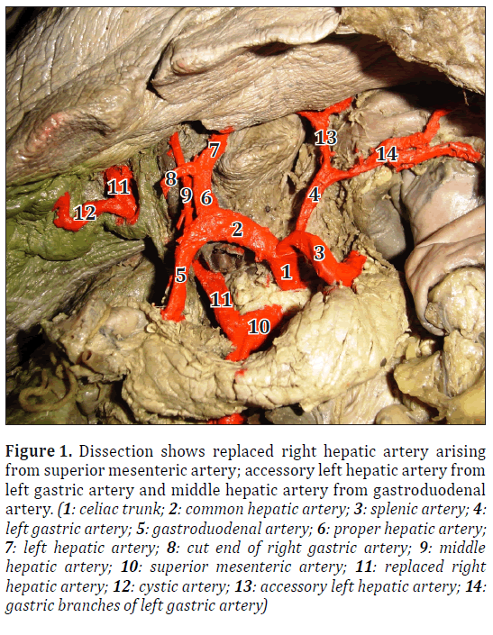
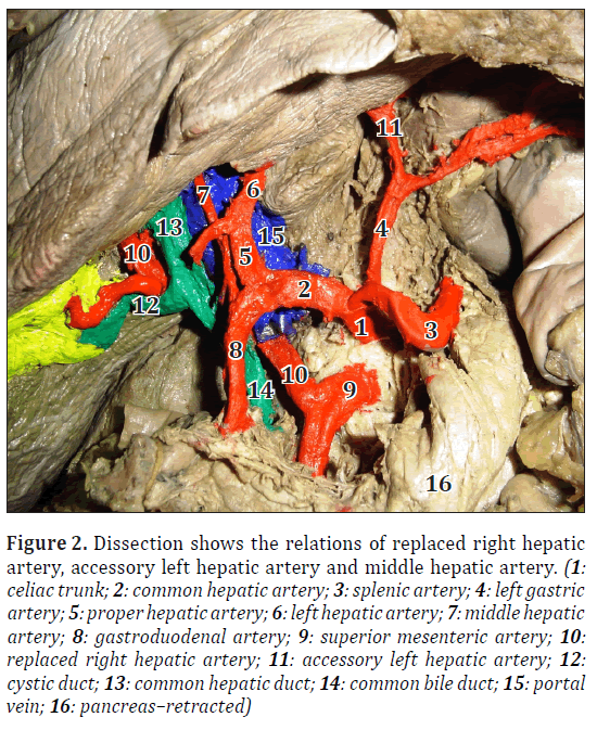
In the present case, the common hepatic artery was observed arising from celiac trunk as one of its three branches. The common hepatic artery divided into gastroduodenal and proper hepatic artery. The proper hepatic artery continued as LHA and the normal RHA was missing. The proper hepatic artery also gave a right gastric artery as usual. The replaced RHA took origin from superior mesenteric artery. It passed upward obliquely from left to right and dorsal to the pancreas, portal vein and common hepatic duct to reach the Calot’s triangle, where it gave cystic artery. Then it passed upward to enter the right lobe of liver (Figures 1, 2).
Figure 1: Dissection shows replaced right hepatic artery arising from superior mesenteric artery; accessory left hepatic artery from left gastric artery and middle hepatic artery from gastroduodenal artery. (1: celiac trunk; 2: common hepatic artery; 3: splenic artery; 4: left gastric artery; 5: gastroduodenal artery; 6: proper hepatic artery; 7: left hepatic artery; 8: cut end of right gastric artery; 9: middle hepatic artery; 10: superior mesenteric artery; 11: replaced right hepatic artery; 12: cystic artery; 13: accessory left hepatic artery; 14: gastric branches of left gastric artery)
Figure 2: Dissection shows the relations of replaced right hepatic artery, accessory left hepatic artery and middle hepatic artery. (1: celiac trunk; 2: common hepatic artery; 3: splenic artery; 4: left gastric artery; 5: proper hepatic artery; 6: left hepatic artery; 7: middle hepatic artery; 8: gastroduodenal artery; 9: superior mesenteric artery; 10: replaced right hepatic artery; 11: accessory left hepatic artery; 12: cystic duct; 13: common hepatic duct; 14: common bile duct; 15: portal vein; 16: pancreas–retracted)
The left lobe of liver was supplied by a normal LHA, which was a continuation of proper hepatic artery. In addition to that, an accessory LHA was found arising from left gastric artery to supply the left lobe. This artery passed from below upwards in the cranial part of the lesser omentum to reach the left end of porta hepatis to enter into the left lobe of liver (Figures 1, 2).
The middle hepatic artery was arising from gastroduodenal artery and coursed anterior to the portal vein, then passed upwards to supply the quadrate lobe.
Discussion
According to Michels, the incidence of replaced right hepatic artery was 18% and accessory left hepatic artery was 11.5%. He also reported that the source of origin of replaced right hepatic artery was from superior mesenteric artery in 12.5%, from celiac trunk 3%, from aorta 2% and from left gastric artery in 0.5% of cases. Moreover, the source of origin of accessory left hepatic artery was from left gastric artery in 11.5% of cases [1]. Molmenti et al. reported that the presence of replaced right hepatic artery in 15-20 % and accessory left hepatic artery in 35% of cases [5]. In the present study also, we found that replaced right hepatic artery was arising from superior mesenteric artery and accessory left hepatic artery was arising from left gastric artery.
Jones and Hardy reported on the origin of middle hepatic artery from gastroduodenal artery in 8.7% [6]. In the present case, a similar finding of middle hepatic artery arising from gastroduodenal artery was encountered.
The aberrant right hepatic artery arising from superior mesenteric artery may have unusual relations in the right free border of the lesser omentum and present hazards while performing cholecystectomy. In ligature of the left gastric artery during gastrectomy, it is wise to look for a large vessel, which sometimes supplies the liver. This may be an accessory or a replaced left hepatic artery from left gastric artery [7]. Accessory left hepatic artery provides a source of collateral arterial circulation in cases of occlusion of the vessels in the porta hepatis. The artery may be injured as it lies in the upper portion of the lesser omentum during mobilization of stomach in gastrectomy and hiatal hernia repair [8,3].
Embryological Significance
At fourth week of development, both dorsal aortae give rise to multiple ventral segmental (omphalomesenteric) arteries. Fusion of the dorsal aortae occurs concurrently with regression of multiple ventral segmental arteries. The celiac axis is derived from the 10th ventral segmental artery and the superior mesenteric artery arises from the 13th segmental artery. The 11th and 12th segmental arteries normally regress.
During early stages of development, there are three hepatic arteries: (1) left hepatic artery arising from the left gastric artery, (2) middle hepatic artery arising from the celiac trunk, and (3) right hepatic artery arising from the superior mesenteric artery. In most cases, the middle hepatic artery is the only one that persists to become the classic proper hepatic artery in the adult. This artery divides into right and left branches, which supply the respective hemi-lobe of the liver [9]. Variations in regression and persistence of these three early arteries account for the so-called accessory and replaced variants [10].
Thus we conclude that the knowledge about the variations in hepatic arterial anatomy is mandatory for surgical gastroenterologists and interventional radiologists for preoperative planning and intraoperative imaging during procedures like liver transplantation, cholecystectomy, gastrectomy, hiatal hernia repair, trans-arterial chemotherapy and hepatic arteriography.
Acknowledgement
We thank the faculty of the Institute of Anatomy, Madras Medical College, Chennai, India for allowing us to perform cadaveric study. We would like to convey our sincere thanks and gratitude to all faculties, Department of Anatomy, Mahatma Gandhi Medical College & Research Institute, Puducherry, India for their help and guidance in continuing this study.
References
- Michels NA. Blood supply and anatomy of the upper abdominal organs with a descriptive atlas. Philadelphia and Montreal, B. Lippincott Company. 1955; 134–183.
- Munson JL, Sanders LE. Cholecystectomy. Open cholecystectomy revisited. Surg Clin North Am. 1994, 74.4; 751–754.
- Standring S, ed. Gray’s Anatomy. 39th Ed., Philadelphia, Elsevier, Churchill Livingstone. 2005; 1218–1230.
- Yang Y, Jiang N, Lu MQ, Xu C, Cai CJ, Li H, Yi SH, Wang GS, Zhang J, Zhang JF, Chen GH. Anatomical variation of the donor hepatic arteries: analysis of 843 cases. Nan Fang Yi Ke Da Xue Xue Bao. 2007; 27: 1164–1166. (Chinese)
- Molmenti EP, Pinto PA, Klein J, Klein AS. Normal and variant arterial supply of the liver and gallbladder. Pediatr Transplant. 2003; 7: 80–82,
- Jones RM, Hardy KJ. The hepatic artery: a reminder of surgical anatomy. J R Coll Surg Edinb. 2001; 46: 168–170.
- Decker GAG, Plessis DJ. Lee Mc Gregor’s Synopsis of Surgical Anatomy. 12th Ed., Bristol, John Wright & Sons Ltd. 1986; 85–93.
- Skandalakis JE. Skandalakis’ Surgical Anatomy. Vol. 2. 1st Ed., Athens, PMP. 2004; 1108–1130.
- Fischer JE, Bland KI. Mastery of Surgery. Vol. 1. 5th Ed., Philadelphia, Lippincott Williams & Wilkins. 2007; 1009–1010.
- Yeo CJ. Shackelford’s surgery of the Alimentary Tract. 6th Ed., Philadelphia, Saunders Elsevier. 2007; 1598–1602.
Narayanaperumal Mugunthan1*, Danabalan Devi Jansirani1, Christilda Felicia2 and Jayaram Anbalagan1
1Department of Anatomy, Mahatma Gandhi Medical College & Research Institute, Pillaiyarkuppam, Puducherry, India.
2SRM Medical College Hospital and Research Centre, SRM Nagar, Potheri, Kattankolathur, Chennai, India.
- *Corresponding Author:
- Dr. Narayanaperumal Mugunthan
Assistant Professor of Anatomy, Mahatma Gandhi Medical College & Research Institute, Pillaiyarkuppam, Cuddlaore-Pondy Main Road, Puducherry – 607402, India
Tel: +91 944 3118932
E-mail: dr.mugunth111 @gmail.com
Date of Received: September 20th, 2011
Date of Accepted: August 18th, 2012
Published Online: December 15th, 2012
© Int J Anat Var (IJAV). 2012; 5: 107–109.
Abstract
The knowledge about the anatomical variations in the arterial supply of liver is mandatory for surgeons. Presence of hepatic arterial variations may lead to surgical injuries in the liver which can be made inadvertently even by the most experienced surgeons. During the routine dissection of a 70-year-old male cadaver, the following variations were observed: Right hepatic artery taking origin as replaced right hepatic artery from superior mesenteric artery; in addition to normal left hepatic artery, an accessory left hepatic artery arising from left gastric artery; middle hepatic artery (artery to quadrate lobe) that usually arises from right or left hepatic artery, in the present case was found to arise from gastroduodenal artery. These aberrant arteries have embryological and clinical significance.
-Keywords
liver, hepatic artery, replaced right hepatic artery, superior mesenteric artery, accessory left hepatic artery
Introduction
The right hepatic artery (RHA) and left hepatic artery (LHA) normally arises from proper hepatic artery. The middle hepatic artery (MHA) arises either from right or left hepatic artery. After its origin RHA crosses anterior to the portal vein from left to right and passes behind the common hepatic duct to enter the Calot’s triangle where it lies to the left of the cystic duct. As it approaches the cystic duct, it gives off the cystic artery and then turns upwards behind the right hepatic duct to enter the right lobe of liver. The LHA takes origin from the proper hepatic artery. It passes anterior and to the left of the portal vein. Then it ascends upwards to reach the left lobe of the liver.
This “text book” arrangement of common hepatic artery arising from celiac trunk leading to proper, left and right hepatic arteries was found only in 55% of cases [1,2]. Variations in the hepatic artery and its branches are exceedingly common.
A vessel which supplies a liver lobe in addition to its usual vessel is defined as an accessory hepatic artery. A replaced hepatic artery is a vessel that does not take origin from an orthodox position and is the sole supply to that lobe [3]. Thus the aberrant arteries may be either replaced or accessory. The common variations include replaced or accessory right hepatic arteries taking origin from superior mesenteric artery in 6.67% of cases and replaced or accessory left hepatic arteries arising from the left gastric artery in 6.41% of cases [4]. These variations are of great importance to surgeons and interventional radiologists.
Case Report
Using conventional dissection techniques, the abdomen was dissected in a 70-year-old embalmed male cadaver for the purpose of teaching first year MBBS students. We observed a replaced RHA taking origin from superior mesenteric artery and an accessory LHA from left gastric artery. MHA (artery to quadrate lobe) was taking origin from gastroduodenal artery. Following fine dissection, the aberrant hepatic arteries were painted and photographed.
In the present case, the common hepatic artery was observed arising from celiac trunk as one of its three branches. The common hepatic artery divided into gastroduodenal and proper hepatic artery. The proper hepatic artery continued as LHA and the normal RHA was missing. The proper hepatic artery also gave a right gastric artery as usual. The replaced RHA took origin from superior mesenteric artery. It passed upward obliquely from left to right and dorsal to the pancreas, portal vein and common hepatic duct to reach the Calot’s triangle, where it gave cystic artery. Then it passed upward to enter the right lobe of liver (Figures 1, 2).
Figure 1: Dissection shows replaced right hepatic artery arising from superior mesenteric artery; accessory left hepatic artery from left gastric artery and middle hepatic artery from gastroduodenal artery. (1: celiac trunk; 2: common hepatic artery; 3: splenic artery; 4: left gastric artery; 5: gastroduodenal artery; 6: proper hepatic artery; 7: left hepatic artery; 8: cut end of right gastric artery; 9: middle hepatic artery; 10: superior mesenteric artery; 11: replaced right hepatic artery; 12: cystic artery; 13: accessory left hepatic artery; 14: gastric branches of left gastric artery)
Figure 2: Dissection shows the relations of replaced right hepatic artery, accessory left hepatic artery and middle hepatic artery. (1: celiac trunk; 2: common hepatic artery; 3: splenic artery; 4: left gastric artery; 5: proper hepatic artery; 6: left hepatic artery; 7: middle hepatic artery; 8: gastroduodenal artery; 9: superior mesenteric artery; 10: replaced right hepatic artery; 11: accessory left hepatic artery; 12: cystic duct; 13: common hepatic duct; 14: common bile duct; 15: portal vein; 16: pancreas–retracted)
The left lobe of liver was supplied by a normal LHA, which was a continuation of proper hepatic artery. In addition to that, an accessory LHA was found arising from left gastric artery to supply the left lobe. This artery passed from below upwards in the cranial part of the lesser omentum to reach the left end of porta hepatis to enter into the left lobe of liver (Figures 1, 2).
The middle hepatic artery was arising from gastroduodenal artery and coursed anterior to the portal vein, then passed upwards to supply the quadrate lobe.
Discussion
According to Michels, the incidence of replaced right hepatic artery was 18% and accessory left hepatic artery was 11.5%. He also reported that the source of origin of replaced right hepatic artery was from superior mesenteric artery in 12.5%, from celiac trunk 3%, from aorta 2% and from left gastric artery in 0.5% of cases. Moreover, the source of origin of accessory left hepatic artery was from left gastric artery in 11.5% of cases [1]. Molmenti et al. reported that the presence of replaced right hepatic artery in 15-20 % and accessory left hepatic artery in 35% of cases [5]. In the present study also, we found that replaced right hepatic artery was arising from superior mesenteric artery and accessory left hepatic artery was arising from left gastric artery.
Jones and Hardy reported on the origin of middle hepatic artery from gastroduodenal artery in 8.7% [6]. In the present case, a similar finding of middle hepatic artery arising from gastroduodenal artery was encountered.
The aberrant right hepatic artery arising from superior mesenteric artery may have unusual relations in the right free border of the lesser omentum and present hazards while performing cholecystectomy. In ligature of the left gastric artery during gastrectomy, it is wise to look for a large vessel, which sometimes supplies the liver. This may be an accessory or a replaced left hepatic artery from left gastric artery [7]. Accessory left hepatic artery provides a source of collateral arterial circulation in cases of occlusion of the vessels in the porta hepatis. The artery may be injured as it lies in the upper portion of the lesser omentum during mobilization of stomach in gastrectomy and hiatal hernia repair [8,3].
Embryological Significance
At fourth week of development, both dorsal aortae give rise to multiple ventral segmental (omphalomesenteric) arteries. Fusion of the dorsal aortae occurs concurrently with regression of multiple ventral segmental arteries. The celiac axis is derived from the 10th ventral segmental artery and the superior mesenteric artery arises from the 13th segmental artery. The 11th and 12th segmental arteries normally regress.
During early stages of development, there are three hepatic arteries: (1) left hepatic artery arising from the left gastric artery, (2) middle hepatic artery arising from the celiac trunk, and (3) right hepatic artery arising from the superior mesenteric artery. In most cases, the middle hepatic artery is the only one that persists to become the classic proper hepatic artery in the adult. This artery divides into right and left branches, which supply the respective hemi-lobe of the liver [9]. Variations in regression and persistence of these three early arteries account for the so-called accessory and replaced variants [10].
Thus we conclude that the knowledge about the variations in hepatic arterial anatomy is mandatory for surgical gastroenterologists and interventional radiologists for preoperative planning and intraoperative imaging during procedures like liver transplantation, cholecystectomy, gastrectomy, hiatal hernia repair, trans-arterial chemotherapy and hepatic arteriography.
Acknowledgement
We thank the faculty of the Institute of Anatomy, Madras Medical College, Chennai, India for allowing us to perform cadaveric study. We would like to convey our sincere thanks and gratitude to all faculties, Department of Anatomy, Mahatma Gandhi Medical College & Research Institute, Puducherry, India for their help and guidance in continuing this study.
References
- Michels NA. Blood supply and anatomy of the upper abdominal organs with a descriptive atlas. Philadelphia and Montreal, B. Lippincott Company. 1955; 134–183.
- Munson JL, Sanders LE. Cholecystectomy. Open cholecystectomy revisited. Surg Clin North Am. 1994, 74.4; 751–754.
- Standring S, ed. Gray’s Anatomy. 39th Ed., Philadelphia, Elsevier, Churchill Livingstone. 2005; 1218–1230.
- Yang Y, Jiang N, Lu MQ, Xu C, Cai CJ, Li H, Yi SH, Wang GS, Zhang J, Zhang JF, Chen GH. Anatomical variation of the donor hepatic arteries: analysis of 843 cases. Nan Fang Yi Ke Da Xue Xue Bao. 2007; 27: 1164–1166. (Chinese)
- Molmenti EP, Pinto PA, Klein J, Klein AS. Normal and variant arterial supply of the liver and gallbladder. Pediatr Transplant. 2003; 7: 80–82,
- Jones RM, Hardy KJ. The hepatic artery: a reminder of surgical anatomy. J R Coll Surg Edinb. 2001; 46: 168–170.
- Decker GAG, Plessis DJ. Lee Mc Gregor’s Synopsis of Surgical Anatomy. 12th Ed., Bristol, John Wright & Sons Ltd. 1986; 85–93.
- Skandalakis JE. Skandalakis’ Surgical Anatomy. Vol. 2. 1st Ed., Athens, PMP. 2004; 1108–1130.
- Fischer JE, Bland KI. Mastery of Surgery. Vol. 1. 5th Ed., Philadelphia, Lippincott Williams & Wilkins. 2007; 1009–1010.
- Yeo CJ. Shackelford’s surgery of the Alimentary Tract. 6th Ed., Philadelphia, Saunders Elsevier. 2007; 1598–1602.


