Assessment of three patients with major vascular variations in head and neck region by imaging and clinical findings
Hanifi Bayarogullari1, Yasar Cokkeser2, Ercan Akbay2* and Murat Altas3
1Department of Radiodiagnostics, Mustafa Kemal University School of Medicine, Antakya, Turkey
2Department of Otolaryngology, Head and Neck Surgery, Mustafa Kemal University School of Medicine, Antakya, Turkey
3Department of Radiodiagnostics and Neurosurgery, Mustafa Kemal University School of Medicine, Antakya, Turkey
- *Corresponding Author:
- Dr. Ercan Akbay
Mustafa Kemal Üniversitesi, Tıp Fakültesi KBB Anabilim Dalı, Serinyol Hatay, Turkey
Tel: +90 505 4975049
E-mail: ercanakbay@yahoo.com
Date of Received: November 21st, 2011
Date of Accepted: August 13th, 2012
Published Online: November 12th, 2012
© Int J Anat Var (IJAV). 2012; 5: 93–95.
[ft_below_content] =>Keywords
internal carotid artery, external carotid artery, vertebral artery, fistula, malformation
Introduction
The purpose of this report is to review the imaging features and clinical significance in variations, especially course of the internal carotid artery (ICA) and major arterio-venous malformations of head and neck region. Such findings are extremely rare but, the awareness of these variations essential for otolaryngologists.
Case Report
Case 1
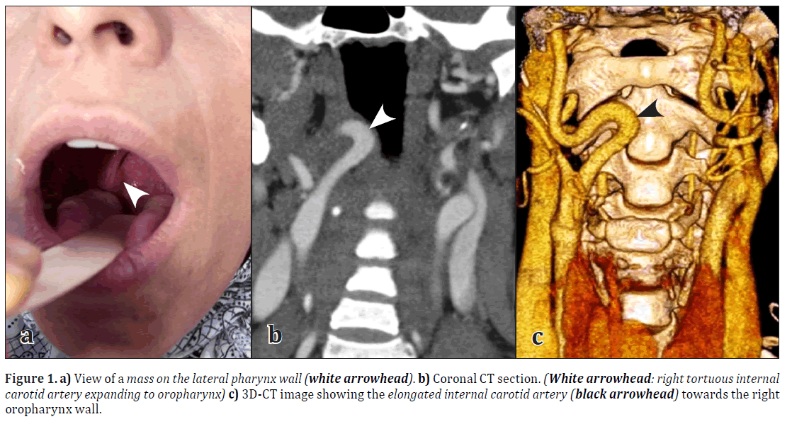
A 30-year-old woman was admitted with a complaint of abnormal sensation on throat and cough present for many years. She was known as suffering from chronic renal failure for 2 years. The physical examination revealed a mass without mucosal abnormality on the right lateral oropharyngeal wall (Figure 1a). The mass showed very little pulsation. Three-dimensional computed tomography (3D-CT) scans showed right tortuous internal carotid artery expanding to oropharynx (Figures 1b, 1c).
Case 2
A 42-year-old man was referred to ENT clinic for evaluation of a mass on the left parotideal region. No other complaint accompanied this swelling. The physical examination revealed a slight mass on the left side of the face. The mass showed very little thrill by auscultation. 3D-CT scans showed an arterio-venous fistula between internal jugular vein and maxillary artery on the left (Figures 2a, 2b). We observed tortuousness at the proximal part of both vessels because of this pathology. No treatment was offered.
Case 3
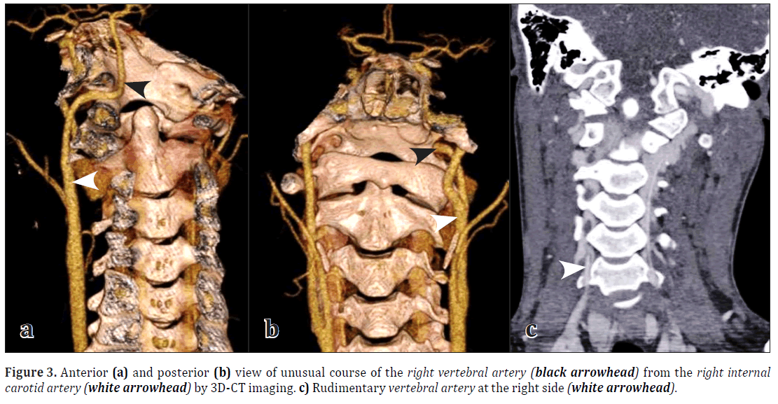
A 17-year-old boy was referred to ENT clinic for consultation from neurology department with a complaint to sudden fall down for 1–2 seconds. The physical examination was normal. 3D-CT scans revealed an unusual origin of the right vertebral artery from the right internal carotid artery (Figures 3a, 3b). At the same side vertebral artery was seen as rudimentary (Figure 3c). We informed the patient about this vascular variation and no treatment offered.
Discussion
Anomalies of the ICA in the head and neck are rarely seen [1,2]. Anatomical variants of the ICA have been reported with a large variability of pattern and degree [3]. In our case 1, there was no mucosal abnormality on the pharyngeal wall. But we have seen the pulsation of the ICA. We have suspected a vascular pathology and then this way confirmed by radiodiagnostic study. The aberrant and tortuous ICA is at high risk of injury during routine surgical procedures if the vessel is running near the pharyngeal wall. Tortuous ICA may cause a several morbidity or mortality during surgeries such as uvulopharyngoplasty, velopharyngeal surgery, routine tonsillectomy and drainage of peritonsillar abscess. These major vascular variations do not need any treatment if the patient does not have central vascular ischemic symptoms [4]. Many times, giving the patients an information form make sure to show it to their doctors, especially before surgical operation is necessary [1].
The second case had an arterio-venous fistula between internal jugular vein and maxillary artery in the left infratemporal region. Most fistulas between major artery and vein are associated with sharp injury. But the patient claimed no history of previous injury or surgery to the neck. This fistula was thought to be congenital in nature. However, the genesis of this vascular variation remains uncertain. During the time the fast growing of left parotid side secondary to tortuousness of proximal part of vessels is possible. If otolaryngologists suspect this variation preoperatively, they can prevent serious hemorrhage and unnecessary parotid surgery.
Third case had a variant vertebral artery in the origin and course. An unusual origin of the vertebral artery from the common carotid artery was reported by Chen et al. [5]. It occurs on the right or left side with complex embryonic mechanisms. Chan et al. discussed a congenital anastomosis between the vertebral artery and internal carotid artery [6]. Multiple variations in the origin of right vertebral artery have been reported in literature [7]. To best of our knowledge, a direct branching of the right vertebral artery from the right internal carotid artery as seen our case 3 has not been yet reported.
References
- Ozcan KM, Ozcan I, Selcuk A, Pasaoglu L, Hatipoglu HG, Dere H. Tortuous internal carotid artery narrowing pyriform sinus: two cases. Clin Imaging. 2008; 32: 220–222.
- Johnson RE, Stambaugh KI, Richmond H, Balbuena L. Tortuous internal carotid artery presenting as an oropharyngeal mass. Otolaryngol Head Neck Surg. 1995; 112: 479–482.
- Agrawal R, Agrawal SK. Dangerous anatomic variation of internal carotid artery – a rare case report. Int J Anat Var (IJAV). 2011; 4: 174–176.
- Okami K, Onuki J, Ishida K, Kido T, Takahashi M. Tortuosity of the internal carotid artery - report of three cases and MR-angiography imaging. Auris Nasus Larynx. 2001; 28: 373–376.
- Chen CJ, Wang LJ, Wong YC. Abnormal origin of the vertebral artery from the common carotid artery. AJNR Am J Neuroradiol. 1998;19:1414-6.
- Chan PN, Yu SCH, Boet R, Poon WS, Ahuja AT. A rare congenital anastomosis between the vertebral artery and internal carotid artery. AJNR Am J Neuroradiol. 2003; 24: 1885–1886.
- Nasir S, Hussain M, Khan SA, Mansoor MA, Sharif S. Anomalous origin of right vertebral artery from right external carotid artery. J Coll Physicians Surg Pak. 2010; 20: 208–210.
Hanifi Bayarogullari1, Yasar Cokkeser2, Ercan Akbay2* and Murat Altas3
1Department of Radiodiagnostics, Mustafa Kemal University School of Medicine, Antakya, Turkey
2Department of Otolaryngology, Head and Neck Surgery, Mustafa Kemal University School of Medicine, Antakya, Turkey
3Department of Radiodiagnostics and Neurosurgery, Mustafa Kemal University School of Medicine, Antakya, Turkey
- *Corresponding Author:
- Dr. Ercan Akbay
Mustafa Kemal Üniversitesi, Tıp Fakültesi KBB Anabilim Dalı, Serinyol Hatay, Turkey
Tel: +90 505 4975049
E-mail: ercanakbay@yahoo.com
Date of Received: November 21st, 2011
Date of Accepted: August 13th, 2012
Published Online: November 12th, 2012
© Int J Anat Var (IJAV). 2012; 5: 93–95.
Abstract
We present three patients with vascular aberrancy in the location and course of the head and neck region accompanied by clinical findings and three-dimensional computed tomography scans. Devastating complications may result from a biopsy or surgical procedures in presence of such variations. Therefore, otolaryngologists should be careful and recognize them.
-Keywords
internal carotid artery, external carotid artery, vertebral artery, fistula, malformation
Introduction
The purpose of this report is to review the imaging features and clinical significance in variations, especially course of the internal carotid artery (ICA) and major arterio-venous malformations of head and neck region. Such findings are extremely rare but, the awareness of these variations essential for otolaryngologists.
Case Report
Case 1
A 30-year-old woman was admitted with a complaint of abnormal sensation on throat and cough present for many years. She was known as suffering from chronic renal failure for 2 years. The physical examination revealed a mass without mucosal abnormality on the right lateral oropharyngeal wall (Figure 1a). The mass showed very little pulsation. Three-dimensional computed tomography (3D-CT) scans showed right tortuous internal carotid artery expanding to oropharynx (Figures 1b, 1c).
Case 2
A 42-year-old man was referred to ENT clinic for evaluation of a mass on the left parotideal region. No other complaint accompanied this swelling. The physical examination revealed a slight mass on the left side of the face. The mass showed very little thrill by auscultation. 3D-CT scans showed an arterio-venous fistula between internal jugular vein and maxillary artery on the left (Figures 2a, 2b). We observed tortuousness at the proximal part of both vessels because of this pathology. No treatment was offered.
Case 3
A 17-year-old boy was referred to ENT clinic for consultation from neurology department with a complaint to sudden fall down for 1–2 seconds. The physical examination was normal. 3D-CT scans revealed an unusual origin of the right vertebral artery from the right internal carotid artery (Figures 3a, 3b). At the same side vertebral artery was seen as rudimentary (Figure 3c). We informed the patient about this vascular variation and no treatment offered.
Discussion
Anomalies of the ICA in the head and neck are rarely seen [1,2]. Anatomical variants of the ICA have been reported with a large variability of pattern and degree [3]. In our case 1, there was no mucosal abnormality on the pharyngeal wall. But we have seen the pulsation of the ICA. We have suspected a vascular pathology and then this way confirmed by radiodiagnostic study. The aberrant and tortuous ICA is at high risk of injury during routine surgical procedures if the vessel is running near the pharyngeal wall. Tortuous ICA may cause a several morbidity or mortality during surgeries such as uvulopharyngoplasty, velopharyngeal surgery, routine tonsillectomy and drainage of peritonsillar abscess. These major vascular variations do not need any treatment if the patient does not have central vascular ischemic symptoms [4]. Many times, giving the patients an information form make sure to show it to their doctors, especially before surgical operation is necessary [1].
The second case had an arterio-venous fistula between internal jugular vein and maxillary artery in the left infratemporal region. Most fistulas between major artery and vein are associated with sharp injury. But the patient claimed no history of previous injury or surgery to the neck. This fistula was thought to be congenital in nature. However, the genesis of this vascular variation remains uncertain. During the time the fast growing of left parotid side secondary to tortuousness of proximal part of vessels is possible. If otolaryngologists suspect this variation preoperatively, they can prevent serious hemorrhage and unnecessary parotid surgery.
Third case had a variant vertebral artery in the origin and course. An unusual origin of the vertebral artery from the common carotid artery was reported by Chen et al. [5]. It occurs on the right or left side with complex embryonic mechanisms. Chan et al. discussed a congenital anastomosis between the vertebral artery and internal carotid artery [6]. Multiple variations in the origin of right vertebral artery have been reported in literature [7]. To best of our knowledge, a direct branching of the right vertebral artery from the right internal carotid artery as seen our case 3 has not been yet reported.
References
- Ozcan KM, Ozcan I, Selcuk A, Pasaoglu L, Hatipoglu HG, Dere H. Tortuous internal carotid artery narrowing pyriform sinus: two cases. Clin Imaging. 2008; 32: 220–222.
- Johnson RE, Stambaugh KI, Richmond H, Balbuena L. Tortuous internal carotid artery presenting as an oropharyngeal mass. Otolaryngol Head Neck Surg. 1995; 112: 479–482.
- Agrawal R, Agrawal SK. Dangerous anatomic variation of internal carotid artery – a rare case report. Int J Anat Var (IJAV). 2011; 4: 174–176.
- Okami K, Onuki J, Ishida K, Kido T, Takahashi M. Tortuosity of the internal carotid artery - report of three cases and MR-angiography imaging. Auris Nasus Larynx. 2001; 28: 373–376.
- Chen CJ, Wang LJ, Wong YC. Abnormal origin of the vertebral artery from the common carotid artery. AJNR Am J Neuroradiol. 1998;19:1414-6.
- Chan PN, Yu SCH, Boet R, Poon WS, Ahuja AT. A rare congenital anastomosis between the vertebral artery and internal carotid artery. AJNR Am J Neuroradiol. 2003; 24: 1885–1886.
- Nasir S, Hussain M, Khan SA, Mansoor MA, Sharif S. Anomalous origin of right vertebral artery from right external carotid artery. J Coll Physicians Surg Pak. 2010; 20: 208–210.


