Bifid rib – a rare incidental observation at autopsy
Santhosh Chandrappa Siddappa*, Viswanathan Karibasappa Gowda and Bande Nawaz
Department of Forensic Medicine, J.J.M. Medical College, Davangere, Karnataka State, India
- *Corresponding Author:
- Dr. Santhosh Chandrappa Siddappa
Associate Professor Department of Forensic Medicine J. J. M. Medical College Davangere 577004 Karnataka State, India
Tel: +91 944 8220096
E-mail: drsan_99@rediffmail.com
Date of Received: July 14th, 2011
Date of Accepted: September 24th, 2011
Published Online: February 5th, 2013
© Int J Anat Var (IJAV). 2013; 6: 22–23.
[ft_below_content] =>Keywords
bifid rib, rib variations, anatomical variation
Introduction
The overall incidence of bifid rib is estimated to be 0.15% to 3.4%. Unusual ribs may be observed as an isolated defect without involvement of any organ or sometimes along with multisystem malformations [1]. Bifid ribs are rare incidental finding at radiography in some symptomatic patients and at autopsy and dissection of cadavers in anatomy department. This case of left 3rd bifid rib is a rare incidental finding at autopsy conducted at mortuary of Department of Forensic Medicine, J. J. M. Medical College, Davangere with anatomical consideration.
Case Report
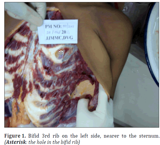
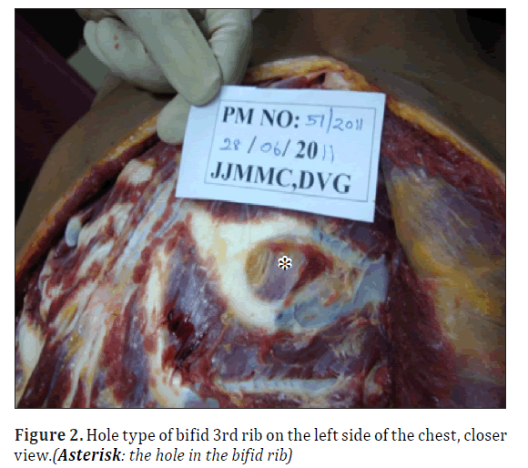
A 16-year-old male committed suicide, and the body was subjected to medico-legal autopsy at our mortuary. Upon detailed dissection of the body with an I-shaped incision from the suprasternal notch to the pubic symphysis and on reflecting the pectoral muscles, the bifid rib was noted on the 3rd rib on the left side of the chest near the costo-condral junction (Figure 1). The 2 cartilaginous division of the rib reunited and articulated with the sternum. The bifid space between the upper and lower divisions of the rib was round and contained intercostals muscles and fasciae. The hole type of bifid rib, where the two cartilaginous divisions reunited and articulated with the sternum was found in this case (Figure 2). The hole of the bifid rib measured 2.3 cm in vertical distance and 2.8 cm in transverse distance of the bifid space. The upper division of the bifid rib measured 0.9 cm and the lower division of the rib measured 1 cm. The upper intercostal space between the 2nd and 3rd was narrowed and it measured 0.5 cm. The lower intercostal space between the 3rd (unusual rib) and 4th rib was widened measuring 2.4 cm.
The 2nd anterior intercostal artery that branched from the internal thoracic artery was the artery supplying the bifid space. The posterior intercostal artery that branched from the thoracic aorta supplied the rest of the intercostal muscles. The venous distribution was accompanying the arterial supply. The intercostal nerve ran along the costal groove of the lower division of the rib and no branch was present in the upper division of the rib. All the other ribs were as usual in morphology.
Discussion
The incidence of bifid rib is 0.15% to 3.4% and it accounts for 20% of all congenital anomalies [2]. In our series out of 600 autopsies conducted for the past 2 years only one case was seen which accounted for 0.16%, which was similar to previously reported incidence.
The bifid rib is more common in males than females and occurs most frequently in 3rd and 4th rib (incidence 3rd = 4th > 5th > 6th > 2nd) [3,4]. The incidence is more common on the right side than on the left side [5]. In our case bifid rib was present in a male individual on the left 3rd rib. The upper intercostal space was narrower than the lower intercostal space in our case, which suggests that the upper division of the bifid was unusual; and the muscles would not be present if the upper division divided from one rib into 2 divisions rather than growing out of the lower division. Most importantly the arterial supply in our case was from 2nd anterior intercostal artery and the intercostal nerve ran along the lower margin of the bifid 3rd rib, which was similar to the previously reported cases [2].
Bifid ribs have been reported as an independent finding when they happen as accidentally or sporadically developed bifid, and are mostly found in elderly cadavers [4,5]. Since the rib originates from the mesoderm, costal defects are associated with malformations in other organs of same mesodermal origin like heart and kidneys [6]. In our case bifid rib was the only incidental finding which was present at autopsy, though a thorough search was made in the heart, kidneys and other organs, where no anomalies or defects were present. If the patient was alive then he could be subjected to genetic studies for the diagnosis of genetic defects and syndromes.
Conclusion
Bifid rib is though an accidental finding; it should not be misdiagnosed as a previous costal fracture or malunion in the cadavers, which may lead to wrong interpretation. This unusual finding further opens up the scope to do a detailed autopsy to check for the other mesodermal anomaly which could coexist with the bifid rib, though it may not be the cause of death. These rarities and anatomical variations will widen the prospects of research in the field of medical science.
Acknowledgements
The authors would to thank our beloved Principal Dr. H. R. Chandrashekar, a well-known pathologist, for his support and encouragement in building up the Mortuary Center at our college.
References
- Kupeli E, Ulubay G. The clinical picture: bony bridge of a bifid rib. Cleveland Clinic Journal of Medicine. 2010; 77: 232–233.
- Song WC, Kim SH, Park DK, Koh KS. Bifid rib: anatomical considerations in three cases. Yonsei Med J. 2009; 50: 300–303.
- Schumacher R, Mai A, Gutjahr P. Association of rib anomalies and malignancy in childhood. Eur J Pediatr. 1992; 151: 432–434.
- Osawa T, Sasaki T, Matsumoto Y, Tsukamoto A, Onodera M, Nara E, Chen JK, Fujimura A, Nozaka Y. Bifid ribs observed in the third and the fourth ribs. Kaibogaku Zasshi. 1998; 73: 633–635.
- Osawa T, Onodera M, Feng XY, Sasaki N, Nagato S, Matsumoto Y, Nara E, Fujimura A, Nozaka Y. Two cases of bifid rib observed in the fourth and the fifth rib. Dental Journal of Iwate Medical University. 2002; 27: 98–103.
- Charles I, Scott J. Pectoral girdle, spine, rib and pelvic girdle. In: Stevenson RE, Hall JG, Goodmann RM, eds. Human Malformations and Related Anomalies. Vol. 2, New York, Oxford University Press. 1993: 655–697.
Santhosh Chandrappa Siddappa*, Viswanathan Karibasappa Gowda and Bande Nawaz
Department of Forensic Medicine, J.J.M. Medical College, Davangere, Karnataka State, India
- *Corresponding Author:
- Dr. Santhosh Chandrappa Siddappa
Associate Professor Department of Forensic Medicine J. J. M. Medical College Davangere 577004 Karnataka State, India
Tel: +91 944 8220096
E-mail: drsan_99@rediffmail.com
Date of Received: July 14th, 2011
Date of Accepted: September 24th, 2011
Published Online: February 5th, 2013
© Int J Anat Var (IJAV). 2013; 6: 22–23.
Abstract
A case of bifid 3rd rib on the left side of the chest found accidentally at autopsy is discussed. The hole type of bifid rib, where the two cartilaginous divisions reunited and articulated with the sternum was found in this case. The 2nd anterior intercostal artery that branched from the internal thoracic artery was the artery supplying the bifid space. The intercostal nerve ran along the costal groove of the lower division of the rib and no branch was present in the upper division of the rib. These rarities and anatomical variations will widen the prospects of research in the field of medical science.
-Keywords
bifid rib, rib variations, anatomical variation
Introduction
The overall incidence of bifid rib is estimated to be 0.15% to 3.4%. Unusual ribs may be observed as an isolated defect without involvement of any organ or sometimes along with multisystem malformations [1]. Bifid ribs are rare incidental finding at radiography in some symptomatic patients and at autopsy and dissection of cadavers in anatomy department. This case of left 3rd bifid rib is a rare incidental finding at autopsy conducted at mortuary of Department of Forensic Medicine, J. J. M. Medical College, Davangere with anatomical consideration.
Case Report
A 16-year-old male committed suicide, and the body was subjected to medico-legal autopsy at our mortuary. Upon detailed dissection of the body with an I-shaped incision from the suprasternal notch to the pubic symphysis and on reflecting the pectoral muscles, the bifid rib was noted on the 3rd rib on the left side of the chest near the costo-condral junction (Figure 1). The 2 cartilaginous division of the rib reunited and articulated with the sternum. The bifid space between the upper and lower divisions of the rib was round and contained intercostals muscles and fasciae. The hole type of bifid rib, where the two cartilaginous divisions reunited and articulated with the sternum was found in this case (Figure 2). The hole of the bifid rib measured 2.3 cm in vertical distance and 2.8 cm in transverse distance of the bifid space. The upper division of the bifid rib measured 0.9 cm and the lower division of the rib measured 1 cm. The upper intercostal space between the 2nd and 3rd was narrowed and it measured 0.5 cm. The lower intercostal space between the 3rd (unusual rib) and 4th rib was widened measuring 2.4 cm.
The 2nd anterior intercostal artery that branched from the internal thoracic artery was the artery supplying the bifid space. The posterior intercostal artery that branched from the thoracic aorta supplied the rest of the intercostal muscles. The venous distribution was accompanying the arterial supply. The intercostal nerve ran along the costal groove of the lower division of the rib and no branch was present in the upper division of the rib. All the other ribs were as usual in morphology.
Discussion
The incidence of bifid rib is 0.15% to 3.4% and it accounts for 20% of all congenital anomalies [2]. In our series out of 600 autopsies conducted for the past 2 years only one case was seen which accounted for 0.16%, which was similar to previously reported incidence.
The bifid rib is more common in males than females and occurs most frequently in 3rd and 4th rib (incidence 3rd = 4th > 5th > 6th > 2nd) [3,4]. The incidence is more common on the right side than on the left side [5]. In our case bifid rib was present in a male individual on the left 3rd rib. The upper intercostal space was narrower than the lower intercostal space in our case, which suggests that the upper division of the bifid was unusual; and the muscles would not be present if the upper division divided from one rib into 2 divisions rather than growing out of the lower division. Most importantly the arterial supply in our case was from 2nd anterior intercostal artery and the intercostal nerve ran along the lower margin of the bifid 3rd rib, which was similar to the previously reported cases [2].
Bifid ribs have been reported as an independent finding when they happen as accidentally or sporadically developed bifid, and are mostly found in elderly cadavers [4,5]. Since the rib originates from the mesoderm, costal defects are associated with malformations in other organs of same mesodermal origin like heart and kidneys [6]. In our case bifid rib was the only incidental finding which was present at autopsy, though a thorough search was made in the heart, kidneys and other organs, where no anomalies or defects were present. If the patient was alive then he could be subjected to genetic studies for the diagnosis of genetic defects and syndromes.
Conclusion
Bifid rib is though an accidental finding; it should not be misdiagnosed as a previous costal fracture or malunion in the cadavers, which may lead to wrong interpretation. This unusual finding further opens up the scope to do a detailed autopsy to check for the other mesodermal anomaly which could coexist with the bifid rib, though it may not be the cause of death. These rarities and anatomical variations will widen the prospects of research in the field of medical science.
Acknowledgements
The authors would to thank our beloved Principal Dr. H. R. Chandrashekar, a well-known pathologist, for his support and encouragement in building up the Mortuary Center at our college.
References
- Kupeli E, Ulubay G. The clinical picture: bony bridge of a bifid rib. Cleveland Clinic Journal of Medicine. 2010; 77: 232–233.
- Song WC, Kim SH, Park DK, Koh KS. Bifid rib: anatomical considerations in three cases. Yonsei Med J. 2009; 50: 300–303.
- Schumacher R, Mai A, Gutjahr P. Association of rib anomalies and malignancy in childhood. Eur J Pediatr. 1992; 151: 432–434.
- Osawa T, Sasaki T, Matsumoto Y, Tsukamoto A, Onodera M, Nara E, Chen JK, Fujimura A, Nozaka Y. Bifid ribs observed in the third and the fourth ribs. Kaibogaku Zasshi. 1998; 73: 633–635.
- Osawa T, Onodera M, Feng XY, Sasaki N, Nagato S, Matsumoto Y, Nara E, Fujimura A, Nozaka Y. Two cases of bifid rib observed in the fourth and the fifth rib. Dental Journal of Iwate Medical University. 2002; 27: 98–103.
- Charles I, Scott J. Pectoral girdle, spine, rib and pelvic girdle. In: Stevenson RE, Hall JG, Goodmann RM, eds. Human Malformations and Related Anomalies. Vol. 2, New York, Oxford University Press. 1993: 655–697.


