Bilateral absence of ovarian artery in a Tanzanian female cadaver: a rare variation
Germanus Urio Kasindye*, Atuganile Samweli Mwasunga and Flora M. Fabian
Department of Anatomy and Histology, International Medical and Technological University, Dar es Salaam, Tanzania
- *Corresponding Author:
- Germanus U. Kasindye
International Medical and Technological University, Faculty of Medicine, Department of Anatomy & Histology P.O. Box 77594 Dar es Salaam, Tanzania
Tel: +255 713042349
E-mail: kasindyeg@yahoo.com
Date of Received: August 22nd, 2011
Date of Accepted: May 30th, 2012
Published Online: November 3rd, 2012
© Int J Anat Var (IJAV). 2012; 5: 73–75.
[ft_below_content] =>Keywords
ovary, bilateral absence of ovarian artery, uterine artery, ovarian branch of uterine artery
Introduction
Ovaries usually are supplied by the gonadal (ovarian) artery and uterine arteries. The ovarian artery usually originates from the abdominal aorta below the renal arteries and then descends to cross the pelvic inlet and supply the ovaries [1]. They anastomose with terminal branches of the uterine arteries [2]. On each side, the vessels travel in the suspensory ligament of the ovary (the infundibulopelvic ligament) as they cross the pelvic inlet to the ovary. Branches pass through the mesovarium to reach the ovary to anastomose with the uterine artery [2]. Early in intrauterine life the ovaries flank the vertebral column inferior to the kidneys, and so the ovarian arteries are relatively short, they gradually lengthen as the ovaries descend into the pelvis [1]. The uterine arteries are branches of the anterior trunk of the internal iliac artery. The uterine artery courses medially and anteriorly in the base of the broad ligament to reach the cervix. Along its course, the vessel crosses the ureter and passes superiorly to the lateral vaginal fornix. Once the vessel reaches the cervix, it ascends along the lateral margin of the uterus to reach the uterine tube where it curves laterally and anastomoses with the ovarian artery [2]. It occasionally supplies branches that may be designated as superior vesical, inferior vesical, ureteral and vaginal arteries [3]. Anatomical variations of ovarian artery have been reported by previous authors; however most of these have shown anatomical variations in the origin and course of the ovarian arteries [4–7]. There has not been any report on the bilateral absence of the ovarian artery have been reported previously. This is therefore considered a rare case of bilateral absence of ovarian artery.
Case Report
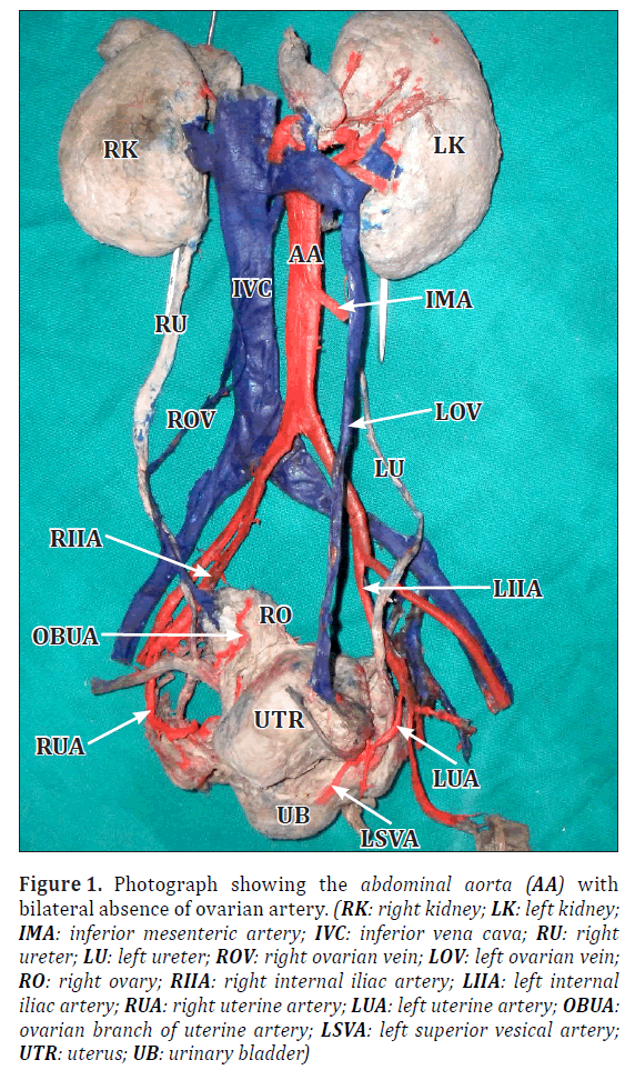
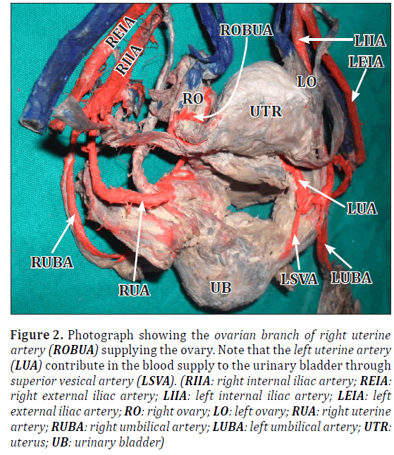
During the anatomical dissection in the Department of Anatomy of International Medical and Technological University (IMTU) we observed bilateral absence of ovarian artery in a 38-year-old Tanzanian female human cadaver. In this case it was observed that in both left and right side there was no ovarian artery supplying the ovaries (Figure 1). Instead both the ovaries were found to receive arterial supply from the ovarian branch of uterine artery (Figure 2). In addition, we observed that the left uterine artery contributed in the blood supply to the urinary bladder, while the right uterine artery didn’t contribute in the blood supply to the urinary bladder (Figure 2). The contribution of uterine artery to supply the urinary bladder occasionally occurs and is regarded as a usual condition.
Figure 1: Photograph showing the abdominal aorta (AA) with bilateral absence of ovarian artery. (RK: right kidney; LK: left kidney; IMA: inferior mesenteric artery; IVC: inferior vena cava; RU: right ureter; LU: left ureter; ROV: right ovarian vein; LOV: left ovarian vein; RO: right ovary; RIIA: right internal iliac artery; LIIA: left internal iliac artery; RUA: right uterine artery; LUA: left uterine artery; OBUA: ovarian branch of uterine artery; LSVA: left superior vesical artery; UTR: uterus; UB: urinary bladder)
Figure 2: Photograph showing the ovarian branch of right uterine artery (ROBUA) supplying the ovary. Note that the left uterine artery (LUA) contribute in the blood supply to the urinary bladder through superior vesical artery (LSVA). (RIIA: right internal iliac artery; REIA: right external iliac artery; LIIA: left internal iliac artery; LEIA: left external iliac artery; RO: right ovary; LO: left ovary; RUA: right uterine artery; RUBA: right umbilical artery; LUBA: left umbilical artery; UTR: uterus; UB: urinary bladder)
Discussion
The absence of ovarian artery either unilateral or bilateral are not common and probably have not been reported by previous authors. Majority of ovarian artery variations that have been reported include variations in the origin and course [5–8]. In the present case there is bilateral absence of ovarian artery, which is extremely uncommon and makes it to be significant (Figure 1). When the ovaries are supplied only by the ovarian branch of uterine artery, any vascular occlusion of this arteries may result in disastrous ischemia of ovaries. Because the ovaries are supplied by ovarian branch of uterine artery, the absence of ovarian artery may be undetectable throughout life of an individual and are usually encountered only during the surgical procedures, angiographic procedures and during cadaveric dissections (Figures 1,2). The knowledge of such variation has clinical importance especially in the field of vascular surgeries and obstetrics and gynecology. Bilateral absence of ovarian artery may cause the potential hazard of ovaries encountered in surgical procedures involving the pelvic organs when the surgeons fails to preserve ovarian branch of uterine artery. So the surgeon must know and take into account the possibility that the anatomic variants should exist as in our case of the bilateral absence of ovarian artery. We believe that, this case will take its place in the literature and play a significant role in the surgical intervention in the abdominal region and also in angiographies. In addition, the observation of left uterine artery to supply the urinary bladder through superior vesical artery (Figure 2) it is regarded as a usual condition, because it occasionally can occur.
Acknowledgements
We (Germanus and Atuganile) would like to take this opportunity to acknowledge with thanks to our supervisor Prof. F. M. Fabian who facilitated a lot in preparation of this case report. Her support and constructive criticism make us to ensure that the quality of the case report is maintained.
We wish to pay special tribute to Mr. K. S. Varma, our assistant supervisor for his great contribution in the image editorial.
We would like to express our sincere thanks to all staff of Gross Anatomy laboratory of IMTU University, Department of Anatomy and Histology, for their technical support and assistance during dissection.
References
- Standring S, ed. Gray’s Anatomy: The Anatomical Basis of Clinical Practice. 40th Ed., Spain, Churchill Livingstone, Elsevier. 2008; 1294.
- Drake RL, Vogl W, Mitchell AWM. Gray’s Anatomy for Students. Philadelphia, Livingstone, Elsevier. 2005; 431.
- Bergman RA, Afifi AK, Miyauchi R. Illustrated Encyclopedia of Human Anatomic Variation: Opus II: Cardiovascular System: Arteries: Pelvis, Uterine Artery. http://www.anatomyatlases.org/AnatomicVariants/Cardiovascular/Text/Arteries/Uterine.shtml (accessed August 2011).
- Cicekcibasi AE, Salbacak A, Seker M, Ziylan T, Buyukmumcu M, Uysal II. The origin of gonadal arteries in human fetuses: anatomical variations. Ann Anat. 2002; 184: 275–279.
- Sulak O, Albay S, Tagil SM, Malas MA. Ovarian arteries with bilateral unusual courses. Saudi Med J. 2005; 26: 1456–1458.
- Rahman HA, Dong K, Yamadori T. Unique course of the ovarian artery associated with other variations. J Anat. 1993; 182: 287–290.
- Nayak S. Abnormal course of left ovarian artery. Int J Anat Var (IJAV). 2008; 1: 4–5.
Germanus Urio Kasindye*, Atuganile Samweli Mwasunga and Flora M. Fabian
Department of Anatomy and Histology, International Medical and Technological University, Dar es Salaam, Tanzania
- *Corresponding Author:
- Germanus U. Kasindye
International Medical and Technological University, Faculty of Medicine, Department of Anatomy & Histology P.O. Box 77594 Dar es Salaam, Tanzania
Tel: +255 713042349
E-mail: kasindyeg@yahoo.com
Date of Received: August 22nd, 2011
Date of Accepted: May 30th, 2012
Published Online: November 3rd, 2012
© Int J Anat Var (IJAV). 2012; 5: 73–75.
Abstract
During dissection of a 38-year-old female human cadaver at International Medical and Technological University (IMTU), Department of Anatomy and Histology the anatomical variation of bilateral absence of ovarian arteries was found. There wasn’t any branch from the abdominal aorta which supplied the ovaries; instead the ovaries were supplied only by the ovarian branch of the uterine artery. Arterial variation of the ovarian artery based on origin and course has been reported previously, but there is no literature which describes bilateral absence of the ovarian artery. Knowledge of this kind of variation will help the surgeon during surgical procedures to be carefully in preserving the ovarian branch of uterine artery that is the sole arterial supplier of the ovaries in the absence of the ovarian arteries.
-Keywords
ovary, bilateral absence of ovarian artery, uterine artery, ovarian branch of uterine artery
Introduction
Ovaries usually are supplied by the gonadal (ovarian) artery and uterine arteries. The ovarian artery usually originates from the abdominal aorta below the renal arteries and then descends to cross the pelvic inlet and supply the ovaries [1]. They anastomose with terminal branches of the uterine arteries [2]. On each side, the vessels travel in the suspensory ligament of the ovary (the infundibulopelvic ligament) as they cross the pelvic inlet to the ovary. Branches pass through the mesovarium to reach the ovary to anastomose with the uterine artery [2]. Early in intrauterine life the ovaries flank the vertebral column inferior to the kidneys, and so the ovarian arteries are relatively short, they gradually lengthen as the ovaries descend into the pelvis [1]. The uterine arteries are branches of the anterior trunk of the internal iliac artery. The uterine artery courses medially and anteriorly in the base of the broad ligament to reach the cervix. Along its course, the vessel crosses the ureter and passes superiorly to the lateral vaginal fornix. Once the vessel reaches the cervix, it ascends along the lateral margin of the uterus to reach the uterine tube where it curves laterally and anastomoses with the ovarian artery [2]. It occasionally supplies branches that may be designated as superior vesical, inferior vesical, ureteral and vaginal arteries [3]. Anatomical variations of ovarian artery have been reported by previous authors; however most of these have shown anatomical variations in the origin and course of the ovarian arteries [4–7]. There has not been any report on the bilateral absence of the ovarian artery have been reported previously. This is therefore considered a rare case of bilateral absence of ovarian artery.
Case Report
During the anatomical dissection in the Department of Anatomy of International Medical and Technological University (IMTU) we observed bilateral absence of ovarian artery in a 38-year-old Tanzanian female human cadaver. In this case it was observed that in both left and right side there was no ovarian artery supplying the ovaries (Figure 1). Instead both the ovaries were found to receive arterial supply from the ovarian branch of uterine artery (Figure 2). In addition, we observed that the left uterine artery contributed in the blood supply to the urinary bladder, while the right uterine artery didn’t contribute in the blood supply to the urinary bladder (Figure 2). The contribution of uterine artery to supply the urinary bladder occasionally occurs and is regarded as a usual condition.
Figure 1: Photograph showing the abdominal aorta (AA) with bilateral absence of ovarian artery. (RK: right kidney; LK: left kidney; IMA: inferior mesenteric artery; IVC: inferior vena cava; RU: right ureter; LU: left ureter; ROV: right ovarian vein; LOV: left ovarian vein; RO: right ovary; RIIA: right internal iliac artery; LIIA: left internal iliac artery; RUA: right uterine artery; LUA: left uterine artery; OBUA: ovarian branch of uterine artery; LSVA: left superior vesical artery; UTR: uterus; UB: urinary bladder)
Figure 2: Photograph showing the ovarian branch of right uterine artery (ROBUA) supplying the ovary. Note that the left uterine artery (LUA) contribute in the blood supply to the urinary bladder through superior vesical artery (LSVA). (RIIA: right internal iliac artery; REIA: right external iliac artery; LIIA: left internal iliac artery; LEIA: left external iliac artery; RO: right ovary; LO: left ovary; RUA: right uterine artery; RUBA: right umbilical artery; LUBA: left umbilical artery; UTR: uterus; UB: urinary bladder)
Discussion
The absence of ovarian artery either unilateral or bilateral are not common and probably have not been reported by previous authors. Majority of ovarian artery variations that have been reported include variations in the origin and course [5–8]. In the present case there is bilateral absence of ovarian artery, which is extremely uncommon and makes it to be significant (Figure 1). When the ovaries are supplied only by the ovarian branch of uterine artery, any vascular occlusion of this arteries may result in disastrous ischemia of ovaries. Because the ovaries are supplied by ovarian branch of uterine artery, the absence of ovarian artery may be undetectable throughout life of an individual and are usually encountered only during the surgical procedures, angiographic procedures and during cadaveric dissections (Figures 1,2). The knowledge of such variation has clinical importance especially in the field of vascular surgeries and obstetrics and gynecology. Bilateral absence of ovarian artery may cause the potential hazard of ovaries encountered in surgical procedures involving the pelvic organs when the surgeons fails to preserve ovarian branch of uterine artery. So the surgeon must know and take into account the possibility that the anatomic variants should exist as in our case of the bilateral absence of ovarian artery. We believe that, this case will take its place in the literature and play a significant role in the surgical intervention in the abdominal region and also in angiographies. In addition, the observation of left uterine artery to supply the urinary bladder through superior vesical artery (Figure 2) it is regarded as a usual condition, because it occasionally can occur.
Acknowledgements
We (Germanus and Atuganile) would like to take this opportunity to acknowledge with thanks to our supervisor Prof. F. M. Fabian who facilitated a lot in preparation of this case report. Her support and constructive criticism make us to ensure that the quality of the case report is maintained.
We wish to pay special tribute to Mr. K. S. Varma, our assistant supervisor for his great contribution in the image editorial.
We would like to express our sincere thanks to all staff of Gross Anatomy laboratory of IMTU University, Department of Anatomy and Histology, for their technical support and assistance during dissection.
References
- Standring S, ed. Gray’s Anatomy: The Anatomical Basis of Clinical Practice. 40th Ed., Spain, Churchill Livingstone, Elsevier. 2008; 1294.
- Drake RL, Vogl W, Mitchell AWM. Gray’s Anatomy for Students. Philadelphia, Livingstone, Elsevier. 2005; 431.
- Bergman RA, Afifi AK, Miyauchi R. Illustrated Encyclopedia of Human Anatomic Variation: Opus II: Cardiovascular System: Arteries: Pelvis, Uterine Artery. http://www.anatomyatlases.org/AnatomicVariants/Cardiovascular/Text/Arteries/Uterine.shtml (accessed August 2011).
- Cicekcibasi AE, Salbacak A, Seker M, Ziylan T, Buyukmumcu M, Uysal II. The origin of gonadal arteries in human fetuses: anatomical variations. Ann Anat. 2002; 184: 275–279.
- Sulak O, Albay S, Tagil SM, Malas MA. Ovarian arteries with bilateral unusual courses. Saudi Med J. 2005; 26: 1456–1458.
- Rahman HA, Dong K, Yamadori T. Unique course of the ovarian artery associated with other variations. J Anat. 1993; 182: 287–290.
- Nayak S. Abnormal course of left ovarian artery. Int J Anat Var (IJAV). 2008; 1: 4–5.


