Bilateral accessory cleidohyoid in a human cadaver
M. Elena Stark1,Bian WU2,Benjamin E. Bluth2 and Jonathan J.Wisco1*
1David Geffen School of Medicine at UCLA, Department of Pathology and Laboratory Medicine, Division of Integrative Anatomy,Los Angeles, CA, USA.
2David Geffen School of Medicine at UCLA, Department of Pathology and Laboratory Medicine, Division of Integrative Anatomy and David Geffen School of Medicine at UCLA,Los Angeles, CA, USA.
- *Corresponding Author:
- Jonathan J. Wisco, PhD
David Geffen School of Medicine at UCLA, Department of Pathology and Laboratory Medicine, Division of Integrative Anatomy,Los Angeles, CA, USA.
Tel: +1 310 825-7880
E-mail: jjwisco@mednet.ucla.edu
Date of Received: July 4th, 2009
Date of Accepted: September 25th, 2009
Published Online: October 10th, 2009
© IJAV. 2009; 2: 122–123.
[ft_below_content] =>Keywords
accessory cleidohyoid,cleidohyoideus accessorius,infrahyoid muscle,anatomical variation
Introduction
Reports of infrahyoid muscle variations abound in the literature. Specifically, anatomical reports mentioning the cleidohyoideus accessorius muscle date back to the 19th century. Several authors have attempted to classify the anatomical variation that constitutes a muscle inserting from the clavicle into the hyoid bone in the presence of an intact omohyoid. In 1897 Le Double described variations of the omohyoid muscle. One such variation was the cleidohyoideus that presented as a double or additional muscle belly inserting into the clavicle and the hyoid bone [1]. In 1912, Loth described cases of infrahyoid muscles originating from the clavicle and inserting into the hyoid bone, classifying them as cleidohyoid muscles [2]. In 1923 Steinbach reviewed the existing literature and classified infrahyoid variations into four categories. One of these categories was that of the “cleidohyoideus accessorius muscle” defined as a “muscle inserting in the clavicle and the hyoid bone when the omohyoid is present” [3]. A more recent literature review by Sasagawa includes ten reports with mention of the cleidohyoideus accessorius muscle [4]. Four of those ten reports state cleidohyoideus accessorius prevalence figures ranging from 0.60 % to 1.04 %. However no details in terms of the unilaterality or bilaterality of the muscle are mentioned. In 1996 in his encyclopedia of human anatomical variation, Bergman illustrated numerous variations of accessory muscles to the omohyoid. One of these variations consists of a fascicle with insertions in the medial clavicular head and the hyoid bone. No data on the prevalence or the characteristics of this variation were reported [5]. Fukuda [6], and Hatipoglu [7] document this variation found unilaterally. This paper describes a case of the cleidohyoideus accessorius muscle found bilaterally during routine dissection of a human cadaver. A brief discussion of the clinical significance of this abnormality is included. To our knowledge, this is the first definitive case of a bilateral cledohyoideus accessorius muscle in the medical literature.
Case Report
During routine anatomical dissection at the David Geffen School of Medicine at UCLA, a rare infrahyoid muscle variation was found in an 88-year-old Caucasian male cadaver. This report describes the bilateral cleidohyoideus accessorius muscle that was found in the presence of an intact omohyoid muscle on both sides.
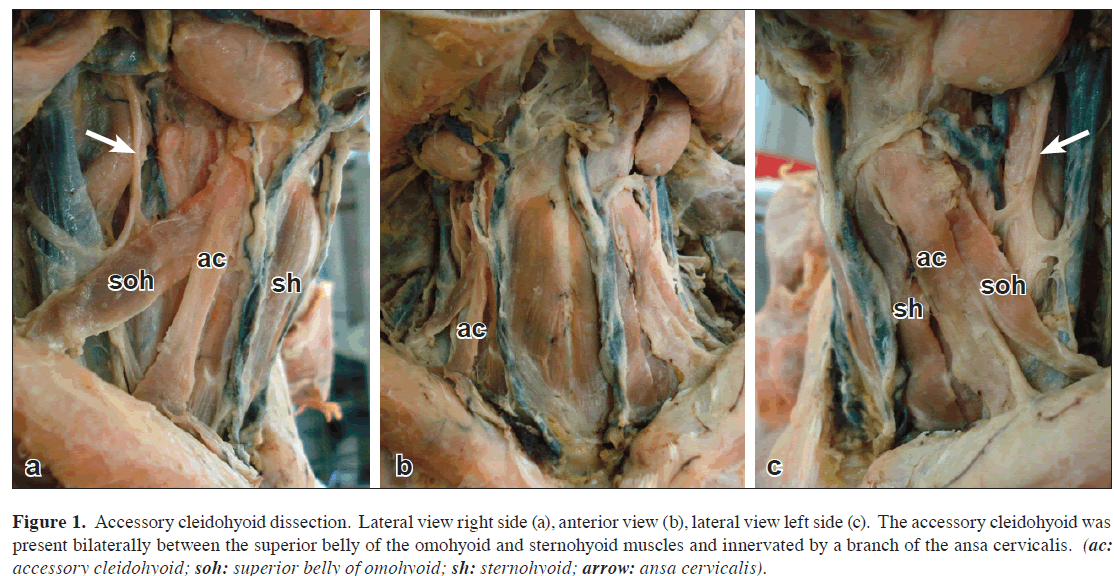
The cadaver exhibited a typical bilateral sternocleidomastoid muscle that was reflected, revealing a typical sternohyoid, sternothyroid, thyrohyoid and omohyoid muscles on both sides. The atypical muscle described in this case report originated from the medial end of the clavicle, coursed parallel and lateral to the sternohyoid muscle and inserted into the hyoid bone. Because this muscle was found in the presence of a typical and intact omohyoid, it was classified as a cleidohyoideus accessorius. From its origin on the clavicle to the hyoid bone the cleidohyoideus accessorius muscle measured 6.4 cm on the left and 6.2 cm on the right (Figure 1). No other muscular or neurovascular abnormalities were observed.
Figure 1: Accessory cleidohyoid dissection. Lateral view right side (a), anterior view (b), lateral view left side (c). The accessory cleidohyoid was present bilaterally between the superior belly of the omohyoid and sternohyoid muscles and innervated by a branch of the ansa cervicalis. (ac: accessory cleidohyoid; soh: superior belly of omohyoid; sh: sternohyoid; arrow: ansa cervicalis).
The ansa cervicalis was found to be typical and it innervated the omohyoid, sternohyoid and sternothyroid muscles as well as the cleidohyoideus accessorius muscle.
Discussion
Multiple cases of atypical infrahyoid muscles have been described. The omohyoid is one of the muscles of the neck with the highest incidence of variations and additional bellies or accessory muscles. Accessory omohyoid muscles originating from the clavicle are much less common than those originating from the scapula or transverse scapular ligament [8-11]. Le Double [1], Loth [2], and Steinbach [3] were the first authors to describe the existence of an infrahyoid muscle originating from the clavicle and inserting into the hyoid bone (cleidohyoid muscle). Steinbach [3] reviewed and categorized cases of cleidohyoid muscles into four categories, one of which included muscles that originate from the clavicle and insert into the hyoid bone, in the presence of an intact omohyoid. The cleidohyoideus accessorius muscle has been described by a number of investigators since [5,6,12]. To our knowledge, this is the first reported case of a bilateral cleidohyoideus accessorius.
Variations of the infrahyoid muscles, including the cleidohyoideus accessorius, are important to consider mainly because of their relevance in diagnostic studies and as surgical landmarks. Knowledge of potential abnormalities in the region is important for any physician or head and neck surgeon.
References
- Le Double AF. Traité des variations du systéme musculaire de l’homme et de leur signification au point de vue de l’anthropologie zoologique. Tome 2. Paris, Libraire C. Rerinwald, Schleicher Fréres. 1897; 129.
- Loth E. Musceln des Halses. Beitrage zur Anthropologie der Negerweichteile. Stuttgart, Streker & Schroder. 1912; 58–73.
- Steinbach K. Uber Varietation der Unterzungenbein- und Brustmuskulatur. Anat Anz. 1923; 15: 488–506.
- Sasagawa I, Takahashi K, Igarashi A, Mori H, Kobayashi K. [A case of an abnormal bundle from the anterior margin of the right and left trapezius and an abnormality in the right omohyoid appearance in a cadaver]. Shigaku. 1982; 70: 439–448. (Japanese)
- Bergman RA, Afifi AK, Miyauchi R. Muscular System; Omohyoideus, Sternohyoideus, Thyrohyoideus, Sternohyoideus (Infrahyoid Muscles). Illustrated Encyclopedia of Human Anatomic Variation: Opus I: Muscular System: Alphabetical Listing of Muscles: O. 2009. http://www.anatomyatlases.org/AnatomicVariants/MuscularSystem/Text/O/14Omohyoideus.shtml (accessed June 2009).
- Fukuda H, Onizawa K, Hagiwara T, Iwama H. The omohyoid muscle: a variation seen in radical neck dissection. Br J Oral Maxillofac Surg. 1998; 36: 399–400.
- Hatipoglu ES, Kervancioglu P, Tuncer MC. An unusual variation of the omohyoid muscle and review of literature. Ann Anat. 2006; 188: 469–472.
- Woods J. Additional Varieties in Human Myology. Proceedings of the Royal Society. 1865; 14: 379–392.
- Woods J. Variations in Human Morphology Observed during the Winter Session of 1866-67. London, King’s College. 1866-67; 15: 518–546.
- Woods J. Variations in Human Morphology Observed during the Winter Session of 1867-68. London, King’s College; 1867-68; 16: 483–525.
- Woods J. On a group of varieties of the muscles of the human neck, shoulder, and chest, with their transitional forms and homologies in mammals. Philosophical Transactions of the Royal Society of London. 1870; 160: 83–116.
- Takano T, Takaya M, Iizuka K, Tomagasawa T, Adachi H. Statistical observation of anomalies of infrahyoid muscles. Iwate Ikadaigaku Kaibougaku Kyousitu Gyouseki Syu. 1955; 2: 113–124.
M. Elena Stark1,Bian WU2,Benjamin E. Bluth2 and Jonathan J.Wisco1*
1David Geffen School of Medicine at UCLA, Department of Pathology and Laboratory Medicine, Division of Integrative Anatomy,Los Angeles, CA, USA.
2David Geffen School of Medicine at UCLA, Department of Pathology and Laboratory Medicine, Division of Integrative Anatomy and David Geffen School of Medicine at UCLA,Los Angeles, CA, USA.
- *Corresponding Author:
- Jonathan J. Wisco, PhD
David Geffen School of Medicine at UCLA, Department of Pathology and Laboratory Medicine, Division of Integrative Anatomy,Los Angeles, CA, USA.
Tel: +1 310 825-7880
E-mail: jjwisco@mednet.ucla.edu
Date of Received: July 4th, 2009
Date of Accepted: September 25th, 2009
Published Online: October 10th, 2009
© IJAV. 2009; 2: 122–123.
Abstract
During routine anatomical dissection of the infrahyoid region, a muscle was found bilaterally originating from the sternal end of clavicle and inserting into the hyoid bone. The muscle coursed parallel and lateral to the sternohyoid muscle. The muscle was found in the presence of an intact omohyoid, thus being classified as an accessory cleidohyoid (cleidohyoideus accessorius) muscle. While other authors have reported the presence of a unilateral cleidohyoideus accessorius muscle, to our knowledge this is the first case of a bilateral cleidohyoideus accessorius muscle in the medical literature. Anatomical variations of the infrahyoid muscles may have functional, diagnostic, surgical and pathological implications.
-Keywords
accessory cleidohyoid,cleidohyoideus accessorius,infrahyoid muscle,anatomical variation
Introduction
Reports of infrahyoid muscle variations abound in the literature. Specifically, anatomical reports mentioning the cleidohyoideus accessorius muscle date back to the 19th century. Several authors have attempted to classify the anatomical variation that constitutes a muscle inserting from the clavicle into the hyoid bone in the presence of an intact omohyoid. In 1897 Le Double described variations of the omohyoid muscle. One such variation was the cleidohyoideus that presented as a double or additional muscle belly inserting into the clavicle and the hyoid bone [1]. In 1912, Loth described cases of infrahyoid muscles originating from the clavicle and inserting into the hyoid bone, classifying them as cleidohyoid muscles [2]. In 1923 Steinbach reviewed the existing literature and classified infrahyoid variations into four categories. One of these categories was that of the “cleidohyoideus accessorius muscle” defined as a “muscle inserting in the clavicle and the hyoid bone when the omohyoid is present” [3]. A more recent literature review by Sasagawa includes ten reports with mention of the cleidohyoideus accessorius muscle [4]. Four of those ten reports state cleidohyoideus accessorius prevalence figures ranging from 0.60 % to 1.04 %. However no details in terms of the unilaterality or bilaterality of the muscle are mentioned. In 1996 in his encyclopedia of human anatomical variation, Bergman illustrated numerous variations of accessory muscles to the omohyoid. One of these variations consists of a fascicle with insertions in the medial clavicular head and the hyoid bone. No data on the prevalence or the characteristics of this variation were reported [5]. Fukuda [6], and Hatipoglu [7] document this variation found unilaterally. This paper describes a case of the cleidohyoideus accessorius muscle found bilaterally during routine dissection of a human cadaver. A brief discussion of the clinical significance of this abnormality is included. To our knowledge, this is the first definitive case of a bilateral cledohyoideus accessorius muscle in the medical literature.
Case Report
During routine anatomical dissection at the David Geffen School of Medicine at UCLA, a rare infrahyoid muscle variation was found in an 88-year-old Caucasian male cadaver. This report describes the bilateral cleidohyoideus accessorius muscle that was found in the presence of an intact omohyoid muscle on both sides.
The cadaver exhibited a typical bilateral sternocleidomastoid muscle that was reflected, revealing a typical sternohyoid, sternothyroid, thyrohyoid and omohyoid muscles on both sides. The atypical muscle described in this case report originated from the medial end of the clavicle, coursed parallel and lateral to the sternohyoid muscle and inserted into the hyoid bone. Because this muscle was found in the presence of a typical and intact omohyoid, it was classified as a cleidohyoideus accessorius. From its origin on the clavicle to the hyoid bone the cleidohyoideus accessorius muscle measured 6.4 cm on the left and 6.2 cm on the right (Figure 1). No other muscular or neurovascular abnormalities were observed.
Figure 1: Accessory cleidohyoid dissection. Lateral view right side (a), anterior view (b), lateral view left side (c). The accessory cleidohyoid was present bilaterally between the superior belly of the omohyoid and sternohyoid muscles and innervated by a branch of the ansa cervicalis. (ac: accessory cleidohyoid; soh: superior belly of omohyoid; sh: sternohyoid; arrow: ansa cervicalis).
The ansa cervicalis was found to be typical and it innervated the omohyoid, sternohyoid and sternothyroid muscles as well as the cleidohyoideus accessorius muscle.
Discussion
Multiple cases of atypical infrahyoid muscles have been described. The omohyoid is one of the muscles of the neck with the highest incidence of variations and additional bellies or accessory muscles. Accessory omohyoid muscles originating from the clavicle are much less common than those originating from the scapula or transverse scapular ligament [8-11]. Le Double [1], Loth [2], and Steinbach [3] were the first authors to describe the existence of an infrahyoid muscle originating from the clavicle and inserting into the hyoid bone (cleidohyoid muscle). Steinbach [3] reviewed and categorized cases of cleidohyoid muscles into four categories, one of which included muscles that originate from the clavicle and insert into the hyoid bone, in the presence of an intact omohyoid. The cleidohyoideus accessorius muscle has been described by a number of investigators since [5,6,12]. To our knowledge, this is the first reported case of a bilateral cleidohyoideus accessorius.
Variations of the infrahyoid muscles, including the cleidohyoideus accessorius, are important to consider mainly because of their relevance in diagnostic studies and as surgical landmarks. Knowledge of potential abnormalities in the region is important for any physician or head and neck surgeon.
References
- Le Double AF. Traité des variations du systéme musculaire de l’homme et de leur signification au point de vue de l’anthropologie zoologique. Tome 2. Paris, Libraire C. Rerinwald, Schleicher Fréres. 1897; 129.
- Loth E. Musceln des Halses. Beitrage zur Anthropologie der Negerweichteile. Stuttgart, Streker & Schroder. 1912; 58–73.
- Steinbach K. Uber Varietation der Unterzungenbein- und Brustmuskulatur. Anat Anz. 1923; 15: 488–506.
- Sasagawa I, Takahashi K, Igarashi A, Mori H, Kobayashi K. [A case of an abnormal bundle from the anterior margin of the right and left trapezius and an abnormality in the right omohyoid appearance in a cadaver]. Shigaku. 1982; 70: 439–448. (Japanese)
- Bergman RA, Afifi AK, Miyauchi R. Muscular System; Omohyoideus, Sternohyoideus, Thyrohyoideus, Sternohyoideus (Infrahyoid Muscles). Illustrated Encyclopedia of Human Anatomic Variation: Opus I: Muscular System: Alphabetical Listing of Muscles: O. 2009. http://www.anatomyatlases.org/AnatomicVariants/MuscularSystem/Text/O/14Omohyoideus.shtml (accessed June 2009).
- Fukuda H, Onizawa K, Hagiwara T, Iwama H. The omohyoid muscle: a variation seen in radical neck dissection. Br J Oral Maxillofac Surg. 1998; 36: 399–400.
- Hatipoglu ES, Kervancioglu P, Tuncer MC. An unusual variation of the omohyoid muscle and review of literature. Ann Anat. 2006; 188: 469–472.
- Woods J. Additional Varieties in Human Myology. Proceedings of the Royal Society. 1865; 14: 379–392.
- Woods J. Variations in Human Morphology Observed during the Winter Session of 1866-67. London, King’s College. 1866-67; 15: 518–546.
- Woods J. Variations in Human Morphology Observed during the Winter Session of 1867-68. London, King’s College; 1867-68; 16: 483–525.
- Woods J. On a group of varieties of the muscles of the human neck, shoulder, and chest, with their transitional forms and homologies in mammals. Philosophical Transactions of the Royal Society of London. 1870; 160: 83–116.
- Takano T, Takaya M, Iizuka K, Tomagasawa T, Adachi H. Statistical observation of anomalies of infrahyoid muscles. Iwate Ikadaigaku Kaibougaku Kyousitu Gyouseki Syu. 1955; 2: 113–124.


