Bilateral additional bellies of the soleus muscle: anatomical and clinical insight
Sarabpreet Singh*, R. K. Suri, Vandana Mehta, Hitendra Loh, Jyoti Arora, Gayatri Rath
Department of Anatomy, Vardhman Mahavir Medical College and Safdarjang Hospital, New Delhi, India.
- *Corresponding Author:
- Dr. Sarabpreet SINGH, MD, MBBS
Department Of Anatomy, Vardhman Mahavir Medical College and, Safdarjang Hospital, New Delhi, 110029, India.
Tel: +91 989 9009497
Fax: +91 011 27654007
E-mail: drsarabpreet@gmail.com
Date of Received: October 14th, 2008
Date of Accepted: January 18th, 2009
Published Online: January 31st, 2009
© IJAV. 2009; 2: 20–22.
[ft_below_content] =>Keywords
soleus, gastrocnemius, tendo calcaneus, tendo-Achilles, fascicle, anlage
Introduction
The soleus muscle arises primarily by two heads, which are united by a tendinous arch. The fibular head of the soleus arises from the head of the fibula and about third of the shaft; the tibial head arises from the soleal line on the tibia. Muscular fibers of the soleus end in a broad aponeurosis and unite with aponeurosis of the gastrocnemius to form tendo calcaneus. It is supplied by branches of popliteal artery and its innervation is derived from tibial nerve [1].
The current study demonstrates presence of additional musculo-tendinous bellies attached to the soleus both proximally and distally.
Injuries of the distal third of the leg pose a clinical challenge due to closeness of the skin and poor circulation [2]. Appropriate coverage of these defects through flaps is being increasingly used by surgeons underlying the importance of a detailed knowledge of this rare musculo-tendinous belly.
To the best of our knowledge, the bilateral presence of additional bellies of soleus muscle has been rarely reported.
Case Report
We observed bilateral additional musculo-tendinous soleus bellies in a 45-year-old male cadaver, during undergraduate dissection session. The additional bellies were cleaned by fine dissection and their attachments were defined. Appropriate photographs were taken.
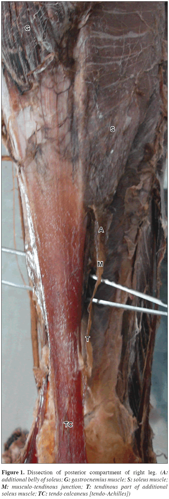
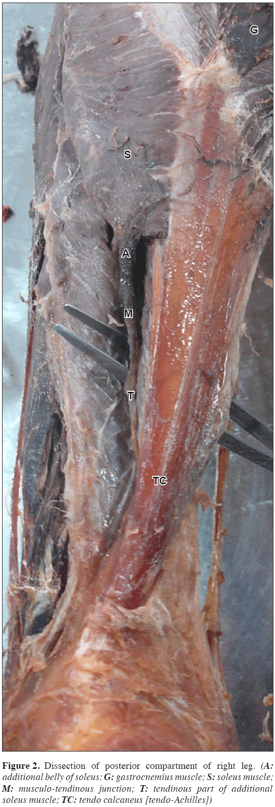
The additional bellies originated from inferolateral end of main soleus muscle (Figures 1 and 2). Both the bellies traversed vertically downwards on the lateral side of tendo-Achilles and tapered into a tendon. The tendon continued longitudinally and medially to insert onto the lateral aspect of the tendo-Achilles. The additional musculo-tendinous belly did not display any bony attachments. There was no evidence of separate vascular or nerve supply to the additional bellies.
The additional belly was longer on left side (8.4 cm) than the right side (7.5 cm). The tendon of both the fasciculi inserted on the tendo-Achilles 7 cm from the calcaneal tuberosity.
Discussion
The present study demonstrates bilateral additional bellies in relation to soleus muscle and deserves special consideration in view of its clinical significance. Though accessory fasciculus, supernumerary fascicles and accessory soleus muscle have been described, the above-mentioned additional bellies are distinct in context of their origin, course and insertion.
An accessory fasciculus is sometimes formed on the anterior surface of the soleus. Its fibers take origin from the fascial covering of soleus and run posteromedially to a bipenniform insertion into a thin lamina that joins with the tendo-Achilles [3]. However, this fasciculus is tightly adhered to the main soleus.
Supernumerary fascicles of muscle have also been described. These are usually reported as thin flat muscles originating from the fibula and soleal line of the tibia or from the deep fascia of the soleus and inserting via a tendon into the calcaneus medial to the tendo-Achilles [4].
In the present case, it cannot be classified as a fasciculus, since it had both muscular and tendinous components. However, it can be considered as a variant of accessory soleus muscle.
The accessory soleus muscle is a congenital anatomical variation that was first described by Cruvelhier in 1843 [4]. According to Petterson et al. (1987), the incidence of the accessory soleus muscle ranges from 0.7 to 5.5% [5].
Five types of insertion of the accessory soleus muscle have been identified: insertion along the tendo-Achilles, tendinous insertion on superior surface of the calcaneus, fleshy insertion on the superior surface of the calcaneus, fleshy insertion on the medial surface of the calcaneus, and tendinous insertion on the medial surface of calcaneus [6].
In a study by Barberini et al (2003), the tendon of accessory soleus was observed to traverse medial to the calcaneal tendon [7]. In the present study, it was a musculo-tendinous slip, originating from the inferolateral aspect of soleus, coursing on the lateral aspect of the tendo-Achilles and fusing distally with the tendon.
Interestingly, the presence of accessory soleus was accompanied by the absence of plantaris muscle [8]. This suggested that the accessory soleus might be a variant of plantaris that may have migrated to the anterior aspect of main soleus muscle. In the present study, the plantaris muscle was also present bilaterally.
The anatomical findings in the present case are in favor of an accessory soleus muscle unusual in its origin and disposition and associated with the presence of plantaris muscle. To the best of our knowledge, such an anomalous accessory soleus muscle has not been reported earlier.
Embryologically, the single anlage of the soleus muscle may undergo early splitting; leading to the development of an accessory muscle [4]. This muscle can derive its blood supply and innervation independent of the soleus muscle or in common with it [9].
Usually, the accessory soleus muscle is asymptomatic and goes unnoticed. However, it can lead to a painful swelling (most common), painless swelling, or association with clubfoot or equines deformity [4]. Painful swelling is thought to be due to an increase in the size of the muscle causing either muscle ischemia [4] or a compressive neuropathy involving the posterior tibial nerve [10].
It is well established that local muscle flaps are easier to perform than microsurgical flaps [9]. The soleus muscle is frequently used to reconstruct soft tissue defects of the lower limb [2]. Beck (2003) reported that the soleus muscle is a valuable tool for flap coverage of wounds of distal third of the leg [9].
Our premise in the present study is that awareness of this soleus muscle variant is imperative as one could diagnose its presence preoperatively by MRI scanning. Thus, this could help the reconstructive surgeon to plan the surgery judiciously by using the accessory muscle as a flap for the coverage of the soft tissue defects of leg.
Conclusion
The present study displays bilateral anomalous pattern of the soleus muscle. The musculo-tendinous belly was uniquely present on the lateral aspect of the soleus muscle fusing both proximally and distally with it. These accessory muscles could be effectively used for flap repairs in the coverage of soft tissue defects of distal third of the leg owing to poor vascularity of this region. The study of soleus muscle anatomy and its variations is of paramount importance to surgeons undertaking reconstructive procedures and to radiologists in interpretations of MRI scans. The occurrence of muscular anomalies as in the current report should not be overlooked because of their propensity to cause misdiagnosis and conflicting interpretations.
References
- Hollinshead WH. Anatomy for Surgeons, Vol 3, The Back and Limbs. 3rd Ed., Philadelphia, Harper and Row Publisher Inc. 1982; 775.
- Fayman MS, Orak F, Hugo B, Berson SD. The distally based split soleus muscle flap. Br J Plast Surg. 1987; 40: 20–26.
- Anson B. Morris’ Human Anatomy. 12th Ed., New York, McGraw-Hill Co., 1966; 592.
- Palaniappan M, Rajesh A, Rickett A, Kershaw CJ. Accessory soleus muscle: a case report and review of the literature. Pediatric Radiol. 1999; 29: 610–612.
- Pettersson H, Giovannetti M, Gillespy T 3rd, Slone R, Springfield D. Magnetic resonance imaging appearance of supernumerary soleus muscle. Eur J Radiol. 1987; 7: 149–150.
- Yu JS, Resnick D. MR imaging of the accessory soleus muscle appearance in six patients and a review of the literature. Skeletal Radiol. 1994; 23: 525–528.
- Barberini F, Bucciarelli-Ducci C, Zani A, Cerasoli D. Unusual extended fibular origin of the human soleus muscle: possible morpho-physiologic significance based on comparative anatomy. Clin Anat. 2003; 16: 383–388.
- Frohse F, Fränkel M. Die Muskeln des menschlichen Beines. In: Bardeleben K, ed. Handbuch der Anatomie des Menschen. Jena, Fischer. 1913; 415–693.
- Beck JB, Stile F, Lineaweaver W. Reconsidering the soleus muscle flap for coverage of wounds of distal third of the leg. Ann Plast Surg. 2003; 50: 631–635.
- Trosko JJ. Accessory soleus: A clinical perspective and report of three cases. J Foot Surg. 1986; 25: 296–300.
Sarabpreet Singh*, R. K. Suri, Vandana Mehta, Hitendra Loh, Jyoti Arora, Gayatri Rath
Department of Anatomy, Vardhman Mahavir Medical College and Safdarjang Hospital, New Delhi, India.
- *Corresponding Author:
- Dr. Sarabpreet SINGH, MD, MBBS
Department Of Anatomy, Vardhman Mahavir Medical College and, Safdarjang Hospital, New Delhi, 110029, India.
Tel: +91 989 9009497
Fax: +91 011 27654007
E-mail: drsarabpreet@gmail.com
Date of Received: October 14th, 2008
Date of Accepted: January 18th, 2009
Published Online: January 31st, 2009
© IJAV. 2009; 2: 20–22.
Abstract
Bilateral additional musculo-tendinous bellies of soleus muscle were encountered during undergraduate gross anatomy teaching program. The additional bellies were found to arise from infero-lateral aspect of the soleus muscle. Distally, this muscle belly tapered into a long tendon measuring 4.3 cm and 4.6 cm on left and right sides, respectively. The tendons on both sides, inserted to the lateral aspect of respective tendo-Achilles. The additional musculo-tendinous bellies had no demonstrable bony attachments and no separate vascular or nerve supply. The clinical relevance of soleus muscle flap in reparative and reconstructive surgeries of distal third of leg is discussed and a possible role of this accessory belly in tendon transfer is being emphasized.
-Keywords
soleus, gastrocnemius, tendo calcaneus, tendo-Achilles, fascicle, anlage
Introduction
The soleus muscle arises primarily by two heads, which are united by a tendinous arch. The fibular head of the soleus arises from the head of the fibula and about third of the shaft; the tibial head arises from the soleal line on the tibia. Muscular fibers of the soleus end in a broad aponeurosis and unite with aponeurosis of the gastrocnemius to form tendo calcaneus. It is supplied by branches of popliteal artery and its innervation is derived from tibial nerve [1].
The current study demonstrates presence of additional musculo-tendinous bellies attached to the soleus both proximally and distally.
Injuries of the distal third of the leg pose a clinical challenge due to closeness of the skin and poor circulation [2]. Appropriate coverage of these defects through flaps is being increasingly used by surgeons underlying the importance of a detailed knowledge of this rare musculo-tendinous belly.
To the best of our knowledge, the bilateral presence of additional bellies of soleus muscle has been rarely reported.
Case Report
We observed bilateral additional musculo-tendinous soleus bellies in a 45-year-old male cadaver, during undergraduate dissection session. The additional bellies were cleaned by fine dissection and their attachments were defined. Appropriate photographs were taken.
The additional bellies originated from inferolateral end of main soleus muscle (Figures 1 and 2). Both the bellies traversed vertically downwards on the lateral side of tendo-Achilles and tapered into a tendon. The tendon continued longitudinally and medially to insert onto the lateral aspect of the tendo-Achilles. The additional musculo-tendinous belly did not display any bony attachments. There was no evidence of separate vascular or nerve supply to the additional bellies.
The additional belly was longer on left side (8.4 cm) than the right side (7.5 cm). The tendon of both the fasciculi inserted on the tendo-Achilles 7 cm from the calcaneal tuberosity.
Discussion
The present study demonstrates bilateral additional bellies in relation to soleus muscle and deserves special consideration in view of its clinical significance. Though accessory fasciculus, supernumerary fascicles and accessory soleus muscle have been described, the above-mentioned additional bellies are distinct in context of their origin, course and insertion.
An accessory fasciculus is sometimes formed on the anterior surface of the soleus. Its fibers take origin from the fascial covering of soleus and run posteromedially to a bipenniform insertion into a thin lamina that joins with the tendo-Achilles [3]. However, this fasciculus is tightly adhered to the main soleus.
Supernumerary fascicles of muscle have also been described. These are usually reported as thin flat muscles originating from the fibula and soleal line of the tibia or from the deep fascia of the soleus and inserting via a tendon into the calcaneus medial to the tendo-Achilles [4].
In the present case, it cannot be classified as a fasciculus, since it had both muscular and tendinous components. However, it can be considered as a variant of accessory soleus muscle.
The accessory soleus muscle is a congenital anatomical variation that was first described by Cruvelhier in 1843 [4]. According to Petterson et al. (1987), the incidence of the accessory soleus muscle ranges from 0.7 to 5.5% [5].
Five types of insertion of the accessory soleus muscle have been identified: insertion along the tendo-Achilles, tendinous insertion on superior surface of the calcaneus, fleshy insertion on the superior surface of the calcaneus, fleshy insertion on the medial surface of the calcaneus, and tendinous insertion on the medial surface of calcaneus [6].
In a study by Barberini et al (2003), the tendon of accessory soleus was observed to traverse medial to the calcaneal tendon [7]. In the present study, it was a musculo-tendinous slip, originating from the inferolateral aspect of soleus, coursing on the lateral aspect of the tendo-Achilles and fusing distally with the tendon.
Interestingly, the presence of accessory soleus was accompanied by the absence of plantaris muscle [8]. This suggested that the accessory soleus might be a variant of plantaris that may have migrated to the anterior aspect of main soleus muscle. In the present study, the plantaris muscle was also present bilaterally.
The anatomical findings in the present case are in favor of an accessory soleus muscle unusual in its origin and disposition and associated with the presence of plantaris muscle. To the best of our knowledge, such an anomalous accessory soleus muscle has not been reported earlier.
Embryologically, the single anlage of the soleus muscle may undergo early splitting; leading to the development of an accessory muscle [4]. This muscle can derive its blood supply and innervation independent of the soleus muscle or in common with it [9].
Usually, the accessory soleus muscle is asymptomatic and goes unnoticed. However, it can lead to a painful swelling (most common), painless swelling, or association with clubfoot or equines deformity [4]. Painful swelling is thought to be due to an increase in the size of the muscle causing either muscle ischemia [4] or a compressive neuropathy involving the posterior tibial nerve [10].
It is well established that local muscle flaps are easier to perform than microsurgical flaps [9]. The soleus muscle is frequently used to reconstruct soft tissue defects of the lower limb [2]. Beck (2003) reported that the soleus muscle is a valuable tool for flap coverage of wounds of distal third of the leg [9].
Our premise in the present study is that awareness of this soleus muscle variant is imperative as one could diagnose its presence preoperatively by MRI scanning. Thus, this could help the reconstructive surgeon to plan the surgery judiciously by using the accessory muscle as a flap for the coverage of the soft tissue defects of leg.
Conclusion
The present study displays bilateral anomalous pattern of the soleus muscle. The musculo-tendinous belly was uniquely present on the lateral aspect of the soleus muscle fusing both proximally and distally with it. These accessory muscles could be effectively used for flap repairs in the coverage of soft tissue defects of distal third of the leg owing to poor vascularity of this region. The study of soleus muscle anatomy and its variations is of paramount importance to surgeons undertaking reconstructive procedures and to radiologists in interpretations of MRI scans. The occurrence of muscular anomalies as in the current report should not be overlooked because of their propensity to cause misdiagnosis and conflicting interpretations.
References
- Hollinshead WH. Anatomy for Surgeons, Vol 3, The Back and Limbs. 3rd Ed., Philadelphia, Harper and Row Publisher Inc. 1982; 775.
- Fayman MS, Orak F, Hugo B, Berson SD. The distally based split soleus muscle flap. Br J Plast Surg. 1987; 40: 20–26.
- Anson B. Morris’ Human Anatomy. 12th Ed., New York, McGraw-Hill Co., 1966; 592.
- Palaniappan M, Rajesh A, Rickett A, Kershaw CJ. Accessory soleus muscle: a case report and review of the literature. Pediatric Radiol. 1999; 29: 610–612.
- Pettersson H, Giovannetti M, Gillespy T 3rd, Slone R, Springfield D. Magnetic resonance imaging appearance of supernumerary soleus muscle. Eur J Radiol. 1987; 7: 149–150.
- Yu JS, Resnick D. MR imaging of the accessory soleus muscle appearance in six patients and a review of the literature. Skeletal Radiol. 1994; 23: 525–528.
- Barberini F, Bucciarelli-Ducci C, Zani A, Cerasoli D. Unusual extended fibular origin of the human soleus muscle: possible morpho-physiologic significance based on comparative anatomy. Clin Anat. 2003; 16: 383–388.
- Frohse F, Fränkel M. Die Muskeln des menschlichen Beines. In: Bardeleben K, ed. Handbuch der Anatomie des Menschen. Jena, Fischer. 1913; 415–693.
- Beck JB, Stile F, Lineaweaver W. Reconsidering the soleus muscle flap for coverage of wounds of distal third of the leg. Ann Plast Surg. 2003; 50: 631–635.
- Trosko JJ. Accessory soleus: A clinical perspective and report of three cases. J Foot Surg. 1986; 25: 296–300.


