Bilateral extensor digitorum brevis manus: case report*
Sofía Mansilla*, Alejandra Mansilla,Alejandro Russo and Eduardo Olivera
Anatomy Department, Facultad de Medicina, Universidad de la República, Montevideo, Uruguay
- *Corresponding Author:
- Sofía Mansilla
Anatomy Department Facultad de Medicina Universidad de la República Montevideo, Uruguay
Tel: +598 (2) 7098369
E-mail: sofiamansillarud@gmail.com
Date of Received: September 29th, 2016
Date of Accepted: December 30th, 2016
Published Online: February 7th, 2017
© Int J Anat Var (IJAV). 2016; 9: 85–87.
[ft_below_content] =>Keywords
bilateral,extensor digitorum brevis manus
Introduction
Anatomic variants of the muscles of the hand, and specially dorsum muscles have important clinical implications. As a matter of fact, these variants are related to surgical procedures such as: muscle transfers, tendon repairs and tendon graft surgeries [1,2].
One of this variant found on the dorsum of the hand is the extensor digitorum brevis manus (EDBM).
Usually not mentioned in classic anatomic textbooks, its first description is acknowledged to Albinus in 1734 [3]. Later, it was McAlister the one who coined the term “Extensor Digitorum Brevis Manus” in 1866 [1]. Depending on its distal insertion, other authors have called this variant “Extensor Inidicis Brevis”, Extensor Digiti III brevis”, “Extensor Medii” and “Anular Brevis” [3]. The purpose of this paper is to report a cadaveric case of EDBM. Anatomic and clinical implications of this variant are discussed here.
Case Report
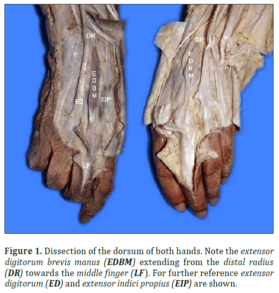
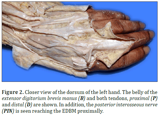
During a routine dissection of a formalin-fixed adult female cadaver, an unusual muscle was found in the dorsum of both hands. The dissection was carried out in the Anatomy Department, Facultad de Medicina, Universidad de la República, Montevideo, Uruguay, by the authors of this paper. In both hands the variant muscle originated via a proximal tendon inserted on the dorsal surface of the distal radius. The muscle belly ran between the third and fourth metacarpal bones. Finally, the distal insertion was bilaterally on the common extensor tendon of the middle finger by means of a tendon, corresponding to a case of EDBM.
The muscle on the left had a length, from proximal to distal insertion, of 8.5 cm. The muscle belly was 4.4 cm long. Each tendon, proximal and distal had a length of 1.0 and 3.1 cm, respectively. The muscle belly width was 1.3 cm, the proximal tendon was 0.4 cm and the distal 0.5 cm. The belly´s thickness was of 0.4 cm, proximal tendon thickness was 0.05 cm and on the distal tendon 0.1 cm.
On the right hand, the muscle´s length was 7.1 cm. Its belly was 4.7 cm long, and the proximal and distal tendons were 0.9 and 1.5 cm long each. The muscle belly width was 0.5 cm, 0.2 cm the proximal tendon and 0.5 cm the distal tendon. Finally the thickness of the belly was 0.5 cm, the proximal tendon was 0.2 cm and 0.1 cm the distal tendon.
The asymmetry between both muscles, suggests that probably the left hand was the dominant one in this case. Both muscles were innervated by the posterior interosseous nerve.
Discussion
The EDBM consists of a very rare variant. Its frequency is estimated of about 4% on the general population [1]. Of these, in only 26% of the cases the presentation is bilateral [3], as reported here. There are no differences related to variables such as ancestry, gender or side [3].
There are two main theories regarding the phylogeny of the EDBM. Firstly, the EDBM is atavistic. In nearly all amphibians, EDBM is present where it mainly controls the extension of the finger of the forelimb. In humans, as its function is taken over by the forearm muscles, the EDBM has been tending to disappear [4]. On the other hand, the second theory states that there are three primordia of the extensor muscles of the forearm: radial, superficial and deep. The last, innervated by the posterior interosseous nerve, originates on the radial side: the abductor pollicis longus and the extensor pollicis brevis, and on the ulnar side: the extensor pollicis longus and the extensor indicis proprius (EIP). According to Bunnel [5] and Souter [6], the EDBM may represent the failure of proximal migration of ulnar element of the deep muscle mass.
The EDBM is classified into three types according to its distal insertion and relation to the EIP [7]. Type I was defined as EDBM with insertion into the dorsal aponeurosis of the index finger combined with absence of EIP. Type II was defined as EDBM inserted on the index finger in presence of EIP. Finally,in type III the EIP inserted on the index finger, but the EDBM inserted on the long finger. Taking this into account, both EDBMs here reported correspond to type III.
From a clinical stand point, the presence of EDBM is usually asymptomatic. Its presence is associated with the so-called “fourth compartment syndrome” [8], leading to dorsal chronic wrist pain. This condition is in part explained by the presence of the EDBM on an inextensible compartment, which also includes: the four tendons of the extensor digitorum, tendon of EIP, posterior interosseous nerve and artery. Thus, particularly during extension motion, the above-mentioned nerve could be compressed, leading to pain within the fourth dorsal compartment. Treatment includes decompression or removal of the muscle [1].
In addition, during clinical examination, this muscle may be confused with a ganglion, synovial cyst or a soft tissue tumor [4]. From a surgical stand point, the EDBM has been suggested as a possible graft for tendon transfers to restore extension function of the hand [9].
In conclusion, we present a case of bilateral EDBM found during a routine dissection of an adult cadaver. Although a rare variant, its presence must not be omitted due to its morphologic, clinical and surgical implications.
References
- Chen K, Gonzalez MH, Mohan V. Extensors of the hand. Curr Orthop Pract. 2013; 24: 189-196.
- Dilip S, Zambre BR. Extensor digitorum brevis manus: a cadaveric study and review. Int J Biol Med Res. 2012; 3: 1952-1954.
- Yammine K. The prevalence of extensor digitorum brevis manus and its variants in humans: a systematic review and meta-analysis. Surg Radiol Anat. 2015; 37: 3-9.
- Dunn W, Adolphus C, Evarts M, Lieutenant C. Extensor digitorum brevis manus muscle: a case report. Clinical Orthopeadics Related Research. 1963; 28: 210-212.
- Bunnel S. Surgery of the intrinsic muscles of the hand other than those producing opposition of the thumb. J Bone J Surg. 1942; 24: 1-31.
- Souter WA. The extensor digitorum brevis manus. Br J Surg. 1966; 53: 821.
- Mahabir RC, Williamson JS, Williamson DG, Raber EL. Extensor digitorum
- Patel MR, Desai SS, Bassini-Lipson L, Namba T, Sahoo J. Painful extensor digitorum brevis manus. J Hand Surg. 1989; 14: 674-678.
- Quadros LS, D´Souza AS. Double-bellied extensor digitorium brevis manus. Int J Anat Res. 2014; 4: 606-608.
Sofía Mansilla*, Alejandra Mansilla,Alejandro Russo and Eduardo Olivera
Anatomy Department, Facultad de Medicina, Universidad de la República, Montevideo, Uruguay
- *Corresponding Author:
- Sofía Mansilla
Anatomy Department Facultad de Medicina Universidad de la República Montevideo, Uruguay
Tel: +598 (2) 7098369
E-mail: sofiamansillarud@gmail.com
Date of Received: September 29th, 2016
Date of Accepted: December 30th, 2016
Published Online: February 7th, 2017
© Int J Anat Var (IJAV). 2016; 9: 85–87.
Abstract
Anatomic variants of the muscles of the hand have important clinical implications. We report a case of bilateral extensor digitorum brevis manus (EDBM), a rare muscle found on the dorsum of the wrist and hand in an adult female formalin-fixed cadaver. In both hands the muscle inserted proximally in the dorsal surface of the distal radius and finished inserting in the common extensor tendon of the middle finger. The anatomic and clinical implications of this variant are discussed here.
-Keywords
bilateral,extensor digitorum brevis manus
Introduction
Anatomic variants of the muscles of the hand, and specially dorsum muscles have important clinical implications. As a matter of fact, these variants are related to surgical procedures such as: muscle transfers, tendon repairs and tendon graft surgeries [1,2].
One of this variant found on the dorsum of the hand is the extensor digitorum brevis manus (EDBM).
Usually not mentioned in classic anatomic textbooks, its first description is acknowledged to Albinus in 1734 [3]. Later, it was McAlister the one who coined the term “Extensor Digitorum Brevis Manus” in 1866 [1]. Depending on its distal insertion, other authors have called this variant “Extensor Inidicis Brevis”, Extensor Digiti III brevis”, “Extensor Medii” and “Anular Brevis” [3]. The purpose of this paper is to report a cadaveric case of EDBM. Anatomic and clinical implications of this variant are discussed here.
Case Report
During a routine dissection of a formalin-fixed adult female cadaver, an unusual muscle was found in the dorsum of both hands. The dissection was carried out in the Anatomy Department, Facultad de Medicina, Universidad de la República, Montevideo, Uruguay, by the authors of this paper. In both hands the variant muscle originated via a proximal tendon inserted on the dorsal surface of the distal radius. The muscle belly ran between the third and fourth metacarpal bones. Finally, the distal insertion was bilaterally on the common extensor tendon of the middle finger by means of a tendon, corresponding to a case of EDBM.
The muscle on the left had a length, from proximal to distal insertion, of 8.5 cm. The muscle belly was 4.4 cm long. Each tendon, proximal and distal had a length of 1.0 and 3.1 cm, respectively. The muscle belly width was 1.3 cm, the proximal tendon was 0.4 cm and the distal 0.5 cm. The belly´s thickness was of 0.4 cm, proximal tendon thickness was 0.05 cm and on the distal tendon 0.1 cm.
On the right hand, the muscle´s length was 7.1 cm. Its belly was 4.7 cm long, and the proximal and distal tendons were 0.9 and 1.5 cm long each. The muscle belly width was 0.5 cm, 0.2 cm the proximal tendon and 0.5 cm the distal tendon. Finally the thickness of the belly was 0.5 cm, the proximal tendon was 0.2 cm and 0.1 cm the distal tendon.
The asymmetry between both muscles, suggests that probably the left hand was the dominant one in this case. Both muscles were innervated by the posterior interosseous nerve.
Discussion
The EDBM consists of a very rare variant. Its frequency is estimated of about 4% on the general population [1]. Of these, in only 26% of the cases the presentation is bilateral [3], as reported here. There are no differences related to variables such as ancestry, gender or side [3].
There are two main theories regarding the phylogeny of the EDBM. Firstly, the EDBM is atavistic. In nearly all amphibians, EDBM is present where it mainly controls the extension of the finger of the forelimb. In humans, as its function is taken over by the forearm muscles, the EDBM has been tending to disappear [4]. On the other hand, the second theory states that there are three primordia of the extensor muscles of the forearm: radial, superficial and deep. The last, innervated by the posterior interosseous nerve, originates on the radial side: the abductor pollicis longus and the extensor pollicis brevis, and on the ulnar side: the extensor pollicis longus and the extensor indicis proprius (EIP). According to Bunnel [5] and Souter [6], the EDBM may represent the failure of proximal migration of ulnar element of the deep muscle mass.
The EDBM is classified into three types according to its distal insertion and relation to the EIP [7]. Type I was defined as EDBM with insertion into the dorsal aponeurosis of the index finger combined with absence of EIP. Type II was defined as EDBM inserted on the index finger in presence of EIP. Finally,in type III the EIP inserted on the index finger, but the EDBM inserted on the long finger. Taking this into account, both EDBMs here reported correspond to type III.
From a clinical stand point, the presence of EDBM is usually asymptomatic. Its presence is associated with the so-called “fourth compartment syndrome” [8], leading to dorsal chronic wrist pain. This condition is in part explained by the presence of the EDBM on an inextensible compartment, which also includes: the four tendons of the extensor digitorum, tendon of EIP, posterior interosseous nerve and artery. Thus, particularly during extension motion, the above-mentioned nerve could be compressed, leading to pain within the fourth dorsal compartment. Treatment includes decompression or removal of the muscle [1].
In addition, during clinical examination, this muscle may be confused with a ganglion, synovial cyst or a soft tissue tumor [4]. From a surgical stand point, the EDBM has been suggested as a possible graft for tendon transfers to restore extension function of the hand [9].
In conclusion, we present a case of bilateral EDBM found during a routine dissection of an adult cadaver. Although a rare variant, its presence must not be omitted due to its morphologic, clinical and surgical implications.
References
- Chen K, Gonzalez MH, Mohan V. Extensors of the hand. Curr Orthop Pract. 2013; 24: 189-196.
- Dilip S, Zambre BR. Extensor digitorum brevis manus: a cadaveric study and review. Int J Biol Med Res. 2012; 3: 1952-1954.
- Yammine K. The prevalence of extensor digitorum brevis manus and its variants in humans: a systematic review and meta-analysis. Surg Radiol Anat. 2015; 37: 3-9.
- Dunn W, Adolphus C, Evarts M, Lieutenant C. Extensor digitorum brevis manus muscle: a case report. Clinical Orthopeadics Related Research. 1963; 28: 210-212.
- Bunnel S. Surgery of the intrinsic muscles of the hand other than those producing opposition of the thumb. J Bone J Surg. 1942; 24: 1-31.
- Souter WA. The extensor digitorum brevis manus. Br J Surg. 1966; 53: 821.
- Mahabir RC, Williamson JS, Williamson DG, Raber EL. Extensor digitorum
- Patel MR, Desai SS, Bassini-Lipson L, Namba T, Sahoo J. Painful extensor digitorum brevis manus. J Hand Surg. 1989; 14: 674-678.
- Quadros LS, D´Souza AS. Double-bellied extensor digitorium brevis manus. Int J Anat Res. 2014; 4: 606-608.


