Bilateral rare variation in origin of biceps brachii short head tendon with clinical implications
Received: 02-Jan-2023, Manuscript No. ijav-20-2815; Editor assigned: 05-Jan-2023, Pre QC No. ijav-20-2815 (PQ); Accepted Date: Jan 24, 2023; Reviewed: 19-Jan-2023 QC No. ijav-20-2815; Revised: 24-Jan-2023, Manuscript No. ijav-20-2815 (R); Published: 31-Jan-2023, DOI: 10.37532/1308-4038.16(1).237
Citation: Billings H. Bilateral rare variation in origin of biceps brachii short head tendon with clinical implications. Int J Anat Var 2023;16(1):240-245.
This open-access article is distributed under the terms of the Creative Commons Attribution Non-Commercial License (CC BY-NC) (http://creativecommons.org/licenses/by-nc/4.0/), which permits reuse, distribution and reproduction of the article, provided that the original work is properly cited and the reuse is restricted to noncommercial purposes. For commercial reuse, contact reprints@pulsus.com
Abstract
While reports of supernumerary heads of the biceps brachii muscle are common, variants of the short head of the biceps brachii tendon are rare, and bilateral presentations have not been previously reported. This case presents novel variants of attachments along a broad aponeurosis present bilaterally that was identified during routine student cadaveric dissection. On the right shoulder, the aponeurosis extended from the coracoid process, where it formed a conjoint tendon with the coracobrachialis, to the lesser tubercle of the humerus where it fused with the tendon of the pectoralis major muscle. On the left shoulder, the aponeurosis extended from the coracoid process to the greater tubercle of the humerus, with an arterial branch passing through the tendon. The presence of a broad aponeurosis of the biceps brachii short head across the shoulder capsule has implications for mobility and surgical approach to the shoulder.
Keywords
Biceps brachii muscle; Anatomical variations; Shoulder; Short head biceps brachin
Introduction
The biceps brachii muscle is an arm flexor muscle with a long head that attaches on the supraglenoid tubercle and a short head that attaches on the coracoid process of the scapula. The tendon of the long head runs within the bicipital (or intertubercular) groove of the humerus, and both heads are supplied by the musculocutaneous nerve. Supernumerary heads of the biceps brachii muscle have been reported previously with occurrences in about 10% to 15% of cadavers examined. In these biceps brachii variants, the innervation is consistently from the musculocutaneous nerve, while arterial blood supply is more variable. In addition to supernumerary heads, there are limited recent reports of variant origins for the long head and short head. In each of these reports, the variation occurred unilaterally. In the case of the variant long head biceps brachii muscle, scar tissue was also present, whereas the variant short head was in a patient symptomatic for shoulder pain. Here, a bilateral variation of the origin of the short head of the biceps brachii is presented with implications for reduced shoulder mobility [1].
Case Report
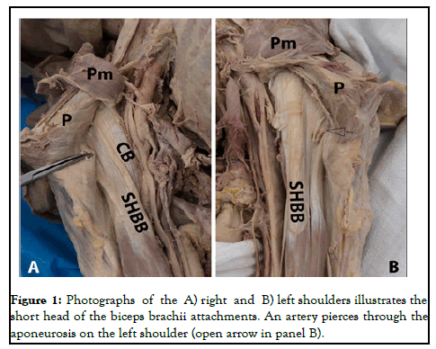
In this case, variants of the Short Head of the Biceps Brachii (SHBB) muscle were identified bilaterally during routine cadaveric dissection in a medical gross anatomy course of a 92 years male who had died from renal cell carcinoma. On the right shoulder, the aponeurotic tendon of the SHBB fused with the tendon of the coracobrachialis at one side and the tendon of the pectoralis major muscle near its insertion on the humerus at the other side. In addition, the SHBB tendon spanned 3.0 cm from the coracoid process to the shoulder capsule where it became indistinct from the shoulder capsule over the lesser tubercle of the humerus. The full length of the muscle was 35.3 cm from origin on the coracoid process to insertion, and 8.1 cm in diameter at its widest point. On the left shoulder, the SHBB tendon formed an aponeurosis fused with the tendon of the long head (Figure 1) [2-4].
The aponeurosis was 5.5 cm wide, spanning from the coracoid process of the scapula, across the intertubercular groove to the greater tubercle of the humerus. An arterial branch pierced through the tendon 2.9 cm from the origin. Additionally, a slip of tendon was present running obliquely to the remaining tendon. This may have been an accessory head, but had been cut by the student dissection team and thus could not be further characterized.
The full length of the muscle from origin on the coracoid process to insertion was 36.7 cm, and the widest point was 7.4 cm diameter [5,6].
Discussion
This case report is unique in the finding of a bilateral variation of the SHBB in which there is fusion of tendons within the shoulder region. There are numerous reports in the literature of accessory heads, but rare reports of fused heads or variations specifically of the SHBB, but none of these variations were present bilaterally. Such a variation has clinical implications in the event of shoulder injury, in which more profound injury may occur through tearing of the fused tendons. Furthermore, the fusion of the aponeurotic tendon of the SHBB on the right shoulder with the shoulder capsule may make the SHBB susceptible to injury in tears of the shoulder capsule itself. Likewise, the distribution of force across a wide aponeurosis, and even shared with other adjacent muscles, would alter the direction of force in the event of humeral or clavicular fracture. The broad aponeurosis would also necessitate modifying surgical approach to the anterior shoulder. Indeed, the one previous report of an aponeurotic tendon of the SHBB, similar to that on the left shoulder of this cadaver, was identified during a deltopectoral approach to the shoulder for hemiarthroplasty. In that previous case, the aponeurotic attachment was not as extensive, or involving other muscles groups, as on the right shoulder in the present case.
Identification of this second case of a similar aponeurotic attachment lends support to the clinical significance of this finding as a variation that is rare but not singularly unique, thus one that may be encountered in additional patients when using the deltopectoral approach.
Conclusion
Based on the fusion of tendons, it may appear that the shoulder would have less range of motion. However, comparisons with the anatomy of the biceps brachii in apes of the Hylobatidae family, which are brachiating apes, provide evolutionary insights into the significance of this variation. These apes, such as gibbons, are reported to have fused biceps brachii muscles rather than two separate heads. Further, they have chains of fused muscles within the upper limb, including attachments between the biceps brachii, coracobrachialis and pectoralis major muscles, as also seen in this case report. It has been hypothesized that these forelimb adaptations help transmit forces of arboreal locomotion. Therefore, rather than limiting range of movement, it is probable that these fused muscles are adaptive for distributing the force of overhead arm movements and permissive of a larger range of motion.
Acknowledgement
The author thanks the West Virginia University Human Gift Registry (WVUHGR) program for access to cadaveric materials used in this study.
References
- Balasubramanian A. Supernumerary head of biceps brachii. Int J Anat Var 2010;3:214-5.
- Sthapak E, Pasricha N, Siddiqui MS. Study on variations in the origin of biceps brachii with special thrust on its innervations. Int J Anat Res 2016;4(1):1994-7.
- Radhika PM, Anupama K, Shetty S. A cadaveric study of accessory heads of biceps brachii in South Indian population. Int J Anat Res 2017;5(1):3491-5.
- Katsuki S, Terayama H, Tanaka R. Variation in origin of the long head of the biceps brachii tendon in a cadaver: A case report. Med 2018;97: 20-3.
- Collett DJ, Karovalia S, Bokor DJ. An unusual variant of the origin of the short head of biceps brachii. Int J Anat Var Vol. 2018;11(4):134.
- Michilsens F, Vereecke EE, D'Aout K, et al. Functional anatomy of the gibbon forelimb: Adaptations to a brachiating lifestyle. J Anat. 2009;215(3):335-54.


