Bilateral supernumerary renal arteries in a single cadaver
Subhasis Chakraborty1, Saktipada Pradhan1, Mithu Paul1 and Sudeshna Majumdar2*
1Department of Anatomy, Nilratan Sircar Medical College, Kolkata, West Bengal, India
2Department of Anatomy, North Bengal Medical College, Sushrutanagar, Darjeeling, West Bengal, India
- *Corresponding Author:
- Sudeshna Majumdar, MS, DNB
Professor and Head Department of Anatomy North Bengal Medical College Sushrutanagar
Darjeeling 734012, West Bengal, India
Tel: +91 943 3007363
E-mail: sudeshnamajumdar.2007@rediffmail.com
Date of Received: January 14th, 2016
Date of Accepted: December 30th, 2016
Published Online: January 24th, 2017
© Int J Anat Var (IJAV). 2016; 9: 64–66.
[ft_below_content] =>Keywords
renal arteries, abdominal aorta, duplication, mesonephric, nephrectomy
Introduction
A single renal artery to each kidney is present in approximately 70% of individuals. Renal arteries take origin from the aorta bilaterally just below the superior mesenteric artery. The arteries vary in their level of origin and obliquity. Near the renal hilum each artery divides into an anterior and a posterior division which divide into segmental arteries supplying the renal vascular segments [1].
Embryologically these arteries are the lateral splanchnic branches of abdominal aorta [2].
One or two accessory renal arteries are present in 20-30% individuals [1]. They frequently arise, (especially on the left side) usually from the aorta, above or below the main artery (the former is slightly more often), enter the kidney above or below the renal hilum; if below, the vessel passes anterior to the ureter and may cause hydronephrosis by obstructing the ureter. On the right side, such artery passes usually anterior to the inferior vena cava. These arteries are regarded as the persistent embryonic lateral splanchnic arteries [1].
The objective of the case report is to bring awareness about the variations in the blood supply of the kidney for any surgical or diagnostic intervention.
Case Report
During routine dissection for undergraduate students, in the department of Anatomy, NRS Medical College, Kolkata, West Bengal, India, bilateral variations were detected in the renal arteries of a 65-year-old male cadaver in December, 2014. The structures concerned were dissected properly for minute observation and relevant photographs were taken.
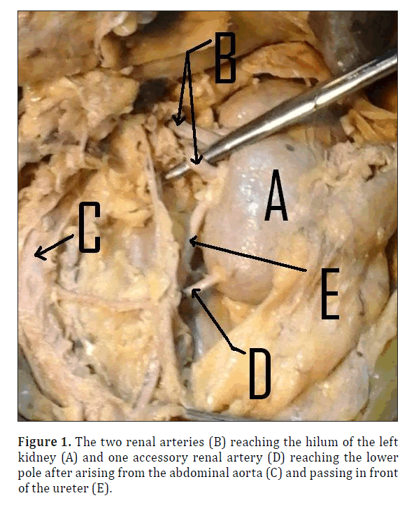
On the left side, there were two renal arteries reaching the hilum of the left kidney from the abdominal aorta. There was one accessory renal artery to reach the lower pole of the right kidney. It arose from the abdominal aorta and passed in front of the ureter (Figure 1).
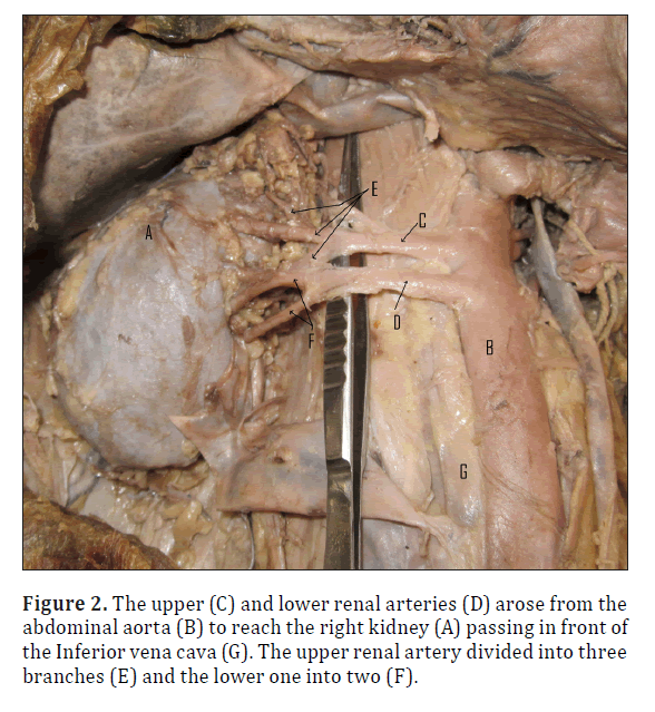
On the right side, there were again two renal arteries arose from the abdominal aorta. They passed in front of the inferior vena cava to reach the right kidney. The upper one divided into three segmental branches. The upper two branches reached the upper pole of the right kidney and the lower one entered the hilum. The inferior renal artery divided into two segmental branches to enter the hilum. So, there were five arteries to enter the right kidney (Figure 2).
Discussion
Embryological Consideration: Embryologically, renal arteries are the lateral splanchnic branches of the dorsal aortae. These lateral splanchnic branches supply the mesonephros, metanephros, gonads (testis and ovary) and the suprarenal gland. All these structures develop, in whole or in part from the intermediate mesenchyme or mesonephric ridge [2]. Accessory renal arteries are common in 20–30% of individuals, usually arising from the aorta above or below the main renal artery. The variation in the number of arteries is because of persistence of lateral splanchnic arteries or due to the persistence of blood supply from lower level than normal [2,3]. The kidneys begin their development in the pelvic cavity. During further development, they ascend to their final position in the lumbar region. When the kidneys are located in the pelvis, they are supplied by the branches of internal iliac or common iliac arteries. While the kidneys ascend to lumbar region, their arterial supply also shifts from common iliac artery to the abdominal aorta [3].
General Discussion: Rusu , in 2006, reported a case with bilateral doubled renal arteries - on the right side superior and inferior hilar renal arteries and on the left side superior hilar and inferior polar renal arteries emerged from the abdominal aorta. Bilateral doubled testicular arteries were found in the same case [4].
Majumdar et al., in 2013, described a case with one accessory renal artery emerged from the left side of the abdominal aorta, passed laterally, above the main left renal artery and reached the upper pole of the left kidney to enter the parenchyma of the organ [5].
According to David Sykes when there were many accessory renal arteries, the superior accessory artery is a separate segmental artery and the inferior accessory artery is a separate lower segmental artery [6]. Supernumerary renal arteries, if unidentified and accidentally injured during renal surgery may cause avascular necrosis of the supplied renal segment [6]. Knowledge of the variations in the renal arteries is important for urologists, radiologists and surgeons in general [3].
Multiple renal arteries are a common finding in renal angiograms and are more common in the aorta and renal vessels in the donor population subjected to angiography, but do not pose any serious risk or contraindication to renal donation [3]. Multiple renal arteries occurred bilaterally in 10.2% of donors and unilaterally in 20.8%, a total incidence of 31%. There was a higher incidence of vascular-related complications following transplantation of kidneys with multiple renal arteries. Attention is drawn to the need for careful technique in identification of multiple renal vessels, especially aberrant vessels, at the time of donor nephrectomy and also to the different techniques available for anastomosis of multiple renal arteries in kidney transplant recipients [3,7]. Besides, the failure to properly anastomose all arteries can lead to graft necrosis, graft rupture, segmental renal infarction, and postoperative hypertension and calyceal fistula formation [3,8].
Knowledge of the varied anatomy of the renal vessels facilitates a safe approach to the kidneys in trauma management. The varied and unpredictable anatomy of the renal vasculature requires prompt change when the normal approach fails to provide access to the vessels. In such cases, the colon should be mobilized promptly. Operative exposure and control of the renal vessels through a transabdominal retroperitoneal (TARP) approach has been advocated for emergency management of renal trauma [3].
Conclusion
This case report will enhance our knowledge regarding the blood supply of kidneys and may be of help to clinicians performing invasive techniques, vascular and other types of renal surgeries and in cases of trauma.
Acknowledgement
We are grateful to Professor (Dr.) Hasi Dasgupta, Dr. Manotosh Banerjee and all other members of the Department of Anatomy, Nilratan Sircar Medical College, Kolkata, India, for their kind co-operation for this study.
References
- Standring S, Healy JC, Borley NR, Collins P, Wigley C, eds . Gray’s Anatomy, The Anatomical Basis of Clinical Practice. 40th Ed., Elsevier Churchill Livingstone. 2008; 1231.
- Williams PL, Bannister HL, Martin MB, Collins P, Dyson M, Dussek EJ, Ferguson W, Glabella JM, eds. Gray’s Anatomy. 38th ., Churchill and Livingstone, London. 1995; 316-318 and 1557- 1558.
- Mir NS1, Ul Hassan A, Rangrez R, Hamid S, Sabia, Tabish SA, Iqbal, Suhaila, Masarat, Rasool Z. Bilateral duplication of renal vessels: anatomical, medical and surgical perspective. Int J Health Sci (Qassim). 2008; 2(2): 179–185.
- Rusu MC. Human bilateral renal and testicular arteries with a left testicular arterial arch around the left renal vein. Rom J Morphol Embryol. 2006;47(2):197-200.
- Majumdar S, Bhattacharya K, Kundu P, Mandal S, Roy H. Case report: Variations in the renal and testicular arteries. Indian Journal of Basic & Applied Medical Research. 2013; 2(6): 502-506.
- Sykes D. The arterial supply of the human kidney with special reference to accessory renal arteries. Br J Surg. 1963 Jan;50:368-374.
- Coen LD, Raftery AT. Anatomical variations of the renal arteries and renal transplantation. Clin Anat.1982;6:425-432.
- Ganesan KS, Huilgol AK, Sundar S, Chandrashekhar V, Prasad S, Raviraj KG. Management of multiple arteries in renal transplantation. Transplant Proc. 1994;26:2101–2102.
Subhasis Chakraborty1, Saktipada Pradhan1, Mithu Paul1 and Sudeshna Majumdar2*
1Department of Anatomy, Nilratan Sircar Medical College, Kolkata, West Bengal, India
2Department of Anatomy, North Bengal Medical College, Sushrutanagar, Darjeeling, West Bengal, India
- *Corresponding Author:
- Sudeshna Majumdar, MS, DNB
Professor and Head Department of Anatomy North Bengal Medical College Sushrutanagar
Darjeeling 734012, West Bengal, India
Tel: +91 943 3007363
E-mail: sudeshnamajumdar.2007@rediffmail.com
Date of Received: January 14th, 2016
Date of Accepted: December 30th, 2016
Published Online: January 24th, 2017
© Int J Anat Var (IJAV). 2016; 9: 64–66.
Abstract
Renal arteries are a pair of lateral branches from abdominal aorta. Normally each kidney receives one renal artery. However, accessory renal arteries can also exist. The normal renal arteries enter the kidney through its hilum whereas the accessory renal arteries might enter the kidney through the hilum or through the surfaces of the kidney. While doing the routine dissection for the undergraduate students, in the dept. of Anatomy of NRS, Medical College of Kolkata, India bilateral supernumerary renal arteries were found in a 65-year-old male cadaver. It is important to be aware that accessory renal arteries are end arteries; therefore, if an accessory artery is ligated or damaged, the part of kidney supplied by it is likely to become ischemic. A sound knowledge of variations of blood vessels is important during operative, diagnostic and endovascular procedures in the abdomen.
-Keywords
renal arteries, abdominal aorta, duplication, mesonephric, nephrectomy
Introduction
A single renal artery to each kidney is present in approximately 70% of individuals. Renal arteries take origin from the aorta bilaterally just below the superior mesenteric artery. The arteries vary in their level of origin and obliquity. Near the renal hilum each artery divides into an anterior and a posterior division which divide into segmental arteries supplying the renal vascular segments [1].
Embryologically these arteries are the lateral splanchnic branches of abdominal aorta [2].
One or two accessory renal arteries are present in 20-30% individuals [1]. They frequently arise, (especially on the left side) usually from the aorta, above or below the main artery (the former is slightly more often), enter the kidney above or below the renal hilum; if below, the vessel passes anterior to the ureter and may cause hydronephrosis by obstructing the ureter. On the right side, such artery passes usually anterior to the inferior vena cava. These arteries are regarded as the persistent embryonic lateral splanchnic arteries [1].
The objective of the case report is to bring awareness about the variations in the blood supply of the kidney for any surgical or diagnostic intervention.
Case Report
During routine dissection for undergraduate students, in the department of Anatomy, NRS Medical College, Kolkata, West Bengal, India, bilateral variations were detected in the renal arteries of a 65-year-old male cadaver in December, 2014. The structures concerned were dissected properly for minute observation and relevant photographs were taken.
On the left side, there were two renal arteries reaching the hilum of the left kidney from the abdominal aorta. There was one accessory renal artery to reach the lower pole of the right kidney. It arose from the abdominal aorta and passed in front of the ureter (Figure 1).
On the right side, there were again two renal arteries arose from the abdominal aorta. They passed in front of the inferior vena cava to reach the right kidney. The upper one divided into three segmental branches. The upper two branches reached the upper pole of the right kidney and the lower one entered the hilum. The inferior renal artery divided into two segmental branches to enter the hilum. So, there were five arteries to enter the right kidney (Figure 2).
Discussion
Embryological Consideration: Embryologically, renal arteries are the lateral splanchnic branches of the dorsal aortae. These lateral splanchnic branches supply the mesonephros, metanephros, gonads (testis and ovary) and the suprarenal gland. All these structures develop, in whole or in part from the intermediate mesenchyme or mesonephric ridge [2]. Accessory renal arteries are common in 20–30% of individuals, usually arising from the aorta above or below the main renal artery. The variation in the number of arteries is because of persistence of lateral splanchnic arteries or due to the persistence of blood supply from lower level than normal [2,3]. The kidneys begin their development in the pelvic cavity. During further development, they ascend to their final position in the lumbar region. When the kidneys are located in the pelvis, they are supplied by the branches of internal iliac or common iliac arteries. While the kidneys ascend to lumbar region, their arterial supply also shifts from common iliac artery to the abdominal aorta [3].
General Discussion: Rusu , in 2006, reported a case with bilateral doubled renal arteries - on the right side superior and inferior hilar renal arteries and on the left side superior hilar and inferior polar renal arteries emerged from the abdominal aorta. Bilateral doubled testicular arteries were found in the same case [4].
Majumdar et al., in 2013, described a case with one accessory renal artery emerged from the left side of the abdominal aorta, passed laterally, above the main left renal artery and reached the upper pole of the left kidney to enter the parenchyma of the organ [5].
According to David Sykes when there were many accessory renal arteries, the superior accessory artery is a separate segmental artery and the inferior accessory artery is a separate lower segmental artery [6]. Supernumerary renal arteries, if unidentified and accidentally injured during renal surgery may cause avascular necrosis of the supplied renal segment [6]. Knowledge of the variations in the renal arteries is important for urologists, radiologists and surgeons in general [3].
Multiple renal arteries are a common finding in renal angiograms and are more common in the aorta and renal vessels in the donor population subjected to angiography, but do not pose any serious risk or contraindication to renal donation [3]. Multiple renal arteries occurred bilaterally in 10.2% of donors and unilaterally in 20.8%, a total incidence of 31%. There was a higher incidence of vascular-related complications following transplantation of kidneys with multiple renal arteries. Attention is drawn to the need for careful technique in identification of multiple renal vessels, especially aberrant vessels, at the time of donor nephrectomy and also to the different techniques available for anastomosis of multiple renal arteries in kidney transplant recipients [3,7]. Besides, the failure to properly anastomose all arteries can lead to graft necrosis, graft rupture, segmental renal infarction, and postoperative hypertension and calyceal fistula formation [3,8].
Knowledge of the varied anatomy of the renal vessels facilitates a safe approach to the kidneys in trauma management. The varied and unpredictable anatomy of the renal vasculature requires prompt change when the normal approach fails to provide access to the vessels. In such cases, the colon should be mobilized promptly. Operative exposure and control of the renal vessels through a transabdominal retroperitoneal (TARP) approach has been advocated for emergency management of renal trauma [3].
Conclusion
This case report will enhance our knowledge regarding the blood supply of kidneys and may be of help to clinicians performing invasive techniques, vascular and other types of renal surgeries and in cases of trauma.
Acknowledgement
We are grateful to Professor (Dr.) Hasi Dasgupta, Dr. Manotosh Banerjee and all other members of the Department of Anatomy, Nilratan Sircar Medical College, Kolkata, India, for their kind co-operation for this study.
References
- Standring S, Healy JC, Borley NR, Collins P, Wigley C, eds . Gray’s Anatomy, The Anatomical Basis of Clinical Practice. 40th Ed., Elsevier Churchill Livingstone. 2008; 1231.
- Williams PL, Bannister HL, Martin MB, Collins P, Dyson M, Dussek EJ, Ferguson W, Glabella JM, eds. Gray’s Anatomy. 38th ., Churchill and Livingstone, London. 1995; 316-318 and 1557- 1558.
- Mir NS1, Ul Hassan A, Rangrez R, Hamid S, Sabia, Tabish SA, Iqbal, Suhaila, Masarat, Rasool Z. Bilateral duplication of renal vessels: anatomical, medical and surgical perspective. Int J Health Sci (Qassim). 2008; 2(2): 179–185.
- Rusu MC. Human bilateral renal and testicular arteries with a left testicular arterial arch around the left renal vein. Rom J Morphol Embryol. 2006;47(2):197-200.
- Majumdar S, Bhattacharya K, Kundu P, Mandal S, Roy H. Case report: Variations in the renal and testicular arteries. Indian Journal of Basic & Applied Medical Research. 2013; 2(6): 502-506.
- Sykes D. The arterial supply of the human kidney with special reference to accessory renal arteries. Br J Surg. 1963 Jan;50:368-374.
- Coen LD, Raftery AT. Anatomical variations of the renal arteries and renal transplantation. Clin Anat.1982;6:425-432.
- Ganesan KS, Huilgol AK, Sundar S, Chandrashekhar V, Prasad S, Raviraj KG. Management of multiple arteries in renal transplantation. Transplant Proc. 1994;26:2101–2102.


