C1 lateral mass screw fixationexposure
Mehmet Senoglu1*, Yakup Gumusalan2
1Kahramanmaras Sutcu Imam University, Faculty of Medicine, Department of Neurosurgery, Kahramanmaras, Turkey.
2Kahramanmaras Sutcu Imam University, Faculty of Medicine, Department of Neurosurgery and Anatomy , Kahramanmaras, Turkey.
- *Corresponding Author:
- Mehmet Senoglu, MD
Assistant Professor of Neurosurgery, Kahramanmaras Sutcu Imam University, Faculty of Medicine, Department of Neurosurgery, 46050 Kahramanmaras, Turkey.
Tel: +90 344 2212337
Fax: +90 344 2212371
E-mail: mehmetsenoglu@hotmail.com
Date of Received: November 4th, 2008
Date of Accepted: January 19th, 2009
Published Online: January 22nd, 2009
© IJAV. 2009; 2: 15–16.
[ft_below_content] =>Keywords
atlas, screw, lateral mass, C1, fixation
Introduction
A number of recent studies have described the feasibility and safety of C1 lateral mass screw placement. It is very important to have an understanding of surrounding anatomy at the C1 lateral mass screw site to avoid injury to the spinal cord, vertebral artery, C2 ganglion or nerve root, internal carotid artery, or hypoglossal nerve [1]. The use of C1 lateral mass screws avoids the technical difficulties in obtaining the required transarticular screw trajectory in patients with unreduced atlanto-axial joints, cervical congenital anomalies, thoracic kyphosis, or obesity [2,3] and offers similar mechanical stability to transarticular screws [4]. It seems to be a safer procedure when compared with transarticular screw fixation in the treatment of atlanto-axial disease [3]. The current safe application of C1 lateral mass requires the guidance of lateral fluoroscopic imaging [2,3,4].
On the lateral mass of a dried human cadaveric cervical spine (C1) the points for screw entry and trajectories for placement of lateral mass screws were determined, as applied in the technique described by Goel and Laheri.
Case Report
In this report, we used an adult C1 spine to describe the surgical technique for placement of C1 lateral mass screws.
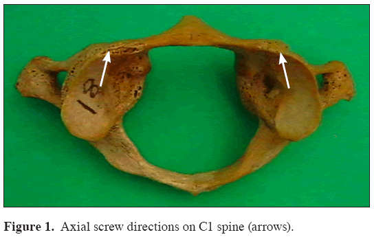
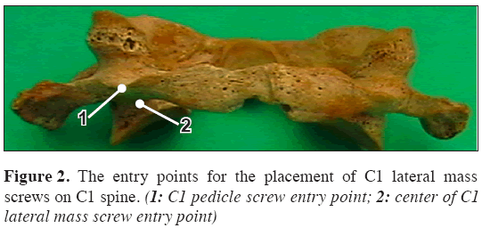
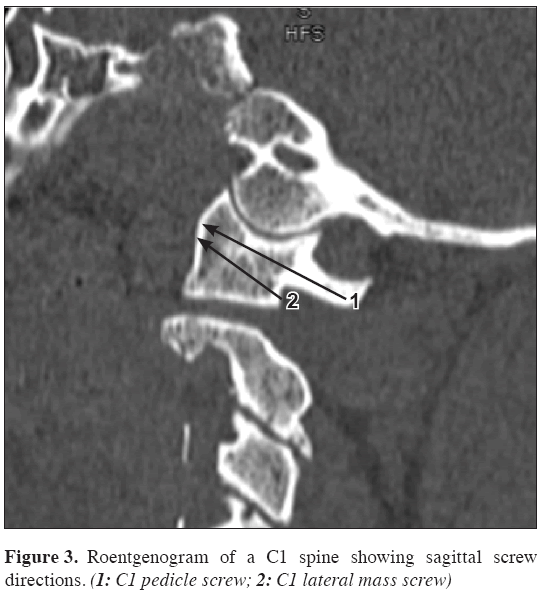
Surgical technique: The patient should be laid in the prone position with the head fixed in a Mayfield head frame. A standard posterior approach should be performed with fixationexposure of the inferior aspect of the C1 arch and the C2 spinous process and lamina. The vertebral artery should be then carefully identified coursing along its groove, usually at 8-12 mm from the midline on the superior aspect of the atlas, and dissected off the bone. There is no need to dissect the artery from the surrounding veins. The medial surface of the C1 lateral mass and the C1 foramen transversarium should be identified using a no 4 Penfield dissector, providing a proper anatomical orientation for the screw entry point and trajectory (Figure 1). Any venous bleeding can be easily stopped using hemostatic agents and gentle packing. An entry point directly on the posterior arch of the atlas, at the midpoint between the medial edge of the foramen transversarium and the medial surface of the lateral mass is then chosen (Figure 2). In some patients a starting point on the lateral mass just below the C1 posterior arch should be preferred because of a too thin posterior arch (Figure 2). A starting point using a 3 mm burr was made on the posterior arch, while the assistant protected the vertebral artery with a Penfield dissector. The C1 lateral mass should then be perforated using a hand-held drill with a 2.5 mm K-wire, under fluoroscopy, stopping just short to the anterior arch cortex, with the drill bit angled medially 5-10 degrees (Figure 1). After drilling the pilot hole and feeling the trajectory with a blunt 1.0 mm probe, the hole should be tapped and a 3.5 mm screw inserted (Figure 3). Generally the screw measures 18-30 mm in length, depending on the size of the lateral mass and whether the starting point is on or below the posterior arch (Figure 3).
Discussion
Goel and Laheri reported on the use of C1 lateral mass screws in 1994, which allowed rigid fixation without the limitation caused by the variable course of the vertebral artery. This technique has been combined with either C2 lateral mass or pedicle screws to provide a safe and effective means for stabilizing the upper cervical spine [5,6]. Biomechanical studies have shown this technique to provide similar stability to the C1-C2 transarticular screw technique [7,8].
This is a technically demanding procedure with significant risk of injury to the neighboring neurovascular structures [4,9]. The hypoglossal nerve and internal carotid artery are at risk on the anterior surface of the lateral mass of the atlas. The hypoglossal nerve lies 2 to 3 mm lateral to the middle of the anterior aspect of the C1 lateral mass. The internal carotid artery varies in location, but can lie within 1 mm of the screw exit point [4,8,9].
The internal carotid artery and hypoglossal nerve lie over the anterior aspect of the lateral mass of the atlas and are at risk from bicortical C1 lateral mass screws. Unicortical screws would reduce the risk of injury to these neurovascular structures; on the other hand, no data are available on the relative strength of unicortical versus bicortical C1 lateral mass screws [8].
Insertion of C1 lateral mass screws is a safe technique for segmental fixation of the C1. These screws can be especially valuable when a multilevel segmental construct that includes the occipitocervical junction or subaxial cervical spine is desired [10].
The authors conclude that C1 lateral mass screw fixation is technically possible for stabilizing the upper cervical spine. A better understanding of the geometry and quantitative dimensions of the C1 lateral mass anatomy is necessary to evaluate the safety and efficacy of this technique.
References
- Christensen DM, Eastlack RK, Lynch JJ, Yaszemski MJ, Currier BL. C1 anatomy and dimensions relative to lateral mass screw placement. Spine. 2007; 32: 844–848.
- Harms J, Melcher RP. Posterior C1-C2 fusion with polyaxial screw and rod fixation. Spine. 2001; 26: 2467–2471.
- Wang M, Samudrala S. Cadaveric morphometric analysis for atlantal lateral mass screw placement. Neurosurgery. 2004; 54: 1436–1439.
- Hong X, Dong Y, Yunbing C, Qingshui Y, Shizheng Z, Jingfa L. Posterior screw placement on the lateral mass of atlas: An anatomic study. Spine. 2004; 29: 500–503.
- Goel A. Treatment of basilar invagination by atlantoaxial joint distraction and direct lateral mass fixation. J Neurosurg Spine. 2004; 1: 281–286.
- Al-Barbarawi M, Sekhon LH. Protection of the C1 posterior arch in atlantal lateral mass fixation: technical case report. J Clin Neurosci. 2005; 12: 59–61.
- Melcher RP, Puttlitz CM, Kleinstueck FS, Lotz JC, Harms J, Bradford DS. Biomechanical testing of posterior atlantoaxial fixation techniques. Spine. 2002; 27: 2435–2440.
- Eck JC, Walker MP, Currier BL, Chen Q, Yaszemski MJ, An KN. Biomechanical comparison of unicortical versus bicortical C1 lateral mass screw fixation. J Spinal Disord Tech. 2007; 20: 505–508.
- Ebraheim NA, Misson JR, Xu R, Yeasting RA. The optimal transarticular C1-2 screw length and the location of the hypoglossal nerve. Surg Neurol. 2000; 53: 208–210.
- Vilela MD, Jermani C, Braga BP. C1 lateral mass screws for posterior segmental stabilization of the upper cervical spine and a new method of three-point rigid fixation of the C1-C2 complex. Arq Neuropsiquiatr. 2006; 64: 762–767.
Mehmet Senoglu1*, Yakup Gumusalan2
1Kahramanmaras Sutcu Imam University, Faculty of Medicine, Department of Neurosurgery, Kahramanmaras, Turkey.
2Kahramanmaras Sutcu Imam University, Faculty of Medicine, Department of Neurosurgery and Anatomy , Kahramanmaras, Turkey.
- *Corresponding Author:
- Mehmet Senoglu, MD
Assistant Professor of Neurosurgery, Kahramanmaras Sutcu Imam University, Faculty of Medicine, Department of Neurosurgery, 46050 Kahramanmaras, Turkey.
Tel: +90 344 2212337
Fax: +90 344 2212371
E-mail: mehmetsenoglu@hotmail.com
Date of Received: November 4th, 2008
Date of Accepted: January 19th, 2009
Published Online: January 22nd, 2009
© IJAV. 2009; 2: 15–16.
Abstract
C1 lateral mass screw placement is a powerful technique for segmental control of the C1 vertebra. In this report, we describe surgical technique for placement of C1 lateral mass screws, on our C1 specimen. C1 lateral mass is anatomically ideally suited for screw fixation to achieve C1-C2 arthrodesis. C1 lateral mass screw fixation is a safe technique and can be performed to achieve rigid and immediate stabilization.
-Keywords
atlas, screw, lateral mass, C1, fixation
Introduction
A number of recent studies have described the feasibility and safety of C1 lateral mass screw placement. It is very important to have an understanding of surrounding anatomy at the C1 lateral mass screw site to avoid injury to the spinal cord, vertebral artery, C2 ganglion or nerve root, internal carotid artery, or hypoglossal nerve [1]. The use of C1 lateral mass screws avoids the technical difficulties in obtaining the required transarticular screw trajectory in patients with unreduced atlanto-axial joints, cervical congenital anomalies, thoracic kyphosis, or obesity [2,3] and offers similar mechanical stability to transarticular screws [4]. It seems to be a safer procedure when compared with transarticular screw fixation in the treatment of atlanto-axial disease [3]. The current safe application of C1 lateral mass requires the guidance of lateral fluoroscopic imaging [2,3,4].
On the lateral mass of a dried human cadaveric cervical spine (C1) the points for screw entry and trajectories for placement of lateral mass screws were determined, as applied in the technique described by Goel and Laheri.
Case Report
In this report, we used an adult C1 spine to describe the surgical technique for placement of C1 lateral mass screws.
Surgical technique: The patient should be laid in the prone position with the head fixed in a Mayfield head frame. A standard posterior approach should be performed with fixationexposure of the inferior aspect of the C1 arch and the C2 spinous process and lamina. The vertebral artery should be then carefully identified coursing along its groove, usually at 8-12 mm from the midline on the superior aspect of the atlas, and dissected off the bone. There is no need to dissect the artery from the surrounding veins. The medial surface of the C1 lateral mass and the C1 foramen transversarium should be identified using a no 4 Penfield dissector, providing a proper anatomical orientation for the screw entry point and trajectory (Figure 1). Any venous bleeding can be easily stopped using hemostatic agents and gentle packing. An entry point directly on the posterior arch of the atlas, at the midpoint between the medial edge of the foramen transversarium and the medial surface of the lateral mass is then chosen (Figure 2). In some patients a starting point on the lateral mass just below the C1 posterior arch should be preferred because of a too thin posterior arch (Figure 2). A starting point using a 3 mm burr was made on the posterior arch, while the assistant protected the vertebral artery with a Penfield dissector. The C1 lateral mass should then be perforated using a hand-held drill with a 2.5 mm K-wire, under fluoroscopy, stopping just short to the anterior arch cortex, with the drill bit angled medially 5-10 degrees (Figure 1). After drilling the pilot hole and feeling the trajectory with a blunt 1.0 mm probe, the hole should be tapped and a 3.5 mm screw inserted (Figure 3). Generally the screw measures 18-30 mm in length, depending on the size of the lateral mass and whether the starting point is on or below the posterior arch (Figure 3).
Discussion
Goel and Laheri reported on the use of C1 lateral mass screws in 1994, which allowed rigid fixation without the limitation caused by the variable course of the vertebral artery. This technique has been combined with either C2 lateral mass or pedicle screws to provide a safe and effective means for stabilizing the upper cervical spine [5,6]. Biomechanical studies have shown this technique to provide similar stability to the C1-C2 transarticular screw technique [7,8].
This is a technically demanding procedure with significant risk of injury to the neighboring neurovascular structures [4,9]. The hypoglossal nerve and internal carotid artery are at risk on the anterior surface of the lateral mass of the atlas. The hypoglossal nerve lies 2 to 3 mm lateral to the middle of the anterior aspect of the C1 lateral mass. The internal carotid artery varies in location, but can lie within 1 mm of the screw exit point [4,8,9].
The internal carotid artery and hypoglossal nerve lie over the anterior aspect of the lateral mass of the atlas and are at risk from bicortical C1 lateral mass screws. Unicortical screws would reduce the risk of injury to these neurovascular structures; on the other hand, no data are available on the relative strength of unicortical versus bicortical C1 lateral mass screws [8].
Insertion of C1 lateral mass screws is a safe technique for segmental fixation of the C1. These screws can be especially valuable when a multilevel segmental construct that includes the occipitocervical junction or subaxial cervical spine is desired [10].
The authors conclude that C1 lateral mass screw fixation is technically possible for stabilizing the upper cervical spine. A better understanding of the geometry and quantitative dimensions of the C1 lateral mass anatomy is necessary to evaluate the safety and efficacy of this technique.
References
- Christensen DM, Eastlack RK, Lynch JJ, Yaszemski MJ, Currier BL. C1 anatomy and dimensions relative to lateral mass screw placement. Spine. 2007; 32: 844–848.
- Harms J, Melcher RP. Posterior C1-C2 fusion with polyaxial screw and rod fixation. Spine. 2001; 26: 2467–2471.
- Wang M, Samudrala S. Cadaveric morphometric analysis for atlantal lateral mass screw placement. Neurosurgery. 2004; 54: 1436–1439.
- Hong X, Dong Y, Yunbing C, Qingshui Y, Shizheng Z, Jingfa L. Posterior screw placement on the lateral mass of atlas: An anatomic study. Spine. 2004; 29: 500–503.
- Goel A. Treatment of basilar invagination by atlantoaxial joint distraction and direct lateral mass fixation. J Neurosurg Spine. 2004; 1: 281–286.
- Al-Barbarawi M, Sekhon LH. Protection of the C1 posterior arch in atlantal lateral mass fixation: technical case report. J Clin Neurosci. 2005; 12: 59–61.
- Melcher RP, Puttlitz CM, Kleinstueck FS, Lotz JC, Harms J, Bradford DS. Biomechanical testing of posterior atlantoaxial fixation techniques. Spine. 2002; 27: 2435–2440.
- Eck JC, Walker MP, Currier BL, Chen Q, Yaszemski MJ, An KN. Biomechanical comparison of unicortical versus bicortical C1 lateral mass screw fixation. J Spinal Disord Tech. 2007; 20: 505–508.
- Ebraheim NA, Misson JR, Xu R, Yeasting RA. The optimal transarticular C1-2 screw length and the location of the hypoglossal nerve. Surg Neurol. 2000; 53: 208–210.
- Vilela MD, Jermani C, Braga BP. C1 lateral mass screws for posterior segmental stabilization of the upper cervical spine and a new method of three-point rigid fixation of the C1-C2 complex. Arq Neuropsiquiatr. 2006; 64: 762–767.


