Comparative study of the fingerprint between two mammalian species and the human
Received: 02-Dec-2022, Manuscript No. ijav-22-5683; Editor assigned: 05-Dec-2022, Pre QC No. ijav-22-5683 (PQ); Accepted Date: Dec 24, 2022; Reviewed: 19-Dec-2022 QC No. ijav-22-5683; Revised: 24-Dec-2022, Manuscript No. ijav-22-5683 (R); Published: 31-Dec-2022, DOI: DOI:10.37532/1308-4038.15(12).230
Citation: Nora Abdulaziz A, Haifa M.A.L.K. Comparative Study of the Fingerprint between Two Mammalian Species and the Human. Int J Anat Var. 2022;15(12):235-239.
This open-access article is distributed under the terms of the Creative Commons Attribution Non-Commercial License (CC BY-NC) (http://creativecommons.org/licenses/by-nc/4.0/), which permits reuse, distribution and reproduction of the article, provided that the original work is properly cited and the reuse is restricted to noncommercial purposes. For commercial reuse, contact reprints@pulsus.com
Abstract
This study aimed to examine the morphological and electronic description of the ventral surface of the finger’s skin or what is known by fingerprint using fingerprint ink, fingerprint scanner and scanning electron microscopy in humans (Homo sapines), baboon (Papio hamadryas) and squirrels (Tamias striatus). The fingerprint is considered as the most common biometric feature. Fingerprints are based on the formation of papillary projections deployed on the surface of the skin take different forms and seem separated by grooves called groove lines.
The present results showed that fingerprints of the five fingers of the human are differentiated from each to another; and this is what has been observed when taking fingerprints of the five fingers using fingerprint ink. But, it was found that all the fingerprints of the baboon monkey are characterized by one form and cannot be classified under any form of known fingerprint types. On the other hand, the ink fingerprinting showed a difficulty of taking a fingerprint for each finger of squirrel because of their hand small size. Thus, this study showed that there is no clear fingerprint in the monkey and squirrels, although the morphological structure of the hand in humans, monkeys and squirrels is similar to the simple differences in the use of hand in dealing with the surrounding environment. The scanning electron microscopy showed that there are numerous differences in the ventral surface of finger’s skin between samples of these three mammalian species.
Keywords
Ossification; Superior sagittal sinus; Parietal lateral lacunae
INTRODUCTION
All apparent parts of the skin of the hand are covered with a series of prominent lines containing ridges and grooves where the fingerprint pattern is given, according on these ridges and grooves, scientists classify fingerprints into many varieties and collected under the main branch of which types of sub-branches to make it easier to track. These ridges and grooves that cover palm skin and fingers in some objects are given by a pattern known as fingerprints. Fingerprints are unique in living organisms and no two individuals can have the same fingerprints. Fingerprints experts classified fingerprints according to specific classifications, based on the shape of the fingerprints and the number of lines in it. There are three major patterns of fingerprints, the first pattern is known as the spiral or whorls; the second is the loops pattern and the third pattern is the arches.
Sheridan [1] pointed out that the location of the fingerprint requires knowledge of fingerprints. There are three types of fingerprints (visible, plastic and latent), it is easy to locate the visible fingerprint because it is visible to the naked eye as a fingerprint impression or blood or ink. Plastic is less common, such as leaving a person on the soap and showing a threedimensional impression. The underlying fingerprint is the most common and takes more effort to locate it because it is not visible as the presence of residues of natural oils on the person’s finger. Also, he showed that there is other types of surfaces are porous surfaces such as wood, the fingerprint is determined using iodine smoke and the latter type is the composite surfaces. The fingerprints are then photographed and uploaded and finally compared using the fingerprint identification system via the computer database.
Haraksim [2] pointed out that the rapid growth of child fingerprints leads to a significant displacement of fine detail points between samples of the same finger acquired a few years apart. This displacement results in a reduction in the accuracy of fingerprint recognition systems when the signal and test sample deviates at the same time. This effect is known as biometric aging.
Si [3] recorded the differences that occur when the fingerprint is taken or printed for the same parts of the finger as they vary in intensity, and the distinctive features and fine details of the fingerprints are scattered. They proposed algorithms to record fingerprint density and relied on fingerprint databases.
Ahmed & Osman [4] pointed out that fingerprints are important biological variables that serve human and genetic morphology as well as forensic medicine. They aimed to analyze the differences in the parameters of finger surface in terms of the density of the ridges of the skin and assess the possibility of application in the identification of sex. The density of fingerprint ridges was assessed for three different regions (radial, zygomatic and proximal) for all ten fingers of each sample. The results showed that fingerprints can be useful for identifying the gender of Sudanese individuals based on the fingerprint intensity of the ridges. Moreover, the density of the ridges can be considered as the morphological characteristic of individual differences in forensic medicine.
Huynh & Halamek [5] provide study in fingerprint analysis, where fingerprints are unique impressions of whorls, arches and loops, and as a result fingerprints are used as means of identifying individuals. Fingerprint samples also have unique chemical contents that can be used to extract valuable information about the persons involved when criminal offenses occur. They included several methods for extracting information on the chemical content of the underlying fingerprints such as spectroscopy, sensors and other biometrics that adopted advanced techniques when analyzing fingerprints based on chemical content.
Oktem [6] examined the sex identification of fingerprints in different populations, examining the density of fingerprints in 118 women and 88 men of students aged between 17 and 28 years using the inking technique. They found that specifically, the fingerprint density in the radial and zygote area was much greater than the end of these regions. They concluded that the density of fingerprint ridges could be used by a forensic examination to determine sex.
Gupta & Gupta [7] explained that algorithm technology is effective for dividing the fingertips from an image and identifying it through the corresponding index, such as the index finger, middle finger, small finger of the right and left hand, and the use of geometric and spatial characteristics to identify the fingertips.
Hamilton [8] pointed out that fingerprints were used by forensic evidence to identify individuals for more than 100 years. Fingerprint recognition quickly what has become the best evidence of forensic detection, fingerprints leave through the ridges of the skin in the surfaces of the internal hands, friction of the ridges of the skin consists of ridges and small grooves. Those interested in fingerprinting study the properties of skin friction, which is the main area of comparison in recognition using a methodology known as analysis, comparison, division and verification, the investigator can compare a marker left at the scene as a known symbol until it reaches the result.
Arora & Garg [9] noted that the extensive use of biological information for the purpose of verifying the identity of a person has raised considerable interest in the reliability and efficiency of biometric systems. Fingerprinting is one of the most promising methods among biometrics techniques and has been used for identity detection since the 19th century. The sample fingerprint is based on a structured device that works in two different ways (registration and identification). The purpose of the registration method is to create a database. In this method, the registered fingerprint is taken and treated in three stages. It is read, processed, and then extracted in the database. In the recognition method, the fingerprint is exposed to the same steps of recording method. Finally, the results are compared with their characteristics.
Zhang & Girault [10] noted that fingerprint analysis is one of the most important means of personal identification and is also evidence found at the crime scene for forensic purposes. It is found that using an electro-mechanical scanning microscope; researchers can visualize the precise composition of human fingerprints on porous and non-porous surfaces by mixing with a silver dye or a precipitated metal technology. The electromagnetic microscope allows chemical activity investigators to take a picture of the fingerprint surface with high accuracy and sensitivity to reveal the underlying fingerprints of the fingers.
Warman & Ennos [11] suggested that the fingerprints improve the grip of the primates, but the efficiency of the ridges depends on the type of behavior of the friction exposed to the skin. The ridges are effective when increasing contact with solids, but friction decreases with rubber materials, reducing the contact area. Their results showed that the fingertips had a behavior on the rubber material more than the solid, the friction decreased at a high natural strength and the friction was high when the finger grip was flat against the plate range and that is when the contact area is greater. Shear stress was greater in high pressures, suggesting a dynamic membrane between the skin and the surfaces. Fingerprints reduce the contact area by one-third compared with flat skin reducing friction.
Kucken & Newell [12] explained that fingerprints have been used as a means of identification for more than 2,000 years and have been scientifically studied by anthropologists and biologists. They tried to demonstrate that the emergence of fingerprint patterns is due to the instability of the twist in the basal cell layer of the embryo’s skin, suggesting that this stress caused by the resistance of grooves and wrinkles in the growth of the layer basal and platelet decline occur during the formation of the ridges.
This study aimed to examine the morphological and electronic description of the ventral surface of fingerprint using ink technique, fingerprint scanner and scanning electron microscopy in humans (Homo sapines), baboon (Papio hamadryas) and squirrels (Tamias striatus).
MATERIALS AND METHODS
Experimental samples: The organisms selected for this study were two species of mammals which found in the Kingdom of Saudi Arabia (baboon and palm squirrel). From 5 to 7 samples of bot species were obtained from farms in Al-Ahsa, Qatif and Dammam for squirrel specimens and from the South Taif for baboon monkeys; considering that these specimens are adult males. The authors have been keen to dissect the lowest number of specimens and select species present in many numbers in our environment in order to preserve the nature of wildlife in Saudi Arabia. As for the human hand was obtained, which is the subject of research from the Department of Anatomy, Faculty of Medicine, Imam Abdulrahman bin Faisal University.
Fingerprinting by ink and scanner: The ventral surface of finger’s skin in the experimental samples was fingerprinting using the fingerprint ink and then photographed. Also, these ventral surfaces of fingers were scanned by digital scanner to diagnose the differentiation of fingerprints between these samples.
Scanning electron microscopy of finger’s skin: The distinctive structures were examined and studied of the visible hand and hand palm (Fingerprint) by using Scanning Electron Microscope which is available in the laboratories of the Institute of Research and Medical Consultancy at the University of Imam Abdulrahman bin Faisal, the parts to be examined were cut into small pieces not more than 0.5 cm × 0.5 cm and install it directly with a primer 4% glutaraldehyde 0.2% dissolve phosphate regulator solution (pH 7.2) was placed in the refrigerator at 4 °C for four hours. Then, the sample well washed three times in a row 0.2 dissolve the phosphate regulator solution and transfer the sample to the secondary fixer 1% osmic acid in a phosphateregulated solution monomer at pH 7.2 and stored in the refrigerator at a temperature 4 °C for four hours. The sample was washed in 0.2 Mall of the phosphate regulator and then remove the water from the sample series with progressive concentrations of alcohol ethyl even absolute alcohol then the sample was dried in the air and put on a small base metal Stubs and proven by material colloidal adhesive is graphic (carbon) and covered with gold and using the air separation unite then examined by using the scanning electron microscopy [13].
RESULTS
Human fingers: The ventral surface skin of the human fingers contains frictions in the form of a series of dark lines of distinctive shape grooves that give the fingerprint unique to the fingers. Fingerprints of the five fingers of the human are differentiated from each to another; and this is what has been observed when taking fingerprints of the five fingers using fingerprint ink. The fingerprint of the thumb, index, middle and ring fingers all take shape with a normal spindle type (Normal whorl) except for the small finger and that’s what the direction of the ridges, the shape of the spherical shape normal type simple in composition and towards it to give a full circle differ in its direction from left to right and vice versa. The small fingerprint takes the ring shape loop different from the rest of the fingerprints, in which they have two rings of semi-detached ridges that give a semi-circular shape (Figures 1-2).
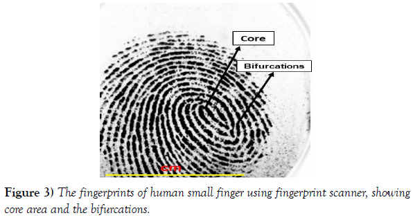
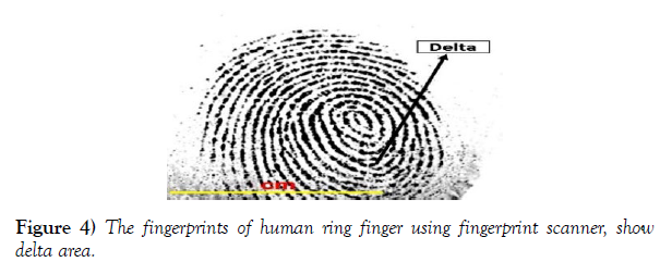
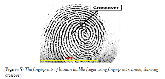
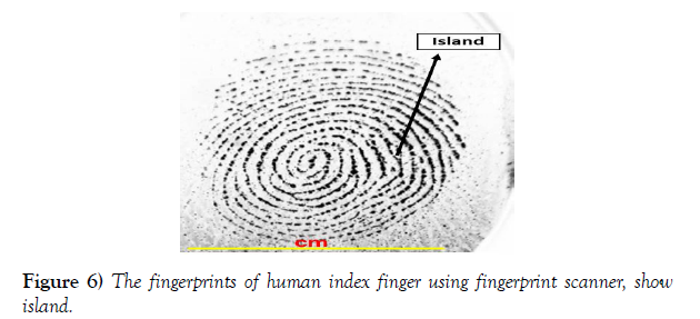
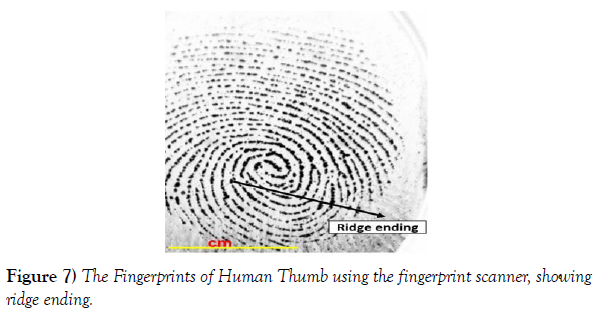
Scanning photographs by scanner showed that all fingerprints of human fingers are more clearly identified. It was found that the small finger clearly characterized by the area of the nucleus or the core area, which usually located in the middle of the fingerprint. The core area is also characterized by the end of the ridges and the shape of the curved ridges. Also there are bifurcations of the ridges, the point that represents the division of the ridge into two parts (Figure 3). Fingerprint of the ring finger characterized by the delta areas, they are usually located in two locations, at the bottom left and on the right side of the fingerprint, forming the center of a series of rectangular ridges (Figure 4). There is a distinct area known as the crossover area, it is observed when taking the fingerprint of middle finger by a fingerprint scanner in which is appeared as two ridges do not reflect each of another (Figure 5). In the index finger, the island area was observed when elongated ridges occupying an intermediate area between these two ridges (Figure 6). The fingerprint of the thumb is characterized by the ridge endings, in which they are the end points of the ridges (Figure 7).
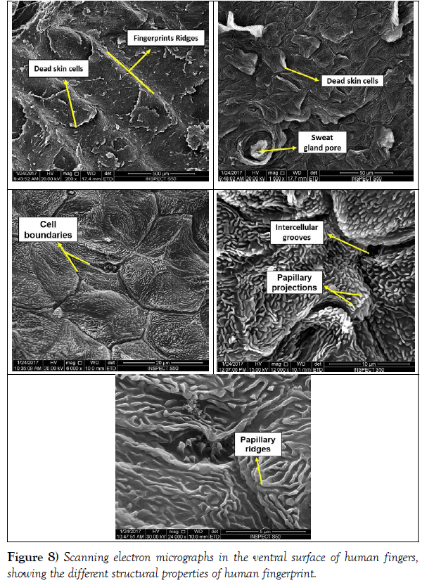
By using the scanning electron microscope, it was found that the ventral skin of the finger consists of fingerprint ridges which take the semi-elongated form. Also, it was noted the presence of dead skin cells. Dead skin cells appeared as regular overlapping layers on the fingerprint, which represent the corneal layer of the skin surface (Figure 8). There is also a distinctive structure on the skin surface known as the pore of sweat gland. It has a rounded shape that works to transfer sweat from the sweat gland to the surface of the skin, and it is surrounded by all dead skin cells (Figure 8). Each dead cell is separated from the other neighboring dead cell by the cell boundaries. It gives the distinctive shape of the overlapping layers of dead cells (Figure. 8). By enlarging the cell boundaries, they appeared as intercellular grooves with the appearance of the papillary projections (Figure 8). The papillary projections are extensions of the dermis on the surface of the skin where they appear when enlarged in the form of heights know as papillary ridges. They are small and numerous and they often increase with age (Figure 8).
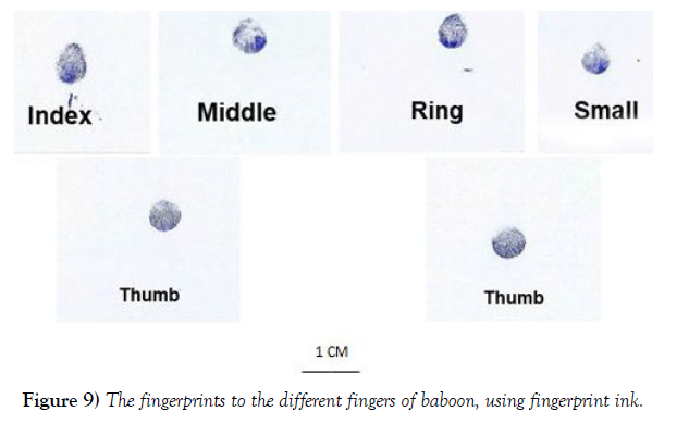
Baboon fingers: The fingerprints of a baboon were taken by using fingerprint ink to distinguish their types, it was observed that all the fingerprints of the baboon monkey are characterized by one form and cannot be classified under any form of known fingerprint types. They are the form of semi-throat located in the middle of the thumbprint of the elongate somewhat in towards one (Figure 9). When examining the fingerprints of the baboon monkey with a fingerprint scanner, the device does not recognize the fingerprints of the baboon monkey.
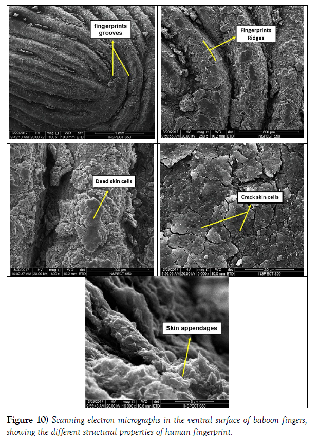
The skin of finger palm in baboon was examined by scanning electronic microscope. It showed the presence of reductions known as fingerprints grooves; it is somewhat cloudy and through which the sweat is distributed and prevents the fingers from being slippery (Figure 10). These grooves give ridges of fingerprints with a distinctive protruding contiguous and slightly curved shape at its top (Figure 10). Thee ridges are covered from all sides with a very thick layer of dead skin cells; it takes the form of a non- regular layer (Figure 10). When examining the other area of the ventral part of baboon finger, a layer of cracked skin cells was observed, this layer does not reach the stage of dead cells until its completely defrauded (Figure 10). When increasing the strength of the electronic magnification, it was found that the ventral surface of the finger is free of the dead and cracked skin cells and contains skin appendages (Figure 10).
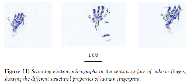
Squirrel fingers: The whole hand fingerprint of squirrel was taken using fingerprinting ink and there’s difficulty of taking a fingerprint for each finger because of the small size of the squirrel hand. It has not seen the emergence of any ridges or represents of the hand fingerprint and this was also observed when we take the squirrel hand fingerprints and fingers by fingerprint scanner (Figure 11).
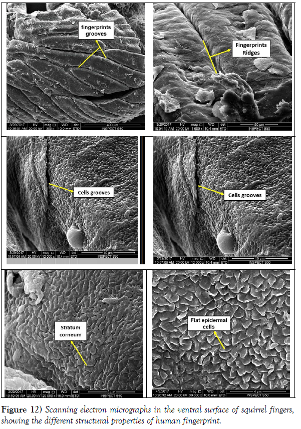
The scanning electron microscope examination to the finger skin of squirrel showed that the fingerprints grooves takes the shape completely submerged and gives the fingerprint its view of the fingerprints ridges with different dimensions (Figure 12). The ridges of fingerprints show overlapping layers of dead skin cells. These cells are large with simple borders and their borders are known as cells grooves (Figure 12). After the enlargement of the dead cells several times, it is evident that they consist of several overlapping and small cells called the stratum corneum and it was found that these cells take a flat shape known as flat epidermal cells (Figure 1-2).
DISCUSSION
The present work studied human, monkey and squirrel fingers to compare between the fingerprint structure of human and these two mammalian species. When fingerprints are taken with fingerprints it turns out that all fingers have a normal spindle-shaped fingerprint except the small finger, It is a form that made us distinguish normal fingerprint fusiform is a simple composition and in the ridges when you follow the direction of those ridges. It was noted that it gives a full circle, while small finger takes the shape of any of the two separate ridges which give form semi-circular. Skin of human fingers is thick and showed a distinctive presence of ridges and grooves, while skin of fingers in squirrel differs from the human finger’s skin and baboon because of its thin and lack of clarity of the skin ridges and grooves. These results is in consistence with the previous studies in which they demonstrated that fingerprints leave through the ridges of the skin in the surfaces of the internal hands; friction of the ridges of the skin consists of ridges and small grooves [12] [8].
Fingerprints of a baboon using a sloop inking cannot be classified under the known fingerprint forms appeared almost throat and has a one-way, while the squirrel did not seen the emergence of ridges or gullies represent the fingerprint. The inking method is the simplest way through which to see characteristic fingerprint for each individual [6] [3]. After taking the fingerprints of several individuals with simple algebra and studying the density of their fingerprints and how they are useful in determining gender by forensic examination, this method is also used to identify the identity of individuals after taking evidence from crime scenes [14] [5].
By using fingerprint scanner for human hand, the device showed the characteristic details of the stamp, as its hills formed areas with different names such as the nucleus located in the middle of the fingerprint and the ramparts of the divided ridges and triangle area, which is the center of a series of triangular ridges and also the crossing area marked by crossing two ridges to each other and finally the area of the island with long distance and the end of the ridges. While the fingerprint of the baboon monkey and squirrel did not show any scientific character using fingerprint scanner. This indicates that the fingerprints of the baboon monkey and the squirrel are only ridges and grooves of a virtual form only and it cannot be recorded and read as in human. These results agree with many other authors [9, 7, 3]. When examining the fingerprints of children, the biometric age resulting from the displacement of the details of the fingerprint is shown. The development of a microscopic image model is the entrance of the biometric chick [2].
When examining the human finger with the scanning electron microscopy, the protruding fingerprints and dead skin cells, which are covered by an overlapping layer (the corneal layer), deriving their characteristic form from the dead cell boundaries, which also play a role in the formation of the cellular grooves. The baboon monkey fingerprint, with its somewhat grooved grooves, gives the ridges long and curved shape when you make and cover a very thick layer of dead cells and a few cracked cells. This differs from the finger grooves in the squirrel in which they are completely hollow and the ridges have different dimensions and are covered with layers of dead skin cells. When the dead cell is enlarged on the fingerprints of the human finger several times, it spreads heavily installation of special known the papillary projections [15]. It represents the basal layer of the epidermis and appears in the form of papillae, while these papillary projections do not appear in the fingerprint of the baboon monkey and squirrel. This confirms that the fingerprint of the baboon monkey and the squirrel is a fingerprint with visible ridges and not an extension of the dermis.
The sweat that emerges from the pores in the fingerprint ridges has a role in sliding the finger pads and controlling the grip as mentioned [11] [16-17]. The friction of hills with different surfaces enhances the function of touch and grip of the primates also facilitates the selection of food through the grip of friction during nutrition. The absence of clear fingerprints in the baboon monkey and palm squirrel, although the anatomical structure of the hand in humans, baboon and squirrels are similar with the different hands used in dealing with the surrounding environment and this reflects that the human hand more sensitive to touch than that of the monkey and squirrel.
ETHICAL CONSIDERATION
The authors ensured that all ethical and other basic principles were considered. A permission to carry-out this study was given by the Institutional Review Board Committee of the Deanship of Scientific Research, with the IRB number (IRB-PGS-2017-10-076).
References
- Sheridan S. Techniques for collecting and analyzing Fingerprints. Forensic Sci. 2013; 1-5.
- Haraksim R, Galbally J, Beslay L. Fingerprint growth model for mitigating the ageing effect on children’s fingerprints matching. Pat Recog. 2019; 88:614-628.
- Si X, Feng J, Yuan B, Zhou J. Dense registration of fingerprints. Pattern Recognit. 2017; 63:87-101.
- Ahmed AA, Osman S. Topological variability and sex differences in fingerprint ridge density in a sample of the Sudanese population. J Forensic Leg Med. 2016; 42:25-32.
- Huynh C, Halamek J. Trends in fingerprint analysis. Trends Analyt Chem. 2016; 82:328-336.
- Oktem H, Kurkcuoglu A, Pelin IC, Yazici AC, Aktas G, et al. Sex differences in fingerprint ridge density in a Turkish young adult population: A sample of Baskent University. J Forensic Leg Med. 2015; 32:34-38.
- Gupta P, Gupta P. An efficient slap fingerprint segmentation and hand classification algorithm. Neurocompiting. 2014; 142:464-477.
- Hamilton I. Fingerprints. Encyclopedia of Forensic Sciences. 2013; 346-351.
- Arora K, Garg P. A Quantitative survey of various fingerprint enhancement techniques. Int J Comput Appl. (0975-8887), 2011; 28(5):24-29.
- Zhang M, Girault HH. SECM for imaging and detection of latent fingerprints. Analyst. 2009; 134:25-30.
- Warman PH, Ennos AR. Fingerprints are unlikely to increase the friction of primate fingerpads. J Exp Biol. 2009; 212(13):2015-2021.
- Kucken M, Newell AC. Fingerprint formation. J Theor Biol. 2005; 235(1):71-83.
- Bancroft JD, Gamble M. Theory and Practice of Histological Techniques. 5th ed. Charchill livingstone, London. 2002.
- Mattson P, Bilous P. Coomassie brilliant blue: an excellent reagent for the enhancement of faint bloody fingerprints. Can Soc Forensic Sci J. 2014; 47(1):20-36.
- Sangiorgi S, Manelli A, Protasoni M, Raspanti M. The collagenic of human digital skin seen by scanning electron microscopy after Ohtani maceration technique. Ann Anat. 2005; 187(1):13-22.
- Adams MJ, Johnson SA, Lefevre P, Levesque V, Hayward V, et al. Finger pad friction and its role in grip and touch. J R Soc Interface. 2012; 10:1-19.
- Smith JM, Smith AC. An investigation of ecological correlates with hand and foot morphology in callitrichid primates. Am J Phys Anthropol. 2013; 152(4):447-458.
Indexed at, Google Scholar, Crossref
Indexed at, Google Scholar, Crossref
Indexed at, Google Scholar, Crossref
Indexed at, Google Scholar, Crossref
Indexed at, Google Scholar, Crossref
Indexed at, Google Scholar, Crossref
Indexed at, Google Scholar, Crossref
Indexed at, Google Scholar, Crossref
Indexed at, Google Scholar, Crossref
Indexed at, Google Scholar, Crossref
Indexed at, Google Scholar, Crossref
Indexed at, Google Scholar, Crossref
Indexed at, Google Scholar, Crossref


