Congenital Malformation of the Aortic Arch and its Branches With no Documented Clinical Symptoms: A Post-Mortem Case Study
Received: 08-Apr-2021 Accepted Date: May 04, 2021; Published: 11-May-2021, DOI: 10.37532/1308-4038.14(5).124-125
Citation: Fulop BD, Tamas A, Reglodi D, et al. Congenital malformation of the aortic arch and its branches with no documented clinical symptoms: a postmortem case study. Int J Anat Var. 2021;13(5):33-34.
This open-access article is distributed under the terms of the Creative Commons Attribution Non-Commercial License (CC BY-NC) (http://creativecommons.org/licenses/by-nc/4.0/), which permits reuse, distribution and reproduction of the article, provided that the original work is properly cited and the reuse is restricted to noncommercial purposes. For commercial reuse, contact reprints@pulsus.com
Abstract
The congenital malformation of the aortic arch and its branches is documented from a 45-year-old male cadaver in our case report. We recorded for the first time a right aortic arch with the following branches: left subclavian artery, common trunk of the carotid arteries, right subclavian artery. The descending aorta was located on the right side pressing the esophagus to the left. Great vessel variations are clinically important for planning surgical procedures and they could be associated with further congenital malformations, commonly with congenital heart disease. Different great vessel variations have a wide range of association with other congenital malformations, therefore, early detection with fetal echocardiography is of great importance. The described great vessel malformation was not associated with cardiac malformations nor did contribute to the death of the person. According to the accessible medical history it did not cause clinical symptoms to the patient.
Keywords
Right aortic arch; Abnormal branching of the aortic arch; Common origin of the carotid arteries; Post-mortem study.
Introduction
Anatomical variations of the aortic arch and its branches could be of clinical interest for planning surgical procedures (e.g. catheterization) and also because they can predict other congenital malformations [1]. The normal development of these vessels initiates from embryonic aortic arches. Normally the left 4th aortic arch connects the aortic sac to the left dorsal aorta to form the definitive aortic arch. The left dorsal aorta continues to the unpaired dorsal aorta, these two structures forming the definitive descending aorta [2]. The branches of a normal left aortic arch could be quite variable. The right brachiocephalic trunk, left common carotid artery, left subclavian artery pattern is the most common, but there are several other branching patterns known, adding up to about 20% of the normal population [3]. Hereby we report the case of a complex malformation of the aorta and its branches without other associated malformations.
Case Report
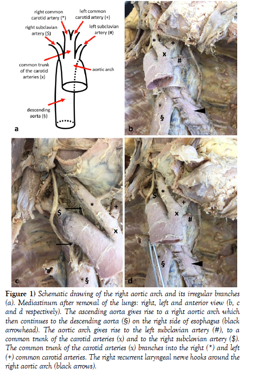
The cause of death of the 45-year-old male cadaver was a consequence of his late-stage T-cell lymphoma. During the dissection, we found a right aortic arch with the descending aorta also placed on the right side of the vertebrae pressing the esophagus to the left side (Figure 1). The first branch of the right aortic arch was the left subclavian artery taking its course in front of the esophagus and trachea. The second branch was the common trunk of the common carotid arteries followed by the right subclavian artery (Figure 1).
Figure 1) Schematic drawing of the right aortic arch and its irregular branches (a). Mediastinum after removal of the lungs: right, left and anterior view (b, c and d respectively). The ascending aorta gives rise to a right aortic arch which then continues to the descending aorta (§) on the right side of esophagus (black arrowhead). The aortic arch gives rise to the left subclavian artery (#), to a common trunk of the carotid arteries (x) and to the right subclavian artery ($). The common trunk of the carotid arteries (x) branches into the right (*) and left (+) common carotid arteries. The right recurrent laryngeal nerve hooks around the right aortic arch (black arrows).
Discussion
We present an anatomical variation where the right aortic arch has the following three branches: left subclavian artery, common trunk of the common carotid arteries, right subclavian artery. We presume that in this case the aortic arch develops from the aortic sac, right 4th aortic arch and right dorsal aorta; the left subclavian artery from the left 4th aortic arch, left dorsal aorta and left 7th intersegmental artery; the right subclavian artery from the right 7th intersegmental artery. The arising of the common trunk of the carotid arteries could be the result of the approach and subsequent fusion of the two 3rd aortic arches.
This branching pattern with a subclavian artery, common trunk of the common carotid arteries and the other subclavian artery is mentioned in the literature with a normal left aortic arch. It could be found in 0.16% of the population based on the work of Natsis et al. [3], although it is not to be confused with the truncus bicaroticus, which additionally contains also the origin of the right subclavian artery and is much more prevalent [4].
Separately both the right aortic arch and this branching pattern (from a left aortic arch) are known anatomical variations, but we have not found the combined appearance of these two congenital variations in the literature.
Clinical importance of great vessel variations is that they could be detected by fetal echocardiography from the 12th gestation week. Associated with other intra- or extracardiac malformations it could be advised to perform prenatal karyotyping or surgery; however, postnatal evaluation is sufficient in the case of the sole presence of a right aortic arch [1].
A right aortic arch is a relatively common variation in the population, continuing in the descending aorta either on the right or on the left side. In the dissected cadaver, it was running on the right side. Białowas et al. [5] has found a 0.01% prevalence of right aortic arch during the dissection of 1700 adult cadavers, however, Achiron et al. [6] described a 0.1%prevalence from 18000 prenatal ultrasound examinations. Right aortic arch could be associated with total situs inversus, or could stand as an individual mirror symptom. In the latter case the branching pattern of the right aortic arch is of clinical relevance - there are two major arising patterns with very different comorbidity ratio. The most typical branching pattern is the left common carotid artery followed by the right brachiocephalic trunk and the aberrantly originating left subclavian artery from a retroesophageal diverticulum. This pattern is rarely linked with congenital heart disease. The second is the mirror image branching, a complete mirroring of the normal anatomical pattern, where a left brachiocephalic trunk is followed by a right common carotid artery and a right subclavian artery. In this case the left dorsal aorta regresses and the right one forms the connection between the right 4th aortic arch and the singular dorsal aorta. This pattern is associated with congenital heart disease in 90% of the cases, often on the ground of a 22q11 microdeletion [7]. Therefore, it was important to evaluate whether further malformations are associated with the presented great vessel variation.
Conclusion
Dissection was carried out in the establishment of the Department of Anatomy, University of Pecs, Medical School. This work was supported by EFOP-3.6.3-VEKOP-16-15 2017-00008 “The role of neuro-inflammation in neurodegeneration: from molecules to clinics”; EFOP-3.6.1-16-2016-00004 “Comprehensive Development for Implementing Smart Specialization Strategies at the University of Pecs”; EFOP-3.6.3-VEKOP-16-2017-00009; New National Excellence Program of the Ministry of Human Capacities UNKP-16-4-IV and UNKP-17-2-II.
REFERENCES
- Zidere V, Tsapakis EG, Huggon IC, et al. Right aortic arch in the fetus. Ultrasound Obstet Gynecol. 2006;28:876-81.
- Sadler TW. Langman’s medical embryology, 12th ed. Wolters Kluwer Health/Lippincott Williams & Wilkins, Philadelphia; 2012.
- Natsis KI, Tsitouridis IA, Didagelos MV, et al. Anatomical variations in the branches of the human aortic arch in 633 angiographies: clinical significance and literature review. Surg Radiol Anat. 2009;31:319-23.
- Müller M, Schmitz BL, Pauls S, et al. Variations of the aortic arch - a study on the most common branching patterns. Acta Radiol. 2011;52:738-42.
- Bialowas J, Hreczecha J, Grzybiak M. Right-sided aortic arch. Folia Morphol. (Warsz) 2000;59:211-6.
- Achiron R, Rotstein Z, Heggesh J, et al. Anomalies of the fetal aortic arch: A novel sonographic approach to in-utero diagnosis. Ultrasound Obstet Gynecol. 2002;20:553-57.
- Hanneman K, Newman B, Chan F. Congenital Variants and Anomalies of the Aortic Arch. Radio Graphics. 2017;37:32-51.


