Corona mortis – a case report with surgical implicationsdistally
Sumathilatha Sakthivelavan1*, Sakthivelavan Duraipandian Sendiladibban2, Sharmila Aristotle3 and Anandarani Velamma Sivanandan1
1Department of Anatomy, Sri Ramachandra Medical college and Hospital, Porur, Chennai, India
2Department of Physiology, Hitech Medical College, Bhubaneshwar, India
3Department of Anatomy, SRM Medical College and Hospital, Porur, Chennai, India
- *Corresponding Author:
- Dr. Sumathilatha Sakthivelavan
Assistant Professor, Department of Anatomy, Sri Ramachandra Medical College and Hospital Porur, Chennai, India
Tel: +91 44 23718875
E-mail: sumathilathads@yahoo.co.in
Date of Received: November 21st, 2009
Date of Accepted: June 30th, 2010
Published Online: August 9th, 2010
© Int J Anat Var (IJAV). 2010; 3: 103–105.
[ft_below_content] =>Keywords
corona mortis, obturator artery
Introduction
Obturator artery is normally a branch of the anterior division of the internal iliac artery. It has marked variations in its origin and course compared to all the other branches of the internal iliac artery. Abnormal or aberrant obturator artery is defined as the origin of obturator artery from the external iliac artery or its branches. It is actually the enlarged pubic branch of the inferior epigastric artery. Accessory obturator artery is the presence of an extra obturator artery in addition to the normal counterpart [1]. When both the normal and accessory obturator arteries are present with rich anastomoses at the obturator canal it is known as “corona mortis” or “crown of death”. It is actually the anastomosis between the pubic rami of the inferior epigastric and the obturator arteries [2]. The observation of corona mortis deserves mention in view of its surgical and radiological significance as partly revealed by its terminology itself.
Case Report
Corona mortis was observed in a 60-year-old female cadaver during dissection of pelvis for a thesis work. The pelvis was separated at the level of L4-L5 articulation. Then the pelvis was bisected longitudinally in the midline to enable detailed dissection. Red lead dye was prepared and injected into the common iliac artery after ligating the femoral artery to ensure the identification of the smaller vessels. Internal iliac artery was traced implicationsdistally to look for the origin of the obturator artery and it was followed till it reached the obturator foramen. External iliac artery and inferior epigastric artery were also dissected accessory obturator artery was identified. It was traced till the obturator foramen.
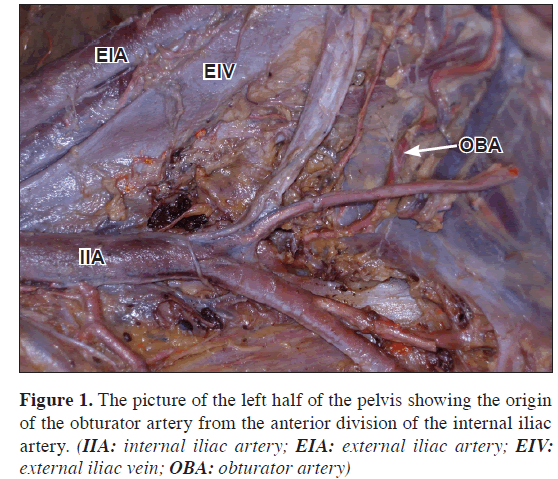
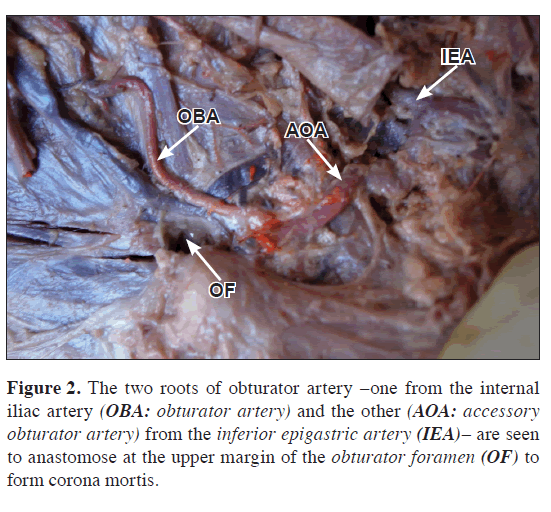
The left pelvic half showed the obturator artery arising from the anterior division of the internal iliac artery and the accessory obturator artery from the inferior epigastric artery (Figure 1). The two arteries were anastomosing at the upper margin of the obturator foramen and passing through the foramen as a single vessel (Figure 2). The average distance between the arch of the lacunar ligament and the anastomotic vessels was 13.1 mm. On the right side, no variations were found.
Discussion
Corona mortis is referred to as arterial or venous communication by various authors. In addition to venous corona mortis, some terms accessory obturator vein, accessory obturator artery, aberrant obturator artery, and anomalous origin of the obturator artery from the external iliac artery were used to refer to the corona mortis by some authors [2].
The accessory obturator artery has been described by various authors and it is found in 30-40% of specimens [3,4]. The high incidence indicates the need to be called as a normal variant rather than as abnormal obturator artery. In a cadaveric study the arterial connection between the internal and external iliac systems was found in 0% of 54 hemipelves [2], in another to be 34% of 55 hemipelves [5] and in an angiographic study to be 28.5% out of 98 hemipelves [6]. The disparity is most probably due to the difference in the size of the vessels considered as corona mortis.
Venous corona mortis has been described in more specimens than the arterial corona mortis in some studies [2,7]. However there was no venous corona noted in our specimen. In another study by Darmanis et al. on 80 pelvic halves, dissection by ilioinguinal approach revealed either venous or arterial corona mortis in 83% of the specimens [8].
The morphological patterns of corona mortis were described by Rusu et al. It was classified in three types as I-III. Our specimen corresponds to type I.3 of this classification where there was anastomosis of the obturator artery and the inferior epigastric artery [9].
The average distance between the arch of the lacunar ligament and the anastomotic vessels was described in a study to be 12.18 ± 3.55 mm [2]. In our specimen it was 13.1 mm which is similar to this finding. This distance is of importance in denoting the risk of anastomosing vessel from getting injured during surgery for femoral hernia. If it is medial to the femoral ring (where the distance from lacunar ligament is less), widening the neck of the hernial sac of femoral hernia during surgery may cause injury to the artery unless noted and ligated before cutting the lacunar ligament [3]. In the presence of corona mortis if the vessels are cut, there will be bleeding from both the ends since both the normal and the accessory obturator arteries are originating from 2 major vessels.
The ilioinguinal approach is appropriate for all fractures including the anterior wall, anterior column, and anterior column with posterior hemi transverse extension. These vessels that form the corona mortis may be disrupted as a result of superior pubic ramus fracture, and during ilioinguinal approach for acetabulum fractures [2]. The surgeon should exercise caution but not alter the surgical approach for fear of excessive hemorrhage [8]. If corona mortis presents, it should be ligated to prevent bleeding, which is difficult to control if it retracts into the pelvis [2].
In case of pelvic fracture, massive extraperitoneal hemorrhage may arise due to the presence of corona mortis. So endovascular specialists managing pelvic injury should keep corona mortis in mind as a potential source of prolonged and dangerous hemorrhage [10].
The obturator artery is formed as a result of uneven growth of the anastomosis between the internal iliac artery and the external iliac artery. All gradations may be found between normal arrangement and complete replacement of the original intrapelvic portion of the obturator artery by the pubic anastomosis. If both the rami are enlarged, then the condition of corona mortis results as seen in our specimen [4].
Corona mortis causes life-threatening emergency only if it is neglected and appropriate precautions are not taken. So every surgeon and endovascular specialists dealing with direct, indirect, femoral, or obturator hernias, superior pubic ramus fractures, and acetabulum fractures needs to be aware of these anastomoses (arterial, venous, or both) and avoid undue hemorrhage.
References
- Moore KL, Agur AMR. Thigh and gluteal region. In: Essential clinical anatomy. 3rd Ed., Philadelphia, Lippincott William and Wilkins. 2007; 332–397.
- Sarikcioglu L, Sindel M, Akyildiz F, Gur S. Anastomotic vessels in the retropubic region: corona mortis. Folia Morphol (Warsz). 2003; 62: 179–182.
- Gray H. The blood-vascular system. In: Anatomy, Descriptive and Surgical. 15th Ed., Pennsylvania, Courage Books. 1901; 564—565.
- Piersol GA. Human Anatomy. 9th Ed., Philadelphia, Lippincott.1930; 813–814.
- Tornetta P, Hochwald N, Levine R. Corona mortis. Incidence and location. Clin Orthop Relat Res. 1996; 329: 97–101.
- Karakurt L, Karaca I, Yilmaz E, Burma O, Serin E. Corona mortis: incidence and location. Arch Orthop Trauma Surg. 2002; 122: 163–164.
- Berberoglu M, Uz A, Ozmen MM, Bozkurt MC, Erkuran C, Taner S, Tekin A, Tekdemir I. Corona mortis: an anatomic study in seven cadavers and an endoscopic study in 28 patients. Surg Endosc. 2001; 15: 72–75.
- Darmanis S, Lewis A, Mansoor A, Bircher M. Corona mortis: an anatomical study with clinical implication in approaches to the pelvis and acetabulum. Clinical Anatomy. 2007; 20: 433–439.
- Rusu MC, Cergan R, Motoc AG, Folescu R, Pop E. Anatomical considerations on the corona mortis. Surg Radiol Anat. 2010; 32: 17–24.
- Daeubler B, Anderson SE, Leunig M, Triller J. Hemorrhage secondary to pelvic fracture: coil embolisation of an aberrant obturator artery. J Endovasc Ther. 2003; 10: 676–680.
Sumathilatha Sakthivelavan1*, Sakthivelavan Duraipandian Sendiladibban2, Sharmila Aristotle3 and Anandarani Velamma Sivanandan1
1Department of Anatomy, Sri Ramachandra Medical college and Hospital, Porur, Chennai, India
2Department of Physiology, Hitech Medical College, Bhubaneshwar, India
3Department of Anatomy, SRM Medical College and Hospital, Porur, Chennai, India
- *Corresponding Author:
- Dr. Sumathilatha Sakthivelavan
Assistant Professor, Department of Anatomy, Sri Ramachandra Medical College and Hospital Porur, Chennai, India
Tel: +91 44 23718875
E-mail: sumathilathads@yahoo.co.in
Date of Received: November 21st, 2009
Date of Accepted: June 30th, 2010
Published Online: August 9th, 2010
© Int J Anat Var (IJAV). 2010; 3: 103–105.
Abstract
Corona mortis is a Latin terminology which means “crown of death”. It indicates the presence of both the normal and variant obturator artery with extensive anastomoses. One such finding was observed in a female cadaver where the obturator artery was arising from the anterior division of the internal iliac artery and also the abnormal obturator artery from the inferior epigastric artery. These two vessels anastomosed at the upper border of the obturator foramen. This is referred to as the origin of the obturator artery by 2 roots in some studies and the abnormal obturator artery is in fact considered as a normal variant due to its common occurrence. Some studies have included such anastomosis between the veins also as corona mortis. The variant vessels are at risk of injury not only in groin surgery but also in orthopedic surgery and pelvic fractures, and are to be dealt with care during surgical procedures.
-Keywords
corona mortis, obturator artery
Introduction
Obturator artery is normally a branch of the anterior division of the internal iliac artery. It has marked variations in its origin and course compared to all the other branches of the internal iliac artery. Abnormal or aberrant obturator artery is defined as the origin of obturator artery from the external iliac artery or its branches. It is actually the enlarged pubic branch of the inferior epigastric artery. Accessory obturator artery is the presence of an extra obturator artery in addition to the normal counterpart [1]. When both the normal and accessory obturator arteries are present with rich anastomoses at the obturator canal it is known as “corona mortis” or “crown of death”. It is actually the anastomosis between the pubic rami of the inferior epigastric and the obturator arteries [2]. The observation of corona mortis deserves mention in view of its surgical and radiological significance as partly revealed by its terminology itself.
Case Report
Corona mortis was observed in a 60-year-old female cadaver during dissection of pelvis for a thesis work. The pelvis was separated at the level of L4-L5 articulation. Then the pelvis was bisected longitudinally in the midline to enable detailed dissection. Red lead dye was prepared and injected into the common iliac artery after ligating the femoral artery to ensure the identification of the smaller vessels. Internal iliac artery was traced implicationsdistally to look for the origin of the obturator artery and it was followed till it reached the obturator foramen. External iliac artery and inferior epigastric artery were also dissected accessory obturator artery was identified. It was traced till the obturator foramen.
The left pelvic half showed the obturator artery arising from the anterior division of the internal iliac artery and the accessory obturator artery from the inferior epigastric artery (Figure 1). The two arteries were anastomosing at the upper margin of the obturator foramen and passing through the foramen as a single vessel (Figure 2). The average distance between the arch of the lacunar ligament and the anastomotic vessels was 13.1 mm. On the right side, no variations were found.
Discussion
Corona mortis is referred to as arterial or venous communication by various authors. In addition to venous corona mortis, some terms accessory obturator vein, accessory obturator artery, aberrant obturator artery, and anomalous origin of the obturator artery from the external iliac artery were used to refer to the corona mortis by some authors [2].
The accessory obturator artery has been described by various authors and it is found in 30-40% of specimens [3,4]. The high incidence indicates the need to be called as a normal variant rather than as abnormal obturator artery. In a cadaveric study the arterial connection between the internal and external iliac systems was found in 0% of 54 hemipelves [2], in another to be 34% of 55 hemipelves [5] and in an angiographic study to be 28.5% out of 98 hemipelves [6]. The disparity is most probably due to the difference in the size of the vessels considered as corona mortis.
Venous corona mortis has been described in more specimens than the arterial corona mortis in some studies [2,7]. However there was no venous corona noted in our specimen. In another study by Darmanis et al. on 80 pelvic halves, dissection by ilioinguinal approach revealed either venous or arterial corona mortis in 83% of the specimens [8].
The morphological patterns of corona mortis were described by Rusu et al. It was classified in three types as I-III. Our specimen corresponds to type I.3 of this classification where there was anastomosis of the obturator artery and the inferior epigastric artery [9].
The average distance between the arch of the lacunar ligament and the anastomotic vessels was described in a study to be 12.18 ± 3.55 mm [2]. In our specimen it was 13.1 mm which is similar to this finding. This distance is of importance in denoting the risk of anastomosing vessel from getting injured during surgery for femoral hernia. If it is medial to the femoral ring (where the distance from lacunar ligament is less), widening the neck of the hernial sac of femoral hernia during surgery may cause injury to the artery unless noted and ligated before cutting the lacunar ligament [3]. In the presence of corona mortis if the vessels are cut, there will be bleeding from both the ends since both the normal and the accessory obturator arteries are originating from 2 major vessels.
The ilioinguinal approach is appropriate for all fractures including the anterior wall, anterior column, and anterior column with posterior hemi transverse extension. These vessels that form the corona mortis may be disrupted as a result of superior pubic ramus fracture, and during ilioinguinal approach for acetabulum fractures [2]. The surgeon should exercise caution but not alter the surgical approach for fear of excessive hemorrhage [8]. If corona mortis presents, it should be ligated to prevent bleeding, which is difficult to control if it retracts into the pelvis [2].
In case of pelvic fracture, massive extraperitoneal hemorrhage may arise due to the presence of corona mortis. So endovascular specialists managing pelvic injury should keep corona mortis in mind as a potential source of prolonged and dangerous hemorrhage [10].
The obturator artery is formed as a result of uneven growth of the anastomosis between the internal iliac artery and the external iliac artery. All gradations may be found between normal arrangement and complete replacement of the original intrapelvic portion of the obturator artery by the pubic anastomosis. If both the rami are enlarged, then the condition of corona mortis results as seen in our specimen [4].
Corona mortis causes life-threatening emergency only if it is neglected and appropriate precautions are not taken. So every surgeon and endovascular specialists dealing with direct, indirect, femoral, or obturator hernias, superior pubic ramus fractures, and acetabulum fractures needs to be aware of these anastomoses (arterial, venous, or both) and avoid undue hemorrhage.
References
- Moore KL, Agur AMR. Thigh and gluteal region. In: Essential clinical anatomy. 3rd Ed., Philadelphia, Lippincott William and Wilkins. 2007; 332–397.
- Sarikcioglu L, Sindel M, Akyildiz F, Gur S. Anastomotic vessels in the retropubic region: corona mortis. Folia Morphol (Warsz). 2003; 62: 179–182.
- Gray H. The blood-vascular system. In: Anatomy, Descriptive and Surgical. 15th Ed., Pennsylvania, Courage Books. 1901; 564—565.
- Piersol GA. Human Anatomy. 9th Ed., Philadelphia, Lippincott.1930; 813–814.
- Tornetta P, Hochwald N, Levine R. Corona mortis. Incidence and location. Clin Orthop Relat Res. 1996; 329: 97–101.
- Karakurt L, Karaca I, Yilmaz E, Burma O, Serin E. Corona mortis: incidence and location. Arch Orthop Trauma Surg. 2002; 122: 163–164.
- Berberoglu M, Uz A, Ozmen MM, Bozkurt MC, Erkuran C, Taner S, Tekin A, Tekdemir I. Corona mortis: an anatomic study in seven cadavers and an endoscopic study in 28 patients. Surg Endosc. 2001; 15: 72–75.
- Darmanis S, Lewis A, Mansoor A, Bircher M. Corona mortis: an anatomical study with clinical implication in approaches to the pelvis and acetabulum. Clinical Anatomy. 2007; 20: 433–439.
- Rusu MC, Cergan R, Motoc AG, Folescu R, Pop E. Anatomical considerations on the corona mortis. Surg Radiol Anat. 2010; 32: 17–24.
- Daeubler B, Anderson SE, Leunig M, Triller J. Hemorrhage secondary to pelvic fracture: coil embolisation of an aberrant obturator artery. J Endovasc Ther. 2003; 10: 676–680.


